Analysis on Synthesis of Silica Nanoparticles and its Effect on Growth of T. Harzianum & Rhizoctonia Species
Abstract
Silica nanoparticles have been prepared by sol-gel method and
peptization process. The silica nanoparticles were obtained by
hydrolysis of tetraethyl-orthosilicate (TEOS) in ethanol but variation
in parameters improved particles size from 290 nm to 85 nm in sol-gel
process and 190 nm by peptization process using nitric acid as
electrolyte. Detailed study on these nanoparticles was done by
characterizing nanoparticles using dynamic light scattering. Scanning
electron microscopy, X-ray diffraction, FTIR analysis and UV-visible
spectrophotometer. Antifungal effect of prepared nano silica was carried
out in Potato dextrose agar (PDA) media using concentration of
nanoparticle at 1wt% against trichoderma harzianum and rhizoctonia
solani.
Keywords: Silica nanoparticles; Temperature; TEOS; DLS; Trichoderma harzianum; Rhizoctonia solani
Introduction
Nanoparticles with size ranging between objects and microparticles
have attracted much attention. These particles with various specialized
functions not only deepen our understanding of nature but also serve the
basic for development of new advance technology. Nanoparticles are
characterized by size-dependent properties both size and surface effects
are important. By controlling these, it is possible in principle to
design materials of required optical, magnetic, elastic, chemical etc.
properties. There is increasing interest in the design and synthesis of
topological structures composed of monocrystals of various size and
shapes [1]. The sol-gel methods are the most general method of synthesis
of silica nanoparticles. Appetence in the sol-gel processing of ceramic
and glass materials started in the half of 1800s by Ebelman and
Graham's researches on silica gels [2]. The sol-gel technique is in
expensive and the silica gels manufactured are non-poisonous matters
[3-8]. Stober supplied monodisperse and nonporous silica spheres with
the hydrolysis of tetraethyl orthosilicate (TEOS) in strongly basic
medium. Stober and Fink promoted chemical reactions which checked up the
growth of spherical silica particles [9]. Bogush and Zukoski procure
monodispersed silica particles with controlled hydrolysis of TEOS in
ethanol [10]. Sung Kyoo Park provided silica nanoparticles from TEOS in
ethanol in order that controlled particle properties using a semi-batch
process [11]. Ryu had prepared amorphous silica by oxidation of silicon
[12-13].
In the present work, we suggested a novel method for preparing
amorphous silica nanoparticles through the sol-gel techniques at various
temperature range from 780C to 650°C to obtain best particle
size even we prepared silica nanoparticles through peptization process
also, using electrolyte nitric acid / hydrochloric acid / sulfuric acid
for comparative study on particle size analysis to obtain best particle
size below 100 nm. Another study on performed for fungal effect on human
body. Fungi are heterotrophic organisms which are able to reproduce
sexually as well as asexually. About 100 infectious fungal agents have
been detected in man. The mycoses or fungal infection can be of various
forms as:
- a) Superficial-seen on the skin, the hair, and nails.
b) Subcutaneous-infection reaching dermis or subcutaneous tissue
c) Systemic-internal organs infected deeply
d) Opportunistic-infection in immunocompromised patients
Several works have been highlighted very high bactericidal efficiency
on different microorganisms but there is no detail explanation for
antifungal testing with silica nanoparticles against fungus that causing
human disease. In this study, the effect of silica nanoparticle was
observed against Trichoderma Harzianum and Rhizoctonia Solani.
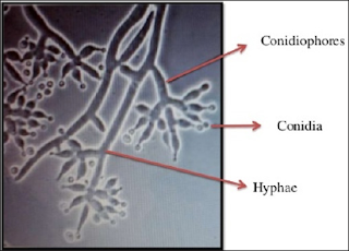
Trichoderma Harzianum
Morphology: Colonies were originally hyaline darkening to
white with green tufts in most species. The form was irregular which
rapidly grew and merged forming green carpet like appearance. The
conidiophores were branched and hyaline. Phialides were divergent and
flask-shaped. Conidia were generally green, smooth or roughened, ranged
in shape from globose to ellipsoidal, and were produced in slimy heads
Conidiophores were highly branched and demonstrated a pyramidal
arrangement.
Pathogenecity: Trichoderma harzianum infections are
opportunistic and develop in immunocompromised patients, such as
neutropenic cases and transplant recipients, as well as patients with
chronic renal failure, chronic lung disease, or amyloidosis.
Peritonitis, pulmonary, perihepatic, and disseminated infections have so
far been reported. A disseminated fungal infection was detected in the
postmortem examination of a renal transplant recipient and confirmed in
culture. The only other reported infection by this fungus caused
peritonitis in a diabetic patient. Trichoderma harzianum were identified
as causative agents of opportunistic fungal infections with increasing
frequency. Trichoderma harzianum isolates are reported predominantly to
cause health problems in humans ranging from localized infections to
fatal disseminated diseases. Thus, it is very necessary to delete the
negative pathogenic aspects of fungi by using proper treatment and care
[14].
Rhizoctonia Solani
Human infections caused by Rhizoctonia are very rare, and first case
report of keratitis by Rhizactonia was reported way back in 1977 [15].
Rhizoctonia solani is a most widely recognized strong saprophyte with a
great diversity of host plants. It is a first ever case of extensive
human mycosis caused by Rhizoctonia solani in a 65-year-old diabetic and
hypertensive farmer, with a history of head injury caused by fall of
mud wall. Necrotic material collected revealed septate fungal hyphae
with bacterial co-infection and after mycological study & diagnosis
it was confirmed the presence of rhizoctonia solani [16]. So, in this
paper we tested anti-fungal effect of silica nanoparticles against these
two fungi i.e. trichoderma harzianum and rhizoctonia solani in PDA
media.
Experimental
Materials
Tetraethyl orthosilicate (TEOS) were purchased from Sigma Aldrich
Chemicals Pvt Limited (India). Ethanol from Fischer Scientific (India),
and Ammonium hydroxide from Qualikems Fine Chemical Pvt. Ltd. (India)
was purchased. Potato dextrose agar (PDA) was purchased from HiMedia
Laboratories Pvt Limited (India). Instruments-UV 1800 Shimadzu UV
spectrophotometer, Malvern instruments-Zetasizer Nano S-90, Shimadzu
8400 spectrophotometer and Bruker D8 Focus X-Ray Diffractometer, Bruker
Universal materials tester (CETR UMT-3) SEM-Zeiss microscopy.
Methods
Synthesis of Silica Nanoparticles through Sol-Gel Method
Silica nanoparticles were prepared according to the well- known
Stober method by hydrolysis and condensation of
tetraethyl-orthosilicate. 8 ml of TEOS was added in a mixture of 100 ml
ethanol with 35 ml of DI water. This solution was stirred for 40 minutes
and pH was maintained at 10 by adding ammonium hydroxide drop-wise. The
synthesis starts with mixing and stirring of the components, requires a
reaction time of about 2hr and is finished by centrifugation at 8000
rpm for 5-10 minutes and overnight of drying at temperatures at 100°C
and calcinated at 650oC for 2 hours. Same process was used to
obtain best particle size value of silica nanoparticle by changing in
different parameters like temperature (78°C, 100°C, 650°C) and TEOS (4
ml,8ml,16ml) concentration.
Synthesis of Silica Nanoparticles through Peptization Process
In a typical peptization process, a specific amount of TEOS (8 mL)
was added to 100 ml of ethanol and 35 ml of DI water under continuous
stirring. After 20 minutes electrolyte i.e., nitric acid or hydrochloric
acid or sulfuric acid was added to this solution and obtained silica
gel containing salt. This silica gel was washed with DI water to remove
salt and obtain a silica wet gel. After that DI water and NaOH was added
to maintain pH at 10. Silica gel was formed, then heated for overnight
at 150°C.
Antifungal Testing
Dissolved 39 gm of PDA in 1000 ml distilled water. Under stirring
this solution was sterilized by autoclaving at 15 psi at 121°C for 15
minutes. Nanoparticles concentration was maintained in the media at 1%.
Cooled it to room temperature prior to dispense. The fungi were
inoculated on separate petriplates containing sterilized media with 1wt%
of nanoparticles. After this placed these petriplates in BOD
(Biochemical oxygen demand) chamber. Growth of fungus was observed for
7-10 days.
Characterization
Prepared nanoparticles were suspended in water and particle size was
measured by Dynamic Light Scattering using Malvern instruments
(Zetasizer Nano S-90). Fourier transform infrared spectroscopy of all
prepared nanoparticles was performed with Shimadzu 8400
Spectro-photometer in powder form. X-Ray Diffraction was carried out
ofall nanoparticles using Bruker D8 Focus in powder form. UV visible
spectrum of prepared nanoparticles have been reported by UV 1800
Shimadzu UV spectrophotometer suspensions form prepared in water and SEM
was performed using Zeiss scanning electron microscope in powder form.
Antifungal effect was observed in PDA (potato dextrose agar) media.
Results and Discussion
Particle size measurement was carried out through dynamic light
scattering. Particle size of prepared silica nanoparticles through
sol-gel method was observed as mentioned in Table 1 and particle size of
prepared silica nanoparticles through peptization process by using
different electrolyte like nitric acid or HCl or H2 SO4
is mentioned in Table 2. Particle size of nano silica at different
concentration of precursor and at different temperature up to 650°C was
measured. To obtain particle size below 100 nm silica nanoparticle was
prepared through two different process- sol gel and peptization process.
Through sol-gel process particle size of nano silica was observed 154
nm at 650°C by 8 ml of TEOS that is minimum particle size as compared to
78°C and 100°C because at higher temperature gel structure breaks.
After that we changed the concentration of TEOS, firstly reduced by %
(4 ml of TEOS),obtained particle size was 260 nm at 650°C.After
increasing the concentration of TEOS twice means 16 ml observed the
particle size was 85 nm at 650°C.
Figure 2: Synthesis procedure of silica nanoparticle through sol-gel process.
Even silica nanoparticle was prepared through peptization process but
obtained particle size was 190 nm, 310 nm and 277 nm using electrolyte
nitric acid, HCl and H2SO4. (Figures 1-7) So
further for all characterization were proceed with silica nanoparticle
prepared through 16 ml of TEOS due to smaller particle size and this
particle size shows better anti-fungal effect as compared to others. XRD
analysis was performed for silica nanoparticle prepared through 16 ml
of TEOS at temperature 78°C, 100°C and 650°C. The X-ray powder
diffraction pattern of the nanoparticles were recorded using CuKa
(1.5406 Ï) radiation at room temperature in the range 20 to 80° in 2θ
scale. Amorphous nature of silica nanoparticle was confirmed by broad
peak in XRD analysis at 20=15° (hkl=100) but XRD peak at 650°C was best
as compared to 78°C and 100°C as shown in Figures 8 & 9.
Figure 3: Synthesis procedure of silica nanoparticle through peptization process.
In SEM image of silica nanoparticle have shown prepared by 16 ml of TEOS
at 650°C. Scanning electron microscopy shows the spherical shape of
silica nanoparticles. UV visible analysis was carried out in the region
of 200-700 nm prepared by 16 ml of TEOS at 650°C and obtained absorption
peak λmaxat 270 nm as shown in Figures 10 & 11. In FTIR
analysis, shows the FTIR spectra of silica nanoparticles prepared
through sol-gel process using 16 ml of precursor at 650°C. In the
spectra of silica, the band around 1070 cm-1corresponds to assymetric stretching vibration of Si-O-Si bond whereas 3300 cm-1 and 1640 cm-1 bands have appeared for H-O-H stretching and bending of absorbed water. Another peak at around 910 cm-1
corresponds to Si-OH bond. Antifungal testing of silica nanoparticle
was carried out in potato dextrose media against fungus- trichoderma
harzianum and rhizoctonia solani as shown in the Figures 12 & 13.
The presence of silica nanoparticle (1wt%) reduced the growth of T harzianum up to 80% as shown in Figure 13(ii). And growth reduction was observed up-to 70% in Rhizoctonia solani as shown in Figure 14.
Conclusion
We successfully prepared silica nanoparticles using sol-gel synthesis
and peptization process. Characterization like DLS, UV spectroscopy,
X-ray Diffraction, SEM was successfully performed. Presence of silica
nanoparticle shows significant growth reduction of both type of fungus-
trichoderma harzianum and rhizoctonia solani. This nanoparticle can be
incorporated in various nanocoating for fungal growth reduction. These
coatings can be used at various places like hospitals, toilets etc.
Acknowledgment
We thank Prof. D.K. Avasthi, Director of Amity Institute of
Nanotechnology, Amity University, Noida, Uttar Pradesh, for his
continuous guidance, motivation and providing all facilities. Our
sincere thanks to Dr.B.K.Goswami, Dr.Neetu Singh and Archana Singh from
Amity Centre for Biomedical & Plant Disease Management for providing
all kind of support for this project.
Dual-Resonance Long Period Grating in Fiber
Loop Mirror Based Platform for Cheap Biomedical
Sample Detection with High Resolution - https://biomedres01.blogspot.com/2020/01/dual-resonance-long-period-grating-in.html


No comments:
Post a Comment
Note: Only a member of this blog may post a comment.