Wednesday, May 17, 2023

Auxiliary Device for Full-Arch Digital Scans in Edentulous Jaws


Auxiliary Device for Full-Arch Digital Scans in Edentulous Jaws

Introduction

Digital impression is not a procedure in itself but represents the first step in a “new” way of conceiving the prosthetic procedure: the digital work-flow. Intra-oral scanning (IOS), Computer Aided Design (CAD) and Computer Aided Milling (CAM) make it possible to avoid the conventional procedures with a significant reduction in production times. Digital technologies are radically revolutionising the prosthetic approach in both clinical and laboratory aspects [1-6]. To date, in implant-supported dental prostheses, scientific literature has validated the use of IOSs for capturing optical impressions for the design and manufacture of short-span restorations such as single crowns (SCs) [7-10] and partial prostheses (PPs) [11-13]. However, in case of long-span restorations, in particular full arches (FAs), IOSs do not yet seem to be sufficiently accurate, as reported by several studies [14,15] and reviews of the literature [16,17]. Although some studies claim that optical systems provide sufficient accuracy in complete-arch impressions, scientific evidence on the intraoral scanning of complete-arches with teeth is lacking and outdated [17]. Elastomeric impressions of complete arches are significantly more accurate than those of optical arches [18] and the precision of intraoral scanners decreases as the distance between each scan body increases [19-21]. As the free scanning area increases, the intrinsic imprecision of the procedure increases, with progressive distortion of the impression and, consequently, a reduction in accuracy.

Edentulous arches, in particular the lower arch, represents still today a challenge for the optical impression: it is confirmed by the evaluation of full arch scans obtained with different scanning systems compared to data obtained with traditional impression. Taking impressions using elastomeric materials to capture dental implants position is still the most widely used technique and remains the gold standard. However, the elastomeric method requires procedural shortcomings and, in addition, the technique is uncomfortable either for the patient or for the clinician [6-8]. Nevertheless, a consensus regarding the implementation of IOS in complete-arch edentulous patients rehabilitated with multiple dental implants has not yet, been established [15].

Obtaining reliable digital scans of arches where there are large homogeneous areas- between implants in edentulous arches- can be challenging, even impossible [20]. Difficulties are due to the absence of anatomic irregularities in the scanned area. In addition, Scan Bodies’ geometry can drive IOS to “interpretation errors”: scanners can recognize different Scan Bodies interpreting them as the same one [21]. Andriessen et al [21] reported that most digital scans of edentulous arches were unusable. The distance between implants also influences the accuracy of the digital scan so that longer the distance with a uniform surface is, more challenging the scanning process become [20,22]. These problems reflect IOS difficulties in matching multiple images captured during the scanning process [23]. Intraoral scanner’ software builds 3D images ‘by best-fit alignment’ of photographic frames collected by camera. The acquisition of larger areas is more challenging and software algorithm processes become more complex [23]. However, despite area’s extention, in edentulous patient, lack of differences in curvature radius leads additional difficulties. Geometric and colour differences (according to the technology used) detected by the scanner will simplify the matching process. The lack of these features in edentulous oral cavity means that digital scanning of edentulous arches is a very difficult tasks in these patients (Table 1).

biomedres-openaccess-journal-bjstr

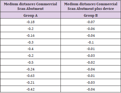
Table 1: Medium distances group A and B.

To solve the problem related to intraoral scan of edentulous arches, several authors suggest several methods with the aim of create artificial anatomical references to compensate the distance between Scan Abutments; in this way, the intraoral acquisition is easier and more accurate. Some authors used auxiliary removable devices interconnecting the scanbodies (with guides, polymer bases obtained through the use of a 3D printer) [24-26]. Methods proposed in literature partially solved the problem but usually require very sophisticated procedures that often are not recommended to the traditional physical impression. To date, therefore, despite advanced technologies, in critical clinical conditions intraoral scanning can lead to non-faithful and inaccurate models.

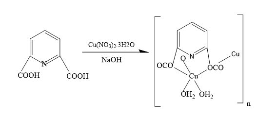
The aim of the study is to show a new concept to improve accuracy of intraoral acquisition in all those critical cases in which the distance between consecutive Scan Abutments affects the trueness of intraoral scans. The procedure outlined in the study has the peculiar property of not requiring complex and laborious steps. The auxiliary device is made of technopolymer material (Polyether ether ketone-PEEK) and it consists in an hexagonal body connected to a settable screw. (Figure 1) The device is designed to be screwed onto a previously drilled Scan Abutment and it does not rest directly on soft tissue so oral mucosa is not covered during the intraoral scan. The chance to successfully acquire oral mucosa with devices assembled on scan bodies avoids the need to acquire two separate impressions, with and without the auxiliary deviceresulting in discomfort for the patient and in extended clinical time.

biomedres-openaccess-journal-bjstr

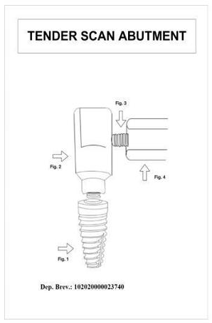
Figure 1: Design of the device.

Materials and Methods

Stereophotogrammetry technology (PIC Camera; PIC DENTAL), thanks to extraoral scanner, allows to gauge the inaccuracy associated with intraoral scanning of an edentulous arches. Gold standard STL file is acquired by indirect method by taking a physical impression, developing the cast, placing PIC Abutments on the laboratory analogues and scanning it with an extraoral scanner. Then, the gold standard is compared to STL file obtained by the direct intraoral scanning of the same arch. The ethics committee examined the research project and gave its approval to the study (ethics committee “Università Federico II” protocol no. 128/21). Patients provided written informed consent to the treatments and to the publication of the study. Written informed consent was obtained from patients for the publication of any potentially identifiable images or data included in this article.

For each of the 11 subject involved in the study three different impressions were carried out. The physical impression was performed according to “open tray splinted” impression technique using polyether material (3M Impregum Penta Soft) mixed thanks to dedicated equipment (Pentamix type). Analog transfers were pooled with self-curing resin and interdental floss. Each impression thus obtained was made into plaster cast using class 4 gypsum mixed by a vacuum- pressure casting machine. Specific targets (PIC Abutments) were placed on each transfert; then, the model was scanned using an extraoral scanner (X5 Dentsply Sirona). EOS provides an STL file where abutments are truthfully setted and we can use it as control (gold standard). Intraoral scanner (Omnicam Dentsply Sirona) is used to record two different digital impressions: the first impression is normally acquired with the only scan abutments seated (Group A) while the second impression involves the experimental device assembled as part of the “scan structure” (Group B). All digital impressions were performed by the same practitioner and all of them have been obtained according to manufacturer’s scanning protocol, starting from the distal area on one side reaching the opposite side through the occlusal area. Using a specific software (Geomagic Control X 2020) we proceed to the alignment and subsequent one-to- one comparison of the STL files obtained from IOS with the STL file obtained from EOS (Gold standard). (Figures 2 & 3) The primary measurement refers to the mean distance (average of the points dismatching between the two STL files) obtained through Geomagic 3D software. P values under 0.05 are expected to be statistically significant. Data provided by the software, expressing mean and standard deviation values either the lowest or highest distances, were summarized using standard descriptive statistic.

The null hypothesis imposes no statistically significant difference between the mean distances Group B- Gold standard and Group A-Gold standard. The comparison between the two different approaches (IOS with and without auxiliary device) is carried out by Student’s t-tests for paired samples. In the current study, the alternative hypothesis does not state direction of deviation therefore “two-tailed” test is adopted. The two-tailed test is a nondirectional hypothesis test, described as a test in which the critical region falls on both sides of the normal distribution. Thus, an alternative hypothesis is accepted in place of the null hypothesis if the calculated value falls in one of the two tails of the probability distribution (Figure 4).

biomedres-openaccess-journal-bjstr

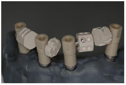
Figure 2: Scan Abutment plus device in peek on the model.

biomedres-openaccess-journal-bjstr

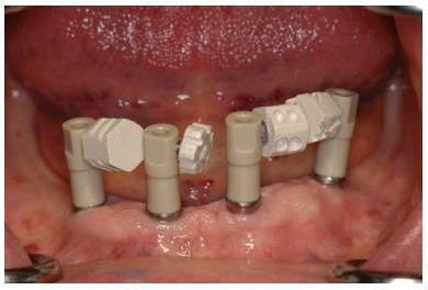
Figure 3: Device in peek in the oral cavity.

biomedres-openaccess-journal-bjstr

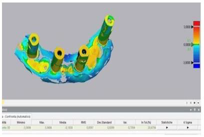
Figure 4: Comparison between stl control and stl with Commercial Scan Abutment.

Results

The two-tailed t-test for paired samples is used to test the null hypothesis that the average distances of the points made using the Scan Abutments according to the standard protocol (group A) are not significantly higher than those obtained using Scan Abutments with the experimental device (Group B). Assuming a difference of 0.1 mm between the two approaches (Group A and Group B) as clinically relevant and a standard deviation of the differences equal to 0.09 (value obtained through the evaluation of preliminary data), a sample size of 11 scans for each protocol will be enough to highlight any difference with a power of 0.9 and a two-tailed significance level of 0.05. Statistical analysis performed by using Spss software showed statistically significant differences (P <.05). Based on the results, the null hypothesis was rejected: group B values, significantly closer to zero, reflect a higher degree in their overlap with the control file. The “color maps” are used to qualitatively compare and evaluate the results: closer is the number to zero, smaller the difference between the two files is. The colors from blue to red indicate in a qualitative way the dimension of the mismatching between the file (A or B) and the gold standard control (Figures 5 & 6).

biomedres-openaccess-journal-bjstr

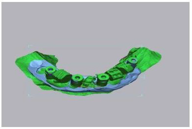
Figure 5: Alignment between stl control and stl with Commercial Scan Abutment plus device.

biomedres-openaccess-journal-bjstr

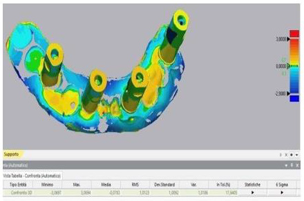
Figure 6: Comparison between stl control and stl commercial Scan Abutment plus device.

Discussion

Evidence suggests that intraoral scanning accuracy varies greatly with interimplant distance, intraoral scanner type, scan body type and operator experience. Several clinical factors can contribute to the global deviations in complete-arch intraoral scanning: the present study shows how the absence of anatomical references in edentulous arches has a negative influence on the accuracy of digital dental scans and how artifical anatomical references can be used to obtain easier and more accurate digital impressions. Limitations of the present study include the small sample size and the use of a single type of intraoral scanner. Comparison of the scans obtained with other intraoral scanners available on the market, still with the help of the device, will be reason for further research.

Conclusion

Based on the results, the null hypothesis was rejected. Full-arch digital implant impression, taken using intraoral scanners, was found to be less accurate than the same impressions recorded by the support of the experimental removable device. The present study shows how the absence of anatomical references in edentulous arches has a negative influence on the accuracy of digital dental scans and how artifical anatomical references intraoral make the acquisition in critical cases easier and more accurate.


For more Articles on: https://biomedres01.blogspot.com/

Tuesday, May 16, 2023

The Influence of Pyramid Structure on Cells


The Influence of Pyramid Structure on Cells

Introduction

Numerous biological effects of pyramid structure (PS) have been reported in recent years. For example, Indian scholars found that the seed germination rate and radicle length of plants under the PS were significantly higher than those of the control group [1-3], and other reported effects included accelerated would healing of mice, improved preservation time of food [4-7], and reduction in the occurrence of tumor in mice accompanied by the improvement in liver enzyme activity along with some other biochemical and physiological indicators, which could explain the retardation of the tumor cell growth [8].Despite the accumulating amount of evidence of biological effects reported, there is very little research investigating the underlying biological mechanisms for the observed phenomena. The PS has been proposed to generate a torsion field [9], which has been extensively studied for its biosocial effects. As a portal to receive, process and integrate information in cells, mitochondria play a critical role when cells were exposed to stimuli including external Qi from a Qigong master [10], Chinese texts with different meaning [11], and torsion field [12]. Our group has previously found that the exposure of cultured mammalian cells to the above stimuli can induce changes in numerous parameters related to the mitochondrial functions, such as the production of ATP, ROS, MMP and mtDNAcn, as well as the cellular oxidant capacity reflected by the reduced GSH.In this study, we used 293T cells to examine any potential responses in the mitochondria for cells grown under PS, with the goal of examining potential underlying biochemical mechanisms of the effects generated by a pyramid. We examined parameters including the growth and vitality of the cells, mitochondrial functions including the production ATP, ROS, MMP, the cellular oxidant capacity reflected by the reduced GSH, as well as cell senescence related parameter such as the telomere length.

Materials and Methods

Cell Culture

The 293T cells was obtained from National Infrastructure of Cell Line Resource (Beijing, China, http://www.cellresource.cn) as visceral cells which grow rapidly. The culture conditions and inoculation process of these cells can refer to our previous articles [12].

Pyramid Structure Model

biomedres-openaccess-journal-bjstr

Figure 1: The copper pyramid model.

The pyramid model used in the study is made of copper (some studies believe that the effect of copper is more obvious than other materials [3]) according to the true proportion and angle of ancient Egyptian pyramids. The copper pyramid model with a square of length 280 mm and height 280 mm (Figure 1).

Experimental Design

Two groups of 293T cells culture were used, including a treatment group (cultured in the pyramid model and named as ‘PS’), and a control group, named CK (away from the pyramid model). 293T cells were seeded at a density of 5~6×10⁵ cells/plate into a 10cm plate, and after a 3h attachment period, plates of the ‘PS’ group were placed in the pyramid model for 24h treatment. The plates for the ‘CK’ group were placed under normal growth conditions in another room about 50 meters away. The cells were collected and subjected to different assays at 4h and 24h respectively during incubation. All experiments were performed in triplicate (Figure 2).

biomedres-openaccess-journal-bjstr

Figure 2: PS: pyramid structure treatment group; CK: control group.

Biochemical Analysis

In the experiment, the detection methods of various biochemical indexes of 293T cells, such as ATP, ROS, MMP, protein, cell viability, SOD and GSH, were unified in our research group [12].

Gene Expression Analysis

Because of using the same research vector (293T cells), the parameters of gene expression analysis such as transcriptome sequencing and mitochondrial DNA quantification can refer to the previous work [12].

Statistical Analysis

Values of different measurements were normalized to a respective mean control value from untreated samples and expressed as percent control. All data are expressed as mean ± standard deviation (SD). They were analyzed by analysis of variance (ANOVA) and Least Significant Difference (LSD) using GraphPad InStat software, where P <0.05 was considered statistically significant.

Results

Effects of Pyramid Structure on Cell Growth

Compared to the control cells, PS did not affect the cell growth and cell viability at 4 hours post treatment and 24 hours (Figure 3), indicating that PS treatment for short period did not affect cell growth evidently (Figure 3).

biomedres-openaccess-journal-bjstr

Figure 3: Effects of PS on 293T cells growth.
(a) Cell Amount;
(b) Cell Viability.

Effects of Pyramid Structure on Mitochondrion

However, the mitochondria were significantly affected by PS as shown in Figure 4. The ATP level of cells in the PS group was upregulated by 20% and 35% at 4h and 24h respectively (Figure 4a) compared to that of the control group. The cellular ROS level was significantly decreased by pyramid at 4h but was not affected at 24h (Figure 4b). MMP level was both upregulated significantly at 4h and 24h (Figure 4c). The activity of GSH, an antioxidant protein, was increased by pyramid at 4h and 24h (Figure 4d), showing a potential role of antioxidant of PS.

biomedres-openaccess-journal-bjstr

Figure 4: Effects of PS on mitochondrion.
(a) ATP level;
(b) ROS level;
(c) MMP level;
(d) GSH level. *,
P<0.05. *Significantly different from the control group.

Effects of Pyramid Structure on mtDNAcn and Telomere Length

Cellular mtDNAcn and telomere length were detected using the qRT-PCR technology and no obvious changes was observed at 4h and 24h in the cells of pyramid group according to the results (Figure 5). All in all, PS modulated the function of mitochondrion in cells and may protect the cells from oxidative damage by upregulating the level of anti-oxidative protein and downregulating ROS level.

biomedres-openaccess-journal-bjstr

Figure 5: Effects of PS on mtDNAcn and telomere length.
(a) mtDNAcn;
(b) Telomere length.

Discussion

In this study, we found that the activity of mitochondria in 293T cells became more active under the influence of PS, and the levels of MMP and ATP were significantly up regulated. At the same time, the overall antioxidant capacity of cells was improved. The PS significantly increased the content of GSH and reduced the level of ROS. No significant changes in cell growth and telomere length were observed in the study, indicating that the PS has a mild effect on cells, which avoids the negative effects caused by strong stimulation. Studies have found that the liver enzyme activity of mice in PS shaped feeding cages is significantly increased [8]. The plasma cortisol content of adult female rats exposed to long-term PS changed significantly, and the antioxidant defense ability of erythrocytes was improved [13,14]. In addition, some researchers found that the PS can accelerate the healing of mouse wounds [4,6], which indicates that the cellular activity of mouse wounds is stimulated, which is very similar to our results. As the “power plant” of cells, mitochondria provide energy for cells, and as a bridge for energy transformation inside and outside cells, they can respond to external stimuli in time. The influence of PS is considered as a torsion field [9], which collects the surrounding fine energy through the spatial layout of the structure itself, and then through the induction of mitochondria in the cell, so as to improve the activity and antioxidant capacity of the cell.

Previous studies mainly focused on the effect of PS on biological system, but there were few studies on the effect at the cell level. In this study, we used biochemical and molecular biological methods to determine that the PS has an obvious promoting effect on cell antioxidant and mitochondrial activity, which is helpful to further study the mechanism of PS effect. The healing effect of this special spatial structure also provides a new way for noninvasive treatment.


For more Articles on: https://biomedres01.blogspot.com/

Monday, May 15, 2023

… And What About those who Disagree with the Measures Against the Pandemic?


… And What About those who Disagree with the Measures Against the Pandemic?

Opinion

In recent months a lively debate is taking place on the effectiveness of the various measures taken by national governments to contain the pandemic. Many have raised questions on the possible side effects of the use of masks in public places, especially in schools, of the side effects of vaccination, and so on. Groups of our fellow citizens are protesting about the extended use of masks and the compulsory nature of vaccination, for example, voicing their opinions in numerous ways. Others are going a step further, urging people towards adopting a stance akin to a (supposed) type of civil disobedience [1]. This category includes some parents who, especially on social networking sites, urge other parents not to send their children to school wearing a mask, vaccinated or having conducted a rapid test. The legitimate question that arises is whether the relevant dialogue and action are subject to certain constitutional limits. One of the fundamental pillars of democracy is that relating to freedom of opinion. Freedom of opinion is intertwined with the right to criticize government action. This criticism can go as far as the ordinary citizen questioning government policy measures. When pandemic issues are raised by journalists in the press, however, it is crucial to ensure the accuracy of the information and to limit fake news. Everyone is free to express his or her opinion, but one must also draw a clear distinction between value judgments and facts. The creation of a climate of misinformation and panic by the media is not in keeping with the social mission of the media, and is likely to cause further problems both for the individual and for society as a whole.

This is even more so the case at this time of extreme difficulty for the population as a whole, when citizens, in a state of restraint and amidst widespread and entirely justified fear of the virus spreading, are regularly informed of developments. Nevertheless, in addition to expressing opinions, many are also taking action inciting civil disobedience. This phenomenon is not unknown: it is an expression of the timeless conflict between natural and statute law. It goes back to antiquity, when Prometheus violated Zeus’ command not to reveal the use of fire to humans. Indeed, Sophocles’s Antigone is the most brilliant example of civil disobedience, as she refuses to comply with King Creon’s order not to bury her brother, Polynices, putting forward her obligation to obey what she considers to be a superior legal system. On the other hand, in Plato’s Crito, Socrates emerges as the father of ‘civil obedience’, as he offers us a prime example of obedience to an unjust, but lawful, sentence [2]. Thoreau is considered by most to be the father of civil disobedience, being the person who introduced the term itself. Back in the mid-19th century he refused to pay taxes and was imprisoned, not because he opposed taxation in general, but because he opposed the US government’s war against Mexico, slavery and the violation of Indian rights [3]. The real father of civil disobedience, however, is John Locke, who pointed out that the existence of arbitrary authority is a prerequisite for disobedience. In this context, he recognized from 1688 the right of Greek Christians to throw off the Turkish yoke imposed on them by force [4].

In this light, the question arises as to whether one has the right not to wear a mask, not to be vaccinated and also to encourage other people to do so, too. Kant could provide us with the answer: according to him, the legal order in a democratic society is a value in itself. “There can be no justified resistance on the part of the people against the legislative authority of the state. A state governed by the rule of law is only strong when there exists universal subordination to its legislative will [...]. The reason why it is the duty of people to tolerate even what is apparently the most intolerable misuse of supreme power is that it is impossible even to conceive of their resistance to the supreme legislation as being anything other than unlawful and liable to nullify the entire legal constitution”.”[5]. It is clearly contradictory, according to Kant, for a legal order to provide the ability to resist its mandates, as this essentially self-negates its supreme authority. This reasoning is only founded when the legal order in question has democratic legitimacy [6]. In a tyrannical regime, where citizens are excluded from the legislative process, there is no issue of obligation of obedience to a heteronomous legal order, and each individual has a right to resist, as well as a right to revolt against it [6]. In a democratic regime, on the other hand, one may express discontent and voice views openly, but has no right not to obey, particularly when the law concerns a fundamental right pertaining to public health. In actual fact, this is not at all an issue of conflict between natural and statute law.

Not complying with rules on hygiene is not related to natural laws. Mask and vaccine deniers are not in agreement with a certain legislative provision, which is based on research data. Let it not be lost on us that, by the same token, they could disagree with the use of masks by doctors in the operating room, the use of gloves by bakers during a pandemic, the use of protective equipment for visitor admission to an intensive care unit, and so on. Such deniers do not argue that wearing a mask and vaccination constitute discrimination against lower social strata – they are essentially projecting a disagreement they have vis-à-vis a particular scientific finding. In the case at hand, and to begin with, scientists ought to make a convincing case to people, based on substantiated arguments. Nevertheless, if a person decides not to wear a mask and does not get vaccinated, without having a medical reason for doing so, then it is wholly reasonable that he or she should be the recipient of the lawful ramifications of the prohibition of entry to the specified area. Therefore, a pupil will be justly denied entry to a classroom, as will a customer to an indoor cinema theatre, a citizen to a public service, to a shop and, even more to the point, to a hospital. Indeed, this is the case as the person in question is unable to support this based on scientific disagreement. Furthermore, if a person is inciting the public to collective disobedience against the use of masks and vaccination, it would be reasonable that he or she should face relevant legal consequences. At this point, it should not be overlooked that any battle of arguments does not place in a vacuum but in the context of a reality which, if ignored, may lead us to very unpleasant surprises [7]. The defiant violation of the use of masks and vaccination is not, at the end of the day, an act of civil disobedience, but rather a manifestation of anti-social autonomy [8].

For the above to become more intelligible, the following conclusions may be drawn:
a) The position “I consider the use of the mask and the enforcement of vaccination in public places problematic: people will not comply and, therefore, this will create more problems” is a constitutionally permissible expression of opinion.
b) The position “I find the use of masks and the imposition of vaccination to be unconstitutional measures” is also a constitutionally permissible expression of opinion.
c) The position of “Don’t wear a mask and don’t get vaccinated” constitutes incitement to disobedience against a specific provision of the law which - especially in the period of the pandemic - goes beyond the permissible limits of freedom of speech.
d) The non-use of masks and the refusal to get vaccinated during a pandemic, where this is required by law, does not constitute civil disobedience in the sense of what has been set out above: it represents illegal and, mainly, anti-social behavior.


For more Articles on: https://biomedres01.blogspot.com/

Saturday, May 13, 2023

Novel Use of GnRH agonist as Neoadjuvant Treatment for Giant Endometrial Polyps


Novel Use of GnRH agonist as Neoadjuvant Treatment for Giant Endometrial Polyps

Introduction

Endometrial polyp is one of the most common structural causes of abnormal uterine bleeding in the reproductive age group. It arises from endometrial overgrowth brought about by a hyper-estrogenic state. Polyps are usually around 2cm in size, and those greater than 4cm are labeled as giant polyps. The cases presented are women in the reproductive age group with fertility problems, having endometrial polyps as large as 10cm occupying the whole uterine cavity, even prolapsing out of the endocervical canal. Conservative management was recommended but due to their size, immediate hysteroscopic removal may not be attainable due to expected difficulty in distending the uterine cavity and possible morbidities like fluid overload and uterine perforation. Although with limited studies, GnRH agonists have been proposed as a neoadjuvant treatment prior to hysteroscopic removal of giant polyps. Given the same mechanism applied with GnRH agonist’s use with myomas, it induces a hypoestrogenic environment at the same time decreasing the vascularity of the endometrial lining that will help significantly decrease the size of the giant polyps, making hysteroscopic removal feasible

Case Report

Case 1

This is a case of Abnormal Uterine Bleeding (AUB) in a 29-yearold, single, nulligravid from Laguna. The patient has unremarkable past medical and family history. She is living in with her 3-year partner and is desirous of pregnancy. Ever since menarche, patient has been experiencing heavy menstrual bleeding soaking 2-3 baby diapers per day, lasting 7-14 days, with no associated symptoms. She was hospitalized at the age of 16 for blood transfusion due to chronic blood loss brought about by heavy menstrual bleeding. Blood dyscrasia was ruled out. At the age of 20, she consulted a government hospital still for persistent heavy menstrual bleeding now associated with dyspareunia and post-coital vaginal bleeding. She was assessed with a cervical mass probably polyp and underwent fractional curettage and polypectomy, which on biopsy revealed polyp. Patient continue to have occasional heavy menstrual bleeding however few months prior to consult in our institution, she had continuous profuse vaginal bleeding, this time associated with an enlarging abdomino-pelvic mass. She was assessed with cervical cancer due to a finding of irregularly shaped cervical mass. A cervical punch biopsy was done which only revealed granulation tissue. She was then referred to our institution for further work up and management.
When we first saw the patient, she was pale but with stable vital signs. Speculum examination revealed a polypoid fleshy mass occupying the upper half of the vaginal canal. On internal examination, patient had a polypoid fleshy mass measuring 8cm x 10cm x 10cm occupying the upper vaginal canal and the cervix was difficult to palpate. The corpus was enlarged to 18-20 weeks size. She was initially diagnosed as a case of to consider cervical cancer and was admitted for blood transfusion and work up. Cervical punch biopsy was repeated and it revealed necrotic and hemorrhagic polypoid lesion. Transvaginal ultrasound done noted an aggregate of polyps at the cervix that arise from the endometrial cavity with attachment at the fundus. Impression was endometrial mass, to consider endometrial pathology with extension to less than 50% of the myometrium, endocervical canal, and lower third of vagina (See Appendix 1, Figure 1). Current working diagnosis was changed to AUB secondary to endometrial mass probably endometrial polyp. Patient was referred to our section for possible hysteroscopic polypectomy. But due to the size of the mass and expected technical difficulty for hysteroscopic removal, we advised pre-treatment with Gonadotropin-Releasing Hormone (GnRH) agonist to be given for 3 to 6 doses every 28 days prior to surgery.

biomedres-openaccess-journal-bjstr

Apendix 1 Figure 1: Endometrial mass and prolapsing cervical mass arising from the endometrium prior to GnRH agonist.

After three doses of GnRH, repeat internal examination showed that the polypoid mass within the vaginal canal disintegrated and was easily removed from its attachment. Patient noted minimal vaginal bleeding with passage of meat like material, probably fragments of the polyp. The corpus size also decreased to 14 to 16 weeks size, from the initial 18-20 weeks size. Additional 3 doses of GnRH agonist were suggested to further decrease the size of the mass and make the hysteroscopy easier. After 6 doses of GnRH agonists with a corpus size of 10-12 weeks, the patient then underwent hysteroscopic polypectomy. Intraoperatively, the polypoid mass was soft and hemorrhagic, but was not friable and non-necrotic, with an aggregate diameter of 17 cm (See Appendix 1, Figures 2 & 3). Cut section revealed multiple cystic spaces all throughout the mass. Final biopsy report showed mixed endocervical-endometrial polyp. Three months post-surgery, there was no recurrence of heavy menstrual bleeding and repeat ultrasound revealed normal uterus with thin endometrium (See Appendix 1, Figure 4). Currently, the patient is already 1 year post op with no recurrence of the heavy menstrual bleeding and is currently for infertility management.

biomedres-openaccess-journal-bjstr

Apendix 1 Figure 2: Endometrial mass and prolapsing cervical mass arising from the endometrium prior to GnRH agonist.

biomedres-openaccess-journal-bjstr

Apendix 1 Figure 3: Endometrial mass and prolapsing cervical mass arising from the endometrium prior to GnRH agonist.

biomedres-openaccess-journal-bjstr

Apendix 1 Figure 4: Post operative ultrasound of patient 3 months after.

Case 2

This is another case of AUB in a 30-year-old, Gravida 2 Para 0 (0020), married, from Mindoro. She has no co-morbids and had 2 previous spontaneous abortions at 12 weeks AOG. She is currently living with her husband of 10 years and is desirous of a successful pregnancy. Four years prior to consult in our institution, patient experienced heavy menstrual bleeding for 7 days, soaking 3 baby diapers per day with blood clots. She was assessed with AUB secondary to thickened endometrium and was admitted for blood transfusion. After 2 years, patient again had recurrence of heavy menstrual bleeding, this time associated with abdominal enlargement. Ultrasound was done and showed myoma uteri. She was again transfused with blood products and advised hysterectomy but patient refused due to financial constraints and desire of future fertility. 3 months prior to consult in our institution, patient again experienced continuous vaginal bleeding and pain with pallor and fatigue. She was rushed again to the local hospital and on assessment, there was a polypoid mass occupying the vaginal canal and was sampled for biopsy. The histopathology result revealed atypical polypoid adenomyoma so the patient was referred to our institution.
On our initial examination, the patient was pale but with stable vital signs. Pelvic examination revealed a hemorrhagic polypoid fleshy mass occupying the whole vaginal canal that easily bleeds in manipulation measuring 6cm x 6cm (See Appendix 2, Figure 1). The cervix was difficult to assess and the corpus was enlarged to 20-22 weeks size. Rectovaginal examination was unremarkable. Transvaginal ultrasound was done which showed an echogenic mass within the endometrial, endocervical and vaginal canal measuring 12.8 x 12.2 x 8.1 cm with multiple irregular cystic spaces, invading >50% of the posterior myometrium (See Appendix 2, Figure 2). The impression was endocervical and endometrial masses consider atypical polyp with >50% myometrial invasion. Repeat biopsy of the prolapsed endometrial mass was done which revealed endometrial polyp. Patient was also referred to our section for possible hysteroscopic polypectomy. But again, due to the size of the mass and expected technical difficulty for hysteroscopic removal, pre-treatment with GnRH agonist prior to surgery was advised. After 2 doses of GnRH, there was significant improvement of patient’s symptoms, but with minimal vaginal bleeding, and passage of meat-like materials. On internal examination, the prolapsing endometrial mass decreased to 2x2cm (See Appendix 2, Figure 3) and the corpus size decreased to 16-18 weeks from the initial 20-22 weeks size. Repeat ultrasound also noted a decrease in the size of the endometrial mass to 10.7 x 11.8 x 5.0 cm (See Appendix 2, Figure 4). After the 3rd dose of the GnRH, the patient will be re-assessed if additional GnRH is needed or if already amenable to hysteroscopic polypectomy.

biomedres-openaccess-journal-bjstr

Apendix 2 Figure 1: Speculum examination of the patient prior to GnRH agonist.

biomedres-openaccess-journal-bjstr

Apendix 2 Figure 2: Ultrasound of the endocervical and endometrial masses prior to GnRH agonist.

biomedres-openaccess-journal-bjstr

Apendix 2 Figure 3: Speculum examination of the patient after 2 doses of GnRH agonist.

biomedres-openaccess-journal-bjstr

Apendix 2 Figure 4: Repeat ultrasound of the patient after 2 doses of GnRH.

Case 3

The last case is a 36-year-old, nulligravid who came in due to abdominal enlargement and inter-menstrual bleeding. Patient has no comorbid but is unable to conceive for 9 years with her husband. 1 year prior to consult, she began to develop intermenstrual bleeding, soaking 3 pads per day lasting 1-2 days associated with abdominal enlargement. On pelvic examination, there is a 3x3cm polypoid mass protruding out the endocervical canal, which seems to be coming from the endometrial cavity and the corpus is enlarged to 18-20 weeks size. Ultrasound showed an endometrial mass measuring 11.0 x 8.2 x 3.2cm with irregular cystic spaces and seems to be contiguous with the endocervical mass (See Appendix 3, Figure 1). Endometrial biopsy was done to rule out malignancy, and the result was endometrial polyp, hyperplastic type. Management was again GnRH for 3 doses and reasses if amenable for hysteroscopy. After her 1st dose of GnRH, the size of the corpus significantly decreased to 14 to 16 weeks size. Patient also reported fragments passage of meat-like material with minimal vaginal bleeding. Repeat ultrasound was done and the size also significantly decreased to 7.5 x 6.5 x 6.1cm (See Appendix 3, Figure 2). Patient is still for completion of 2 more doses of GnRH prior to re-assessment.

biomedres-openaccess-journal-bjstr

Apendix 3 Figure 1: Ultrasound of the endocervical and endometrial masses prior to GnRH agonist.

biomedres-openaccess-journal-bjstr

Apendix 3 Figure 2: Repeat ultrasound of the patient after 1 dose of GnRH.

Discussion

Endometrial polyp is one of the most common structural causes of AUB among the reproductive age group. It could present as intermenstrual or heavy menstrual bleeding and the amount of bleeding is usually proportional to the size of the endometrial polyp: the larger the size, the more debilitating the symptoms. Aside from bleeding, it is also associated with infertility just as the cases presented. Endometrial polyps are formed from localized overgrowths of endometrial tissue composed of variable amount of glands, stroma, and blood vessels covered by epithelium. The stroma of a polyp is composed of fibroblast like spindle cells and large blood vessels with thick walls. Commonly, the size of a polyp is less than 2cm, however, polyps greater than 4 cm are called giant polyps, as shown in the cases above, whose sizes range from 10 to 12 cm. The development of endometrial polyps has been explained by a number of molecular mechanisms such as monoclonal endometrial hyperplasia, over-expression of endometrial aromatase and gene mutations [1]. It usually arises because of estrogen hypersensitivity in some areas of the endometrium, probably caused by hyperactivation of the beta or the alpha estrogen receptor during the first phase of the cycle. Furthermore, they do not shed with menstruation because the estrogen related inflammation block the apoptosis via bcl-2 gene expression. Giant polyps, on the other hand are known to occur due to unbalanced estrogen levels. This estrogen related polyp growth occurs due to angiogenic growth factors’ deregulation, produced under hormone control inside the polyp, within a short time and few cycles [2].
Histopathological examination must be performed on all resected polyps to rule out endometrial polyps (0.5%–3%) with malignant foci. Lasmar BP reported that endometrial polyps larger than 15 mm are associated with hyperplasia and Wang J et al. identified that polyps measuring more than 10 mm are associated with malignancy [3]. The 3 cases of giant polyps presented all underwent endometrial biopsy to rule out the possibility of malignancy prior to choosing conservative treatment, which was advocated since all 3 cases have issues with infertility. For the management of endometrial polyps, hysteroscopic resection is the preferred method [4]. However, in some instances like in giant polyps, immediate hysteroscopic removal may not be feasible. This could be due that giant polyps occupy the entire uterine cavity that distending the cavity during hysteroscopy for visualization of the polyp’s attachment may not be done. Aside from failed removal of the polyp, morbidities like fluid overload and uterine perforation can be encountered. Although there is limited evidence with the use of GnRH agonist in endometrial polyps, it’s use was suggested as it may significantly decrease the size of the polyps to make the hysteroscopic resection achievable.
GnRH agonists has revolutionized the management of many conditions in gynecology based on the discovery that when administered in a non-pulsatile fashion, they down regulate pituitary GnRH receptors, and therefore decrease the production of FSH and LH. It has been widely used for submucus myomas to decrease its size and make its hysteroscopic removal manageable with less morbidity. It used as a neo-adjuvant therapy prior to hysteroscopy to decrease bulk of the myoma and induce a state of hypoestrogenism. Pretreatment with GnRH agonists improves the hemoglobin level, reduces blood loss during surgery, and reduces uterine and leiomyoma volumes in as much as 40-50%, with most of the reduction occurs in the first 12 weeks [5]. This is especially helpful in infertile patients because this prevents destruction of the surrounding endometrial tissue [6]. Given the same concept for myomas, GnRH can then be used as pre-treatment for giant polyps since they have characteristic cytogenetic rearrangements similar to uterine leiomyoma [2]. As said previously, polyps contain estrogen as well as progesterone receptors, and the concentrations of these receptors are significantly higher in the glandular epithelium of endometrial polyps than those in the normal endometrium [7].
Hormonal inhibition of the endometrium can induce mucosal thinning and decreased bleeding. The effect of GnRH agonist can extend up to 1 month from the last dose and this could be due to persistent hypoestrogenism and consequent inhibition of regeneration of viable endometrial cells [5]. In the cases presented, GnRH agonist has significantly decreased the size of giant polyps even after 1 dose. All of the patients have reported vaginal bleeding along with passage of meat-like materials, which were believed to be fragments of the polyp spontaneously sloughing off from its attachment due to devascularizing effect of the GnRH agonist. None of the cases developed anemia or needed blood transfusion despite the said vaginal bleeding. The 1st case even successfully underwent hysteroscopic polypectomy after 6 doses of GnRH with no morbidity encountered and no residuals after, given the initial size of her polyp. With this in mind, there is room for GnRH agonists in treating endometrial polyps, specifically the giant polyps, where immediate hysteroscopic removal may tend to be difficult or may lead to failure, more so, morbidity. The cases have shown the effect of GnRh agonists in significantly decreasing the size of the polyps probably by inducing a hypoestrogenic state and at the same time deceasing the vascularization of the polyps causing it to spontaneusly slough off from its base. This will facilitate complete hysteroscopic removal of the giant polyp at the same time decreasing chances of morbidity. Although further studies are needed to strengthen its use in endometrial polyps, this may open an area for more research work.

Conclusion

Endometrial polyps, specifically the giant polyps, can be an incapacitating cause of abnormal uterine bleeding, which are often associated with infertility. In such cases, conservative management in the form of hysteroscopic removal is encouraged. However, given the size of a giant polyp, there will be technical difficulty in doing its resection. Use of GnRH agonist as neoadjuvant treatment for giant polyps can be advised. Using the same concept of GnRH agonist in shrinking myomas, it can induce a hypoestrogenic state causing devascularization of the endometrial lining. This will help significantly decrease the size of polyp, allowing complete hysteroscopic removal. However, further research studies are still suggested to strengthen its neoadjuvant use with giant endometrial polyps.


For more Articles on: https://biomedres01.blogspot.com/

The Skin Aging Process and Anti-Aging Strategies

  The Skin Aging Process and Anti-Aging Strategies Introduction Appearances play a vital role in society as it is how one communicates to ot...