Showing posts with label Open access clinical and medical journal. Show all posts

Saturday, June 25, 2022

Phototheraputic Keratectomy for a Patient with Corneal Intraepithelial Dysplasia

Phototheraputic Keratectomy for a Patient with Corneal Intraepithelial Dysplasia

Introduction

Intraepithelial neoplasia and squamous cell carcinoma of the cornea and conjunctiva are the most common tumors of the ocular surface [1] with a reported incidence of 0.13 to 1.9/100,000, depending on geographic location [2]. This tumor is classified as an in situ form, known as conjunctival and/or Corneal Intraepithelial Neoplasia (CIN), or invasive squamous cell carcinoma. CIN encompasses entities that were previously referred to as dysplasia and carcinoma in situ. Corneal epithelial dysplasia occurs in elderly patients. Although the etiology remains unknown, it has been suggested that excess ultraviolet exposure and human papilloma virus infection may cause the lesion [3,4]. The pathogenesis of corneal epithelial neoplasia usually commences at the limbal region in that abnormal epithelium grows towards the central cornea and often associates with neovascularization into the lesion. The most common treatment for conjunctival and corneal neoplasia is radical excision in combination with cryotherapy [5]. Newer chemotherapeutic modalities, such as mitomycin C (MMC) [6] 5-fluorouracil (FU) [7] and interferon,8 are now being used to avoid the operating room altogether and decrease the potential risk of limbal stem cell loss and scarring. There has been only one case of recurrent corneal intraepithelial dysplasia treated with Phototherapeutic Keratectomy (PTK) [8,9]. In this report, we describe a case of primary corneal intraepithelial dysplasia after PTK.

Case Report

A 68-year-old woman presented with blurred vision in the right eye for 6 months before the initial visit to the clinic. We did not obtain a medical history of the etiologic factors associated with induction of the neoplasm. Her Corrected Distance Visual Acuity (CDVA) was 20/40 and 20/20 in the right and left eyes, respectively. Central pachymetry measured 564 μm in the right eye and 530 μm in the left eye. Biomicroscope demonstrated plaque-like isolated epithelial corneal lesions extending from the paracentral cornea into the central visual axis in her right eye. The cornea of her left eye also showed translucent epithelial corneal lesions at the paracentral cornea, but no symptoms occurred. The cornea was avascular, and the corneal stroma seemed to be uninvolved. The anterior chamber was quiet, and the remainder of the ocular examination was unremarkable. Anterior Segment Optical Coherence Tomography (AS-OCT) (CASIATM, Tomey Corporation, Nagoya, Japan) was performed and demonstrated thickening and highly increased reflectivity of the corneal epithelial layer extending from the paracentral to central cornea of the right eye. In accordance with the thickening lesions, anterior corneal steepening was observed in the keratometric map using AS-OCT. Based on these morphological characteristics, we diagnosed corneal intraepithelial dysplasia in both eyes in this patient.

We conducted PTK with the NIDEK EC-5000 excimer laser system and used the following parameters: wavelength, 193 nm; fluency, 165 mJ/cm2; repetition rate, 40 Hz; ablation zone diameter, 7.0 mm; transition zone, 1.0 mm; and ablation depth, 200 μm based on the AS-OCT. We used the transepithelial technique for removal of the corneal epithelium. No histological examination was performed on the ablated tissue after PTK. Postoperatively, steroidal (0.1% fluorometholone) and antibiotic (1.5% levofloxacin) medications were topically administered 4 times daily for 1 week after insertion of a soft contact lens, and the dose was steadily reduced thereafter. The patient returned at the 1-month follow-up examination with subjective improvement in vision and no subjective complaints. The preceding epithelial corneal lesions had diminished. Her CDVA had improved to 20/20 in the affected eye, with mild punctate epithelial erosions at the inferior cornea. AS-OCT was again performed, demonstrating normal epithelium without the lesions previously observed. The anterior corneal astigmatism also improved to 1.2 diopters in the AS-OCT keratometric map. The CDVA remained at 20/20, and no recurrence of epithelial corneal lesions occurred at the 6-month follow-up.

Discussion

Current treatment modalities include excision with or without adjuvant cryotherapy, topical chemotherapy (e.g., MMC, 5-FU, or interferon), radiation therapy, and in extreme cases, exenteration of the orbit [10]. Surgical excision alone of CIN has been associated with higher rates of recurrence, ranging from 17% to 24% for dysplasia and from 30% to 41% for squamous cell carcinoma [2,11]. In the current case, the corneal lesion was diagnosed as corneal intraepithelial dysplasia of the ocular surface before treatment with PTK. Although histological examination might be helpful in determining the confirmed diagnosis, a dysplastic tissue sample was not obtained because of excimer laser ablation. To our knowledge, this is the first reported case of PTK in primary corneal intraepithelial dysplasia. Our case demonstrated isolated neovascularized corneal epithelial dysplasia compared to a previous case report by Dausch, et al [9]. We conclude that PTK is an appropriate technique for mild to moderate cases of corneal intraepithelial dysplasia. Excimer laser ablation may be effective in the treatment of corneal intraepithelial dysplasia. High-resolution AS-OCT is emerging as an important noninvasive technique that can help diagnose and evaluate the efficacy of treatment. AS-OCT was performed to look for evidence of recurrence in the present case. We are conducting imaging using an AS-OCT described in many patients.

For more Articles on : https://biomedres01.blogspot.com/

Friday, June 24, 2022

Diaphragmatic Eventration in Nf1 Microdeletion Syndrome: A Rare Association Unmasked by Pregnancy

Diaphragmatic Eventration in Nf1 Microdeletion Syndrome: A Rare Association Unmasked by Pregnancy

Introduction

Neurofibromatosis type 1 (NF1), also known as von Recklinghausen disease, is a neurocutaneous condition with an estimated prevalence between 1 in 2000 to 1 in 4000, characterized by a great variability in its clinical presentation. Clinical hallmarks such as café au-lait macules, neurofibromas, axillar or inguinal freckling, optic pathway glioma and iris hamartomas (Lisch nodules) represent the main NF1 diagnostic criteria set by the U.S. National Institutes of Health (NIH) in 1988 [1]. Beside these typical signs, the possible association with skeletal, endocrine, cardiovascular and oncologic complications make NF1 a multi-systemic disorder [2]. A similar broad spectrum of clinical manifestation is caused by mutations occurring in the Neurofibromin on co suppressor gene, on chromosome band 17q11.2, and inherited in an autosomic dominant manner [3]. In about 5–10% of cases takes place a large deletion involving the NF1 gene and its flanking regions which results in the “NF1 microdeletion syndrome” [4]. Despite the lack of genotype–phenotype correlations in NF1, patients with Type 1 microdeletion (1.4 Mb encompassing about 14 genes) typically present a serious illness, mainly characterized by dysmorphic facial features and developmental delay [5]. During pregnancy, NF1 women should be monitored because they are more prone to complications such as hypertension, preeclampsia, fetopelvic disproportion, poor fetal growth and oligohydramnios [6]. Furthermore, pregnancy for NF1 women is related to an increased number and size of cutaneous neurofibromas [7]. Regarding pregnancy in women with NF1 microdeletion, no specific literature data are available, possibly due to their low fitness and to the rarity of the condition.

Diaphragmatic defects have never been described before as a pregnancy complication in NF1 women. Similar conditions are also unusual in general population, with only 56 reported cases until 2018 [8]. The increased abdominal pressure experienced by women during pregnancy might represent a risk factor for unmasking hidden congenital diaphragmatic defects (CDD). Among these defects, diaphragm eventration (DE) represents an uncommon finding, defined as the upward displacement of a portion or the entire diaphragm, otherwise intact. Congenital DE (CDE) diagnosis is often an incidental finding in adults, as it can remain completely asymptomatic. Even if scarcely described first presentation of CDD during pregnancy, owing to the risk of rupture, represents a lifethreatening complication both for mother and fetus [9]. Here we report a case of (CDE) complicating the pregnancy of a 30-year-old patient with NF1 Type 1 Microdeletion Syndrome.

Case Report

The patient is a 30-year-old pregnant woman, affected by NF1 type 1 microdeletion syndrome, followed since the age of 24 at our Clinical Reference Centre for NF1. She has undergone numerous clinical and instrumental evaluations over the years; the main clinical issues that have emerged are summarized here: mild intellectual disability, typical NF1 cutaneous features (cafè au laits spots and diffuse cutaneous neurofibromas) and two plexiform neurofibromas on the scalp and on the left hemiabdomen. Furthermore, multiple spinal neurofibromas were reported at the age of 26, involving all the conjugation foramina, especially in the thoracic and lumbar tracts of the spine. At the age of 28 due to a suspected diagnosis of pheochromocytoma, the patient underwent left adrenalectomy; histological examination diagnosed a ganglioneuroma and steroid replacement therapy was therefore introduced. In addition, a nonfunctioning pituitary microadenoma was detected on routine brain magnetic resonance imaging (MRI), steady in size on control MRI in the following years. In view of these comorbidities, as the patient got pregnant, she was followed by a multidisciplinary team, consisting of gynecologists, endocrinologists, neurologists and medical geneticists. Regular ultrasound evaluations were performed throughout the entire pregnancy. The patient and her partner decided not to proceed with any invasive tests for prenatal diagnosis.

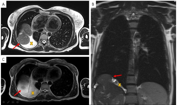
The pregnancy progressed regularly. In preparation of spinal anesthesia, a spine MRI was performed to monitor the wellknown spinal neurofibromas that appeared to be steady in size and number. Serendipitously, three round-shaped images at the base of right chest were reported and interpreted as possible diaphragmatic hernia (DH) with partial displacement of the liver to the chest. A right-sided pleural effusion was also reported, which retrospectively appeared to be present and unchanged since the previous control. An echocardiogram and an abdominal ultrasound were performed as follow-up investigations. The former reported mild mitral and tricuspid insufficiency, but no pericardial effusion was observed. The second examination showed a moderate pleural effusion on the right side with thin fibrinous septae and gross outpouchings of the liver parenchyma, the largest of almost 4cm. Thus, the previous suspicion of DH was confirmed. In view of the numerous comorbilities, a caesarean section was performed and the patient gave birth to a female newborn. Genetic NF1 analysis performed after birth revealed the transmission of the maternal NF1 microdeletion to the baby. Since both the mother and the newborn were in good health, they were discharged five days after delivery, once DH-related complications were excluded. Computed Tomography (CT) of the upper abdomen performed two months later showed three diaphragmatic bulges, with a maximum diameter of 4.2cm on the VIII segment of the liver and of 4.8-2.8cm on the VII segment. The right-sided pleural effusion was still present and, as the diaphragm was not clearly visible over hepatic protrusions and appeared worsen from previous imaging, the hypothesis of hernias was corroborated (Figure 1). Therefore, surgical correction of the defect was planned after the weaning of the child. The patient underwent video-assisted thoracoscopic surgery (VATS), which revealed an intact but very thin diaphragm, consistent with a diagnosis of DE. There was no evidence of occult neurofibromas nearby, nor adjacent lesions that might be the very first cause of the diaphragmatic eventration. In addition, a voluminous pleuro-pericardial cyst was found, explaining the pleural effusion previously reported. Finally, a diaphragm plication and cyst excision were performed. The removed tissue was biopsied and this examination revealed the presence of a cystic wall, covered by a single stratified cubic-cylindrical epithelium. No atypia was found. The post-operative course was uneventful.

Figure 1: Chest MR Imaging:

A. Two round shape images with partial liver shift in right pleural space (red arrow) and surrounding fluid collection (yellow X);

B. Coronal plane;

C. MR imaging three years before pregnancy.

Discussion

Diaphragmatic weakness became clinically relevant during pregnancy, when the rising abdominal pressure pushes the diaphragm upwards while the muscle itself contracts downwards. In our patient, these two opposing forces probably enlarged a preexisting diaphragmatic defect, resulting in liver herniation. To our knowledge this is the first reported case of a patient with NF1 microdeletion showing a DE and a pericardial cyst, three extremely rare conditions. Fortunately, the liver was the only organ involved in herniation and the patient remained asymptomatic, even during pregnancy. As mentioned, among NF1 population, patients with type 1 microdeletion generally display a more severe phenotype. This is possibly due to the deletion not only of the NF1 gene, but also of its flanking genes, which could partially influence the clinical manifestation of the disease. In particular, NF1 type 1 microdeleted patients are more likely to develop malignant peripheral nerve sheath tumors (MPNSTs), cardiovascular anomalies [5] and connective tissue abnormalities [10]. Although the precise molecular basis of connective tissue involvement in NF1 is still unclear, neurofibromin has been shown to play a regulatory role in mesenchymal stem cell differentiation [10]. Moreover, during embryogenesis, this protein also takes part to axons’ elongation in order to ensure the correct activity of the nervous system [11]. The involvement of NF1 gene in proper neuronal and mesenchymal development must be particularly stressed as neurons and connective tissue, together with several other structures from different embryonic origins, participate to the complex sequence of events that leads to diaphragm development [12]. In particular, CDE has been related to defects in migration and proliferation of muscle fibres, two steps leaning on the regulatory action of connective tissue cells [13].

Whether the association between NF1 and CDE is causal or is in fact a possible associated complication, the management of our case demonstrates the crucial importance of a multidisciplinary approach to pregnant women affected by a rare disease. NF1 microdeletion syndrome is associated with numerous comorbidities, most of which are just barely known. Particularly, knowledge about pregnancy-related complications in NF1 microdeleted women is still lacking and even more about possible urgent events; as a consequence, no specific management guidelines are available yet. As shown by our case report, pregnancy in these women can turn out to be a challenging moment and should be carefully supervised, as it represents an event in which the delicate equilibrium of such fragile patients might unbalance, possibly leading to unexpected, unknown and potentially serious complications. In our opinion the complexity of this condition, the unpredictability of complications’ onset and severity can be properly managed only by gaining a wider and multifaceted vision of the disease, which can be obtained only by means of a dedicated multidisciplinary team. A well-coordinated multi-specialized equipe could represent the primary step towards widening our perspective of the condition and towards implementing tools for personalized care and follow-up, with the ultimate aim of ensuring early diagnosis and prompt management of possible upcoming emergencies, especially during pregnancy in these vulnerable women.

For more Articles on : https://biomedres01.blogspot.com/

Wednesday, June 22, 2022

Restorative Bio-Compatible Prosthetic Epoxy-Zeolite Composites: Strength and Resistance

Restorative Bio-Compatible Prosthetic Epoxy-Zeolite Composites: Strength and Resistance

Introduction
The injection of inorganic binders into the epoxy resin is a known method of obtaining coatings, adhesives, compounds and repair compositions. Epoxy compositions with building binders are used as the basis for the production of self-leveling floors, repair compositions and industrial compounds [1-22]. Attempts to use epoxy compounds for implantation and exo-prosthetics [7-9,23,24] have become popular recently. Epoxy glass and carbon plastics, as well as composites with biocompatible fillers, were able to compete with traditional metal and other (ceramic, wood) materials. In particular, due to porosity, nerves, oxones and body cells can grow through the surface of epoxy materials (which is impossible for tantalum and titanium implants) [23,24]. And in exo-prosthetics (manufacturing of external prostheses), epoxy composites have a number of advantages - in particular, the possibility of manufacturing in the field, replaceability, ease [8,9,23,24]. An interesting potential filler for biocompatible innovative epoxy composites is zeolite and its varieties. There are researches on the effect on epoxy polymers of montmorillonite [14,15,25], clays and brick dust [3,16], aluminum oxide [10,20,21], pyrophyllite [11], brick powders [3,16], natural zeolites [18], bentonite [22], copper [26,27]. These issues are dealt with by teams led by Danchenko & Barabash [19-21], Erdogan [18], Choi & Leе [15], Burmistrov & Mostovoy [16], Starokadomsky [3,10], Borisov and others [25], Kahramanov & Allahverdieva [26,27]. The aim of the work was to establish the possibilities of zeolite as a potentially enhancing eco- The Composites Department in Chuiko ISC has 30 years of experience in polymer research with alumina and silica, as evidenced by our works [1-13,19]. Also, we have experience in the creation of prosthetic [5,8,9] and biocompatible [7,11-13] compounds and fiberglass compatible and cheaper epoxy filler. The selected concentration of 50 wt% is practically convenient because it forms a convenient viscous composition, and also allows you to mix components without precision scales (which is important in the field conditions). At the same time, epoxy-ceolite-copper composite was investigated in order to further create electro- and thermally conductive materials for electronics and medicine (prosthetics).

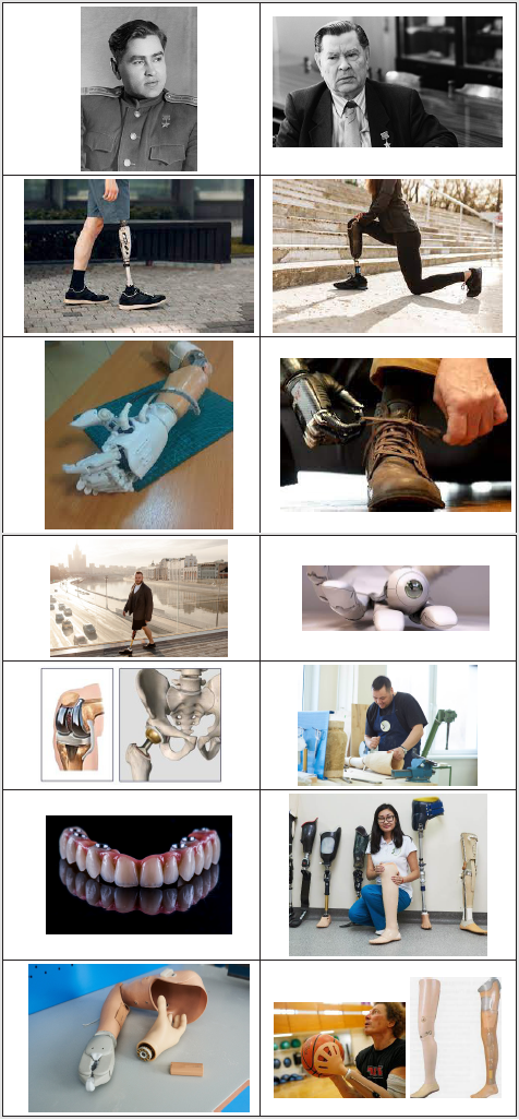
Figure 1 Pilot, Hero of the Soviet Union A. Maresyev (noncolored photos), in 1943-1946, on a military plane, flew 86 sorties with prostheses instead of amputated legs. Сolored photo - examples of modern prostheses (see [28,29] and Wikipedia.org). Prosthetics has a long history. Even medieval Caribbean pirates also often had wooden legs and iron hooks instead of hands and fingers, and there have been attempts at dental prosthetics since primitive times. However, real shifts in science and practice began in the last century. Already in the 2nd World War, there are vivid examples of the effectiveness of prosthetics for maintaining extreme loads. So, the Soviet pilot A. Maresyev (Figure 1), after being wounded and amputated of his legs, returned to the sky and flew with prostheses. In total, during the war, he made 86 sorties, shot down 10 enemy (german) aircraft: three before being wounded and seven after. A. Maresyev (1916-2001) lost his legs at the age of 37, but with prostheses he lived an active life for another 59 years, and died almost at 90 years old in the new XXI-th century. He became the prototype for the hero of the soviet bestseller “The Story of a Real Man” [28,29].

Figure 1: Pilot, Hero of the Soviet Union A. Maresyev (non-colored photos), in 1943-1946, on a military plane, flew 86 sorties with prostheses instead of amputated legs. Сolored photo - examples of modern prostheses (see [28-29] and Wikipedia.org).

Methods and Reagents

Required Reagents and Method of Obtaining Composites

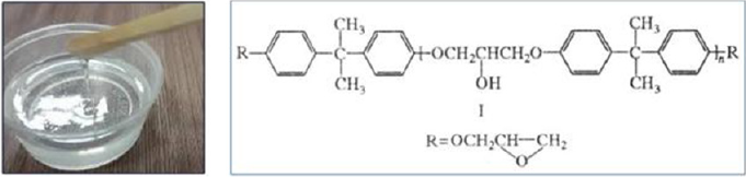
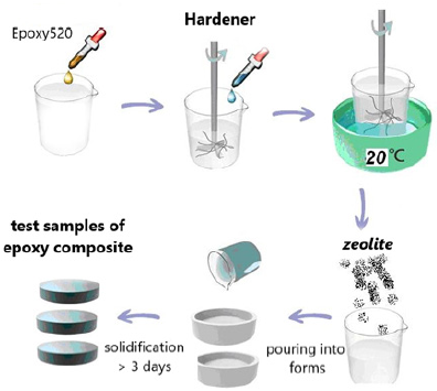
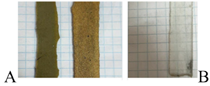
The Czech cold-hardening epoxy resin “Epoxy520” (Figure 2) was used for the works, which was cured with PEPA polyamine in a ratio of 5:1. The filler was added immediately after mixing these components, after which the composition was homogenized and immediately formed into samples (Figure 3, Table1). As a filler were used nano- and micro-sized zeolite particles (manufactured by PE “Eco Instinct”) for which sieving up to 100 μm was performed. Its supramolecular structure provides for the presence of micropores and internal nanopores (Figure 4). Visually, it is a gray powder converting the epoxy composition to a viscous ocher mass (Figure 4). Curing them gives wood-like biocompatible composites (Figure 5). Of the strength properties, we were interested in those that practically determine the applicability of these compositions. These are adhesion, strength (and modulus of elasticity) in compression, bending and abrasion, microhardness. Determination of strength and stability was performed according to methods corresponding to standards (GOST or ASTM). Shear adhesion tests (GOST 14760- 59) were performed on steel tear plates with a bonding area of 3cm2 and are presented in kgf. The corresponding value of adhesive strength can be obtained by dividing the obtained index by three. For compression tests (taking into account GOST 4651- 2014; ISO 604: 2002), cylindrical samples (diameter d= 6.5mm, height h=12 +-1mm) were taken and compressed on a Louis Shopper press machine until complete destruction. The machine provided compression of the sample with a given constant speed of movement of the active gripper, measurement of the load with an error of no more than ± 5% of the measured value. According to the test results, the strength was calculated: f = P / s (P - load in kgf, s - area equal to 0.332cm2) and module E: E = f / e (e - elongation equal to the length of the rectilinear section of the diagram in cm, divided by 10). Abrasion was performed by passing composite cylinders (diameter 6.5cm) on the surface of emery P60 at a distance of 10cm in both directions 40 times. Weight loss in mg and mm was determined. Abrasion resistance was calculated as the inverse of the abrasion mass of the sample. Abrasion resistance was determined by the empirical (derived from experiments) formula W = 1 × (m / mH) / P = m / mH × P, where mH / m characterizes the increase in mass (density) of the sample after filling.

Figure 2: Epoxy-resin, typical shape and formula.

Figure 3: A typical scheme for obtaining epoxy-filled composites.

Figure 4: View of zeolite powder (A), zeolite structure (B), epoxy-zeolite (C) & epoxy-zeolite-Cu and (D) compositions

Figure 5: View of polymerized composites - epoxy-zeolite & epoxy-zeolite-Cu with (A) and without zeolite (B)

Table 1: Description and marking of samples.

Strength of Composite Materials

Adhesion Strength of the Epoxy-Composite to Steel

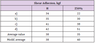
Shear adhesion to steel tends to be enhanced after the addition of 50% by weight of the zeolite. This can be seen from Table 2, from the results of which the most informative are the maximum obtained values (Table 2 d) and the modified average value (not taking into account the smallest indicators out of the series - in this case a) for C50%).

Table 2: Load values when testing adhesion to steel (bonded area 3 cm2).

Mechanical Abrasion of the Epoxy Composites

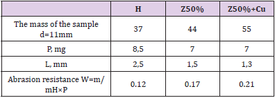
As can see (Table 3), the injection of fillers leads to a marked increase in abrasion resistance. This is also noticeable while estimating abrasion in millimeters: the filled samples lose much less in size than the H-polymer sample. The increase in abrasion resistance (especially after the addition of copper, table 3) is quite natural, given the abrasive resistance of inorganic filler particles. Here need to notice also that the density of the sample naturally increases, especially after the addition of copper - even more (see the masses of the samples, Table 3).

Table 3: Abrasion of samples (emery P60, 40 passes in both directions on 10cm).

Compressive Strength and Young Modulus of Composites

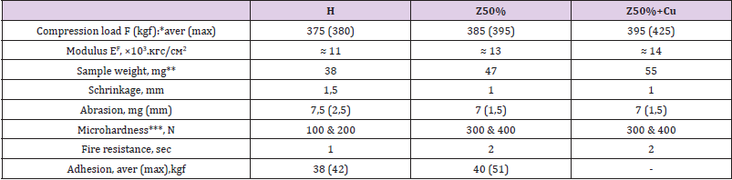
Table 4 demonstrate that 50 wt% zeolite makes it possible to increase the compressive strength and Young’s modulus. When copper powder is added to the composition, the effect is enhanced - in particular, the maximum value obtained is increased by 20% (425 instead of 380) compared to the H-polymer. The filling also changes the nature of the destruction from plastic (the sample crumples like plasticine) to brittle (cracks along the puncture lines). We can notice from Table 5, hat process of filling gives approximately double growth of microhardness. However, the filling increases the fragility of the samples - they crack already when hemispherepunch immersing to 20μm (Table 5) – while unfilled withstanding immersion up to 60μm. From the summary Table 6 you can see the overall effect of zeolite on the mecha parameters of polyepoxide. From Table 6 it is seen that the introduction of 50 wt% zeolite makes it possible to increase the strength and Young’s modulus under compression, microhardness, adhesion and fire resistance. This reduces shrinkage (an undesirable component of polymer synthesis) and abrasion wear. The addition of micro-nanoparticles of copper sometimes enhances the effect of increasing strength (for example, in compression).

Table 4: Values of load F in compression (cylindrical specimens with a diameter d=6.5cm, height 11-12cm), Young’s modulus E (*estimated) and the nature of the destruction of cracks when gripping.

Table 5: Microhardness (in H) of the surface of the composites, when the hemisphere is immersed by 10-30 microns. The # symbol indicates brittle cracking of the sample at the time of testing.

Table 6: All investigated parameters of strength of composites (* - aver (max) means Average and Maximal obtained value; ** - cylinder at h = 11mm, d = 6,5mm; *** - at 10 and 20μm immersion of steel hemisphere).

Resistance and Swelling of the Epoxy Composite in Aggressive Environments

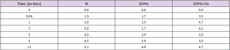
Endurance in 60% H2O2: Hydrogen peroxide, even at low concentrations (2-5%) is a substance quite aggressive against polymers (which is why it is used as a bleach in cosmetics). When the concentration of H2O2 reaches 30-40% and especially 50- 60%, its destructive effect is multiplied. At concentrations of 50- 60% peroxide easily corrodes organic tissues (in particular, forms severe burns on the skin). Polyepoxides are not significantly stable in 50-60% hydrogen peroxide. Therefore, the dynamics of their swelling and destruction in it can be tracked quite quickly (within 1-2 weeks) - in contrast to many other aggressive sopluk (acids, gasoline, alkalis). This makes the peroxide a convenient medium for evaluating the effect of the filling on the stability of the composite. From Table 7 it can be seen that the zeolite gives the composite greater resistance to peroxide. This can be seen from the decrease in the degree of swelling at all stages of exposure. The addition of copper powder in the composition somewhat weakens the positive effect of the filling, bringing the swelling index to that for the unfilled (H) sample (Table 7, Figure 6).

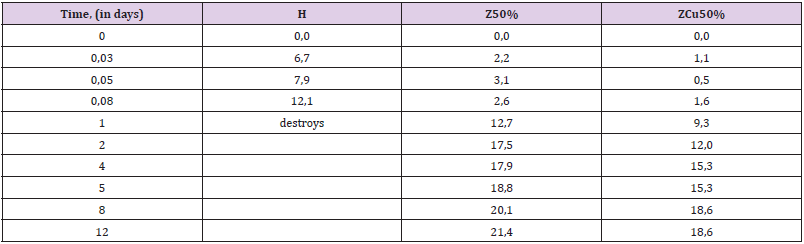
Resistance in a Mixture of Acetone: Ethylacetate (1 : 1): Acetone and acetone-contained solvents are very aggressive media for polyepoxides [3-9]. The unfilled polymer (especially freshly made) destructs in acetone solvents in a matter of days and sometimes hours. At the same time, the filling of epoxides can significantly increase their resistance to these mediums [3-11]. This can be seen from the results obtained. The unfilled sample is strongly swollen already in the first hours of endurance, and at the end of 1 day of endurance completely destroys (scatters in solvent). After the introduction of zeolite, the charge does not destroy, and swells much more slowly than the H-polymer. The addition of copper powder enhances this effect (Table 8).

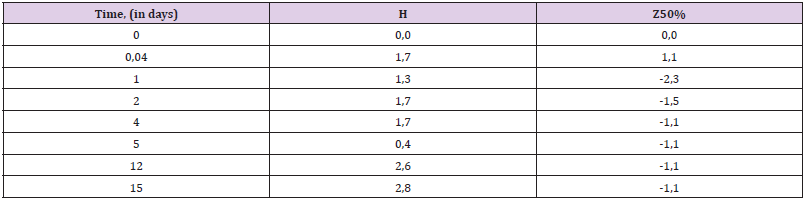
Water Absorption of Composites: Polyepoxides are generally quite stable in water. A water absorption rate of up to 1-2% during the month of exposure is considered normal. However, often even this figure needs to be improved. In addition. Epoxy products (coatings, parts, structures or ships) are often in the water for many years, and they need high water resistance. From our research we can conclude that the addition of zeolite makes the composite somewhat more resistant to water in the first half of the exposure (Table 9). Filling even causes the effect of slight weight loss, which can be caused by the phenomena of leaching of substances from the structure of the zeolite filler.

Table 7: The degree of swelling of the samples in 60% Н2О2.

Table 8: Swelling degree of samples in acetone: ethylacetate mixture.

Table 9: The degree of swelling of the samples in H2O.

Thermo-Oxidative Destruction of Polymer Powders without and with Filler

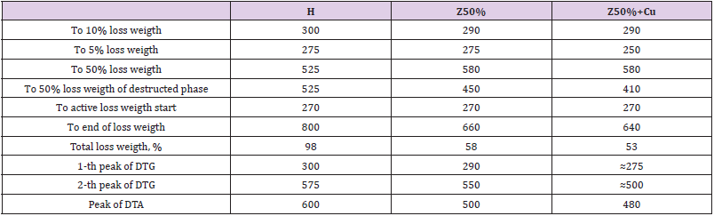
A significant increase in fire resistance (Table 6) gives reason to expect certain changes in resistance to destructive thermal oxidation (DTO). A typical thermogram of unfilled polyepoxide is presented in Figure 9H. It shows that the maximum weight loss due to destructive thermal oxidation (DTO) occurs at 300 оС. The temperature of the 5% weight loss of the H-polymer, respectively, is 275 oC, and 10% 300 oC (Figure 9H. TG curve). After that, the DTO processes are significantly slowed down, forming a cycle of primary destruction processes. Starting from 450 оС, secondary processes (called burnout of coke residue) are activated. They also have their peak activity but occur with a more active and stable (without sharp differences) dynamics of heat release and weight loss (Figure 9). Half (50%) of weight loss occurs at 525 оС. Secondary DTO processes are completed only at the approach of 800 оС, when almost 100% of the sample burns out (Figure 9H). With the injection of 50 wt% zeolite, you can expect some changes in the thermal oxidation of the sample. Indeed, the primary DTO is less active. Thus, on the DTG curve at least at -0.1 (whereas for H-polymer at -0.2) and the DTA curve does not rise above 0.8. The maximum of the primary DTO is shifted toward 290 оС, ie at a lower temperature than the DTO of the H-polymer. Also, at lower temperatures (than for H-polymer) there is a secondary DTO, which according to the DTG curve is completed much faster - at 670 оС (H-polymer at 780 оС). At the same time, only up to 60% of the sample burns out, obviously it is 50% of the polymer and another 10% of the zeolite mass. When adding 10-13 wt.% Micro nano-particles of copper in the composite with zeolite, the nature of the thermal decomposition changes markedly. Now we actually have a single DTO process that is made up of sequential processes. All destructive processes are completed in record time - up to 630 оС. From Figure 9 and Table 10 it is seen that the filling generally impairs the resistance of polyepoxide to thermal oxidative destruction.

Table 10: Stat-parameters of DTO processes, according to the obtained thermograms.

Conclusion

1) The injection of zeolite (in the amount of 50 wt%) is promising for the production of bio-eco-compatible composites for industrial and biomedical use, with enhanced strength and stability characteristics. The addition of micro-nanoparticles of copper in the composition in some cases can enhance the quality and strengthening effects of polymerized composites.

2) It is experimentally shown that the introduction of zeolite can dramatically increase the stability in an aggressive solvent (a mixture of acetone: ethyl acetate). Unlike unfilled (which completely destroys in 1 day of endurance), composites don`t destroy in it, and swell much more slowly. Also, the filling (after 2-3 days of exposure) increases the resistance to swelling in highly concentrated (60%) H2O2 peroxide: after 5 days of exposure, the unfilled swells by almost 5%, while the composite with zeolite - less than 3%.

3) It is established that the zeolite filling gives a double increase in microhardness and a slight increase in compressive strength and modulus. The addition of micro-nanoparticles of copper sometimes enhances the effect of increasing strength (for example, in compression). However, the filling increases the fragility of the samples.

4) It is shown that the filling doubles the fire resistance (2sec, whereas unfilled - 1sec). Thermogravimetry shows that the filling does not increase the resistance to primary thermooxidation of the epoxy polymer (passes at 290 oC instead of 300 oC in the unfilled) but makes it less active. Also, at lower temperatures are the processes of secondary thermal oxidation (at 670 oC instead of 780 oC in the H-polymer). The positive effect of the filler is reflected in an almost twofold reduction in the burn-up mass (up to 50-60% of the sample instead of 98% in the unfilled one).

For more Articles on : https://biomedres01.blogspot.com/

Thursday, June 2, 2022

Phototheraputic Keratectomy for a Patient with Corneal Intraepithelial Dysplasia

Phototheraputic Keratectomy for a Patient with Corneal Intraepithelial Dysplasia

Introduction
Intraepithelial neoplasia and squamous cell carcinoma of the cornea and conjunctiva are the most common tumors of the ocular surface [1] with a reported incidence of 0.13 to 1.9/100,000, depending on geographic location [2]. This tumor is classified as an in situ form, known as conjunctival and/or Corneal Intraepithelial Neoplasia (CIN), or invasive squamous cell carcinoma. CIN encompasses entities that were previously referred to as dysplasia and carcinoma in situ. Corneal epithelial dysplasia occurs in elderly patients. Although the etiology remains unknown, it has been suggested that excess ultraviolet exposure and human papilloma virus infection may cause the lesion [3,4]. The pathogenesis of corneal epithelial neoplasia usually commences at the limbal region in that abnormal epithelium grows towards the central cornea and often associates with neovascularization into the lesion. The most common treatment for conjunctival and corneal neoplasia is radical excision in combination with cryotherapy [5]. Newer chemotherapeutic modalities, such as mitomycin C (MMC) [6] 5-fluorouracil (FU) [7] and interferon,8 are now being used to avoid the operating room altogether and decrease the potential risk of limbal stem cell loss and scarring. There has been only one case of recurrent corneal intraepithelial dysplasia treated with Phototherapeutic Keratectomy (PTK) [8,9]. In this report, we describe a case of primary corneal intraepithelial dysplasia after PTK.

Case Report

A 68-year-old woman presented with blurred vision in the right eye for 6 months before the initial visit to the clinic. We did not obtain a medical history of the etiologic factors associated with induction of the neoplasm. Her Corrected Distance Visual Acuity (CDVA) was 20/40 and 20/20 in the right and left eyes, respectively. Central pachymetry measured 564 μm in the right eye and 530 μm in the left eye. Biomicroscope demonstrated plaque-like isolated epithelial corneal lesions extending from the paracentral cornea into the central visual axis in her right eye. The cornea of her left eye also showed translucent epithelial corneal lesions at the paracentral cornea, but no symptoms occurred. The cornea was avascular, and the corneal stroma seemed to be uninvolved. The anterior chamber was quiet, and the remainder of the ocular examination was unremarkable. Anterior Segment Optical Coherence Tomography (AS-OCT) (CASIATM, Tomey Corporation, Nagoya, Japan) was performed and demonstrated thickening and highly increased reflectivity of the corneal epithelial layer extending from the paracentral to central cornea of the right eye. In accordance with the thickening lesions, anterior corneal steepening was observed in the keratometric map using AS-OCT. Based on these morphological characteristics, we diagnosed corneal intraepithelial dysplasia in both eyes in this patient.

We conducted PTK with the NIDEK EC-5000 excimer laser system and used the following parameters: wavelength, 193 nm; fluency, 165 mJ/cm2; repetition rate, 40 Hz; ablation zone diameter, 7.0 mm; transition zone, 1.0 mm; and ablation depth, 200 μm based on the AS-OCT. We used the transepithelial technique for removal of the corneal epithelium. No histological examination was performed on the ablated tissue after PTK. Postoperatively, steroidal (0.1% fluorometholone) and antibiotic (1.5% levofloxacin) medications were topically administered 4 times daily for 1 week after insertion of a soft contact lens, and the dose was steadily reduced thereafter. The patient returned at the 1-month follow-up examination with subjective improvement in vision and no subjective complaints. The preceding epithelial corneal lesions had diminished. Her CDVA had improved to 20/20 in the affected eye, with mild punctate epithelial erosions at the inferior cornea. AS-OCT was again performed, demonstrating normal epithelium without the lesions previously observed. The anterior corneal astigmatism also improved to 1.2 diopters in the AS-OCT keratometric map. The CDVA remained at 20/20, and no recurrence of epithelial corneal lesions occurred at the 6-month follow-up.

Discussion

Current treatment modalities include excision with or without adjuvant cryotherapy, topical chemotherapy (e.g., MMC, 5-FU, or interferon), radiation therapy, and in extreme cases, exenteration of the orbit [10]. Surgical excision alone of CIN has been associated with higher rates of recurrence, ranging from 17% to 24% for dysplasia and from 30% to 41% for squamous cell carcinoma [2,11]. In the current case, the corneal lesion was diagnosed as corneal intraepithelial dysplasia of the ocular surface before treatment with PTK. Although histological examination might be helpful in determining the confirmed diagnosis, a dysplastic tissue sample was not obtained because of excimer laser ablation. To our knowledge, this is the first reported case of PTK in primary corneal intraepithelial dysplasia. Our case demonstrated isolated neovascularized corneal epithelial dysplasia compared to a previous case report by Dausch, et al [9]. We conclude that PTK is an appropriate technique for mild to moderate cases of corneal intraepithelial dysplasia. Excimer laser ablation may be effective in the treatment of corneal intraepithelial dysplasia. High-resolution AS-OCT is emerging as an important noninvasive technique that can help diagnose and evaluate the efficacy of treatment. AS-OCT was performed to look for evidence of recurrence in the present case. We are conducting imaging using an AS-OCT described in many patients.

Patient Consent
Written consent to publish this case report has been obtained from the patient.

For more Articles on : https://biomedres01.blogspot.com/

Thursday, May 26, 2022

Marine Algal Bioactive Metabolites: Effects and Occurrence

Marine Algal Bioactive Metabolites: Effects and Occurrence

Mini Review
When the algal density reached over the baseline level and caused harmful effects, these algal blooms are defined as harmful algal blooms (HABs) Hallegraeff [1]. Over the past several decades, marine algal bioactive metabolites have become a concern for the environment and human health. The contact (e.g., ingestion) of these metabolites results in an alternation of cellular enzyme functionality and causes cell deformation and mortality in the worst cases. Marine diatoms, dinoflagellates, and cyanobacteria are the known producers of these harmful metabolites. Bioactivity and the occurrence of these algal metabolites will be reviewed herein. Marine diatoms were the known producer of domoic acid. In 1987, ingestion of cultured blue mussels (Mytilus edulis) containing domoic acid (DA) caused food poisoning that killed three people and sickened >100 others Bates, et al. [2,3]. The structure of DA was determined to be an analogue of glutamic acid (Wright et al., 1989), and known producers of DA are species of a marine diatom genus Pseudo-nitzschia Jeffery, et al. [4]. DA is a neurotoxin that causes neuronal degeneration and necrosis in specific hippocampus regions, leading to amnesic shellfish poisoning. Several reports on the accumulation of DA in various organisms. However, DA can be degraded through the process of frozen storage and cooking, suggesting the low stability of this compound (as cited in Jeffery, et al. [4]).

Marine dinoflagellates are known producers of a series of bioactive metabolites that are classified into five major groups by their bioactivities after ingestion of toxin-containing fish and shellfish. These bioactivities including paralytic shellfish poisoning (PSP), diarrhoeic shellfish poisoning (DSP), neurologic shellfish poisoning (NSP), azaspiracid shellfish poisoning (AZP), and ciguatera fish poisoning (CFP) metabolites. PSP in humans is caused by the ingestion of seafood containing a group of alkaloids, including saxitoxin and its analogues (Cusick, et al. [5]). The pharmacological action of the PSP toxin is characterized as the blockage of the voltage-gated sodium channel (VGSC), leading to numbness and respiratory paralysis that could be fatal. Marine dinoflagellates (e.g., Alexandrium) and freshwater cyanobacteria (e.g., Dolichospermum) are the known producers of PSP toxins. Diarrhea, nausea, vomiting, liver necrosis, cardiac muscle damage, and abdominal pain are the know symptoms of DSP, caused by the ingestion of bivalves containing lipophilic metabolites, including okadaic acid (OA) and dinophysistoxins (DTX) (Reguera, et al. [6]). Marine species of Dinophysis and Prorocentrum are the and pectenotoxins (PTX) are two groups of lipophilic toxins that are structurally distinct from OA and DTX but possess similar bioactivities. However, some YTX and PTX can cause liver necrosis and cardiac muscle damage without diarrhea (Domínguez, et al. [7]). The common origins of YTX are marine species of Prorocentrum, and PTX are produced by Dinophysis.

The Florida and Gulf of Mexico coastal red tide former, Karenia brevis (syn. Gymnodinium breve and Ptychodiscus breve), is the common producer of lipophilic brevetoxins (PbTX) that cause NSP (Baden, et al. [8]). Structurally, PbTX can be divided into two groups, one with 10 cyclic rings and the other with 11 rings. The toxicity of NSP toxins was caused by opening the VGSC, leading to nausea, vomiting, paralysis, seizures, and coma. The aerosolization of PbTX caused by wave action leads to asthmalike symptoms in humans. The next dinoflagellate toxin group is AZP toxins, including azaspiracids (AZA). To date, over 50 AZA were isolated from species in marine dinoflagellate Azadinium, and contaminated seafood (Twiner, et al. [9,10]). The ingestion of AZA caused nausea, vomiting, diarrhea, and stomach cramps (Twiner, et al. [9,10]), while the mechanism of action has not been elucidated. The final dinoflagellate toxin group is CFP that caused by lipophilic ciguatoxins (CTX). The common origin of CTX is Gambierdiscus toxicus, while species in Prorocentrum were also reported as producers (Friedman, et al. [11]). CTX found in the Pacific region (n=13) had a different number of cyclic rings than CTX in the Caribbean region (n=14). CTX mechanism of action is similar to PbTX; however, some CTX had a greater affinity for VGSC than PbTX. Cyanobacteria are prolific bioactive secondary metabolite producers. To date, 157 known bioactive classes have been identified. Four (i.e., microcystins, saxitoxin, anatoxin-a, and cylindrospermopsins) of these known bioactive classes were listed in the EPA Contaminant Candidate List 4 (CCL4).

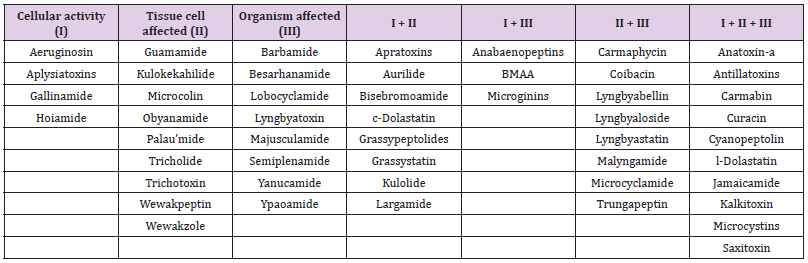
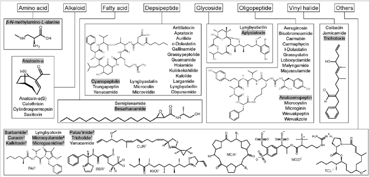
In the author’s previous effort, the 157 known bioactive classes have been reclassified to 55 structurally unique bioactive classes based on similarities of their structure and biological activity (Huang, et al. [12]). This effort was necessary because some metabolites share similar chemical structures and bioactivity but have been named differently. Therefore, a classification system was proposed to include both the original class names and reclassified class names. For example, lyngbyaureidamides have a similar structure as anabaenopeptins. Thus, when describing this compound, the proposed description will be anabaenopeptinlyngbyaureidamides. Fifty of 55 classes, including isomers/ synonyms, have been described from the marine environment (Table 1). Three of the four EPA CCL4 listed classes have been described from marine cyanobacteria: microcystins, anatoxin-a, and saxitoxin. Thirty of these 55 marine secondary groups were originally isolated from benthic cyanobacteria (e.g., Lyngbya and Moorea), with some compounds were elucidated from marine invertebrates (e.g., Dolabella). Over the past three decades, the knowledge of marine cyanobacterial metabolites has increased tremendously (Huang, et al. [13-15]). Their structures belong to eight main groups: amino acids, alkaloids, fatty acids, depsipeptides, glycosides, oligopeptides, vinyl halides, and other structures (Figure 1). Some cyanobacterial bioactive metabolites are chlorinated and brominated. The bioactivity of these compounds started from enzyme inhibition and VGSC blockage, causing organ bleeding and swelling, eventually, the death of the organisms (Table 1).

Table 1: Summary of marine cyanobacterial metabolites bioactivities. The column with “I + II”, “I + III”, “II + III”, and “I + II + III” presented a combination of bioactivity has been found. For example, compounds in column “I + II” contain both cellular and tissue cell activity.

Figure 1:Summary of cyanobacterial bioactive compound general structures. These compounds can be classified into eight groups: amino acids, alkaloids, fatty acids, depsipeptides, glycosides, oligopeptides, vinyl halides, and other structures. In addition, the structures of gray-highlighted compounds are shown. Among these classes, anatoxin-a(S), calothrixins, microguanidines, microviridins, and cylindrospermopsins have not been reported in the marine ecosystem.

Algal blooms will likely increase in frequency and intensity due to climate change and anthropogenic nutrient input. Accompanied with this trend, the frequency of toxin-producing algae cooccurrence will also be increased. Unfortunately, most researchers and monitoring programs only focus on certain toxins of interest, while other toxin classes with similar or higher toxicity are unstudied. Therefore, if two toxins carry similar toxicity co-exist, the estimate of the bloom might toxicity not be accurate. Further, HAB co-occurrence could result in synergistic effects caused by multiple bioactive metabolites that possess different known bioactivities. For example, when an enzyme inhibitor co-occurs with a linear cytotoxic oligopeptide, this inhibitor can deactivate the enzyme digesting ability on the oligopeptide, leading to more damage caused by the oligopeptide. Thus, a reevaluation of the monitoring protocols to include strategies for toxin co-occurrence is needed.

For more Articles on : https://biomedres01.blogspot.com/

Tuesday, May 24, 2022

Recurrent ADEM Mimicking Young Stroke


Recurrent ADEM Mimicking Young Stroke

Introduction

ADEM, also known as post-infectious encephalomyelitis, is a demyelinating CNS disorder that usually follows the occurrence of infection, or more infrequently, after the administration of a vaccination [1-3]. ADEM should be suspected when one or more of the following features are present such as a multifocal and polysymptomatic initial presentation, the presence of signs and symptoms suggestive of meningoencephalitis, encephalopathy, bilateral optic neuritis, cerebrospinal fluid (CSF) fluid pleocytosis along with the typical magnetic resonance imaging (MRI) picture [4,5]. It affects children more than adults but can affect anyone. Although it is monophasic by definition, relapsing forms of ADEM have also been recognized.

Case Report

A 30-year male, right-handed, presented with a history of fever for 7 days, holocranial episodic acute severe headache for 3 days, which was associated with nausea, photophobia, and phonophobia. Headache was short-lived but was recurrent, it persisted for around 2-3 hours and get relieved. No triggering factor for this exacerbation. No associated neck pain. The patient also had 1 episode of involuntary body movements, frothing from the mouth, urinary incontinence with loss of consciousness for 1 day. H/O altered sensorium with the irrelevant talk was present. A few hours later, the Patient complaint of Weakness of the left upper and lower limb with facial deviation to the right side. Weakness was to the extent he was unable to lift hand and legs above the bed. On examination, patient was conscious but altered sensorium, left UMN facial nerve palsy, left hemiparesis (Grade- 3/5), brisk deep tendon reflexes, bilateral Extensor plantar reflex, no meningeal signs, no involvement of bowel/bladder, and the sensory deficit was there. Systemic examinations were normal. Blood picture and biochemical investigations were within normal limits. Magnetic resonance imaging (MRI) brain showed focal hyperintense lesions (FLAIR and T2) in the subcortical white matter of right basal ganglia, right corona radiate, left parietal- temporal- occipital region [(Figure 1)-MRI July 2018], and bilateral cerebellar hemisphere and no restriction in DWI &meningeal enhancement in a gadoliniumbased MRI contrast study.

biomedres-openaccess-journal-bjstr

Figure 1: Magnetic resonance imaging brain showing focal hyperintense lesion ( FLAIR& T2) in cerebral cortex and subcortical white matter indicating demyelination of brain in the first episode (July 2018).

Cerebrospinal fluid (CSF) study was done which shows Sugar 45mg/dl, protein 248mg/dl, cell count 25/cmm (99% lymphocytes), negative for Gram-stain or Ziehl–Neelsenstain, and no growth in culture, viral encephalitis panel, and TB PCR was negative. Keeping diagnosis of ADEM, he put on intravenous immunoglobulin (IVIG) for 5 days then oral prednisolone and antiepileptic, and was discharged in stable condition. After 3 months, in November 2018, he was again admitted with a similar severe headache, left hemiparesis grade 2/5, left UMN type facial palsy with left focal seizure and secondary generalization, and global aphasia. MRI brain showed focal hyperintense, demyelinating lesions [(Figure 2)-Nov 2018], in the same brain territory as in the previous episode [(Figure 1)-MRI July 2018]. Immunological markers: Antinuclear antibody, antineutrophil cytoplasmic antibody, anticardiolipin antibody, lupus anticoagulant were negative. Serum Lactic acid and PBF for Sickle cell were normal. MR Venography and CT angiography brain plus neck vessel were normal. His treatment was IVIG for 5 days, followed by oral Prednisolone in the tapering dose and antiepileptics and the patient showed improvement. After follow-up on 1 month, clinically/MRI brain was normal (Figure 3).

biomedres-openaccess-journal-bjstr

Figure 2: Magnetic resonance imaging brain showing focal hyperintense lesion ( DWI& T2) in cerebral cortex and subcortical white matter indicating demyelination of brain in the second episode (Nov 2018).

biomedres-openaccess-journal-bjstr

Figure 3: MRI brain after 1 month showed disappearance of lesions.

Discussion

The index case presented with recurrent episodes of headache, seizures, encephalopathy, a focal neurological deficit in form of hemiparesis and aphasia. The differential diagnosis of recurrent CNS lesions is considered [6]:
1) Recurrent Meningo Encephalitis
2) Reversible cerebral vasoconstriction syndrome(RCVS)
3) MELAS
4) Primary CNS vasculitis/Angiitis
5) Recurrent CNS inflammatory demyelinating disorder like NMO, MS, ADEM
Of the above, Systemic diseases like MELAS were excluded as, S. Lactic acid was normal, no family history, and no multiorgan manifestations like myopathy, hearing impairment, short stature, dementia, and Recurrent meningoencephalitis was excluded as there was no stiff neck or signs of meningismus and CSF HSV PCR was negative. Multiple sclerosis (MS) and ADEM, the two major CNS inflammatory demyelinating diseases, are difficult to differentiate in the initial episode. MS is a continuous demyelinating disease with a characteristically relapsing-remitting course [6]. Although recurrence is characteristic of MS, a second ADEM is described. Furthermore, there are no definite guidelines to differentiate MS and ADEM [6].
Recurrent ADEM (RADEM) is defined as the occurrence of a new episode with a recurrence of the first symptoms and signs, 3 or more months after the first ADEM event and after at least 1 month completing therapy, without a new central nervous system (CNS) lesion (clinical or neuroimaging). The incidence of the second episode has occurred in 10–18% of cases [7]. The category of recurrent ADEM was eliminated in the 2013 criteria, and replaced by the term multiphasic disseminated encephalomyelitis (MDEM), describing 2 episodes consistent with ADEM, separated by at least 3 months [8]. In our patient, 2 episodes of headache, seizures, encephalopathy, a focal neurological deficit in form of hemiparesis and aphasia separated by a period of 3 months, elevated CSF protein, an absent oligoclonal band in CSF, same territory MRI lesions, and complete clinic neuroradiological recovery clinch to the diagnosis RADEM. In previous studies, there are only a few adult RADEM have been reported [8-10]. However, more than two ADEM should be suspicious for MS. Neuropsychiatric features may be the main presentation of a relapse. Since recurrent ADEM is a corticosteroid-responsive condition, awareness and early diagnosis are mandatory [10].

Conclusion

RADEM can be diagnosed clinically, initiated treatment at the earliest because it is most important for the outcome.

For more Articles on :https://biomedres01.blogspot.com/

Pain, And Mental Health Features Before and During COVID-19 Pandemic Social Isolation in Brazil

Pain, And Mental Health Features Before and During COVID-19 Pandemic Social Isolation in Brazil

Introduction
The necessity of being confined at home as a consequence of the outbreak of COVID-19 caused great involvement and concerns worldwide, changing drastically people daily life and quality of life [1], the economy and the nations’ health systems, especially during the first wave of the pandemic. As a consequence of the Social Isolation (SI), a negative impact in the general health due to sedentary behaviors [2-6], pain [6,7] and lead to psychological conditions, such as anxiety and stress [6,8,9] would be possibly expected. The study of SI impact has been investigated on pain conditions and mental health. A longitudinal study about SI in people seeking treatment for pain management investigated the SI interference on pain and provided evidence that individuals with higher levels of SI status presented greater impact on pain levels and decreased physical function [10]. Another study investigated whether SI predicts the clinical outcomes of disability, anxiety, depression and pain in people with low back pain [11].

Emotional support, to participate in social activities, and satisfaction are inherent to human beings [10] and negative social experiences can induce profound effect on our emotional wellbeing [12]; loneliness on a daily basis, shows an up regulation of pro-inflammatory response genes, which may contribute to the risk of inflammatory diseases in individuals who experience chronically high levels of subjective SI [13]. The degree of SI can induce a higher or lower prevalence of physical or mental health problems [12]. Considering that the first wave of COVID-19 caused great public concern, because the population experienced the fear of the unknown, the fear of death and the amount of daily information about the pandemic “numbers”, the aim of this study was to use the validated questionnaire “Physical exercise level before and during social isolation” (PEF-COVID19) [14], to identify in Brazil and its mains macro-regions, the changes in the level of pain and psychological impact as stress and anxiety during the first wave of COVID-19 outbreak. Our hypothesis was that COVID-19 lockdown will result in major levels of self-related pain, anxiety and stress.

Materials And Methods

A self-administered questionnaire delivered through the e-mail and social media was conducted in this observational crosssectional study. The data collection happened in Brazil from 21st April until May 3rd 2020. Ethical approval was obtained from of the Hospital Universitário Pedro Ernesto (HUPE), Universidade do Estado do Rio de Janeiro (UERJ), Plataforma Brasil with the number CAAE 30649620.1.0000.5259.

Sample

Brazilian population over 18 years old was invited to participate of this study. The responders who agreed to participate, after an explanation of the consent form in the beginning of the survey, with ≥18 years old, were included. At any time, the responder could give up concluding the survey without any penalty or constraints. Brazilians were divided according to the macro-region where they lived during the survey application. Survey. The questionnaire PEFCOVID19 was created to assess the levels of physical exercise and psychological impact of general population before and during the social isolation due to the COVID-19 pandemic. The psychometric properties of this instrument were previously described [14], and it reports the validity indexes and the test-retest reliability and feasibility, being considered a valid and reliable instrument.

The PEF-COVID19 was divided into four sections,

(I) Subject’s characterization with demographic, anthropometric and health status questions;

(II) Physical exercise performed or not, pain, anxiety and stress before COVID-19;

(III) Confinement situation update;

(IV) Physical exercise performed or not, pain, anxiety and stress during COVID-19 [14].

Data Collection

The questionnaire was distributed using social and network media (Whatsapp, Facebook, Messenger, Linkedin), and email inviting people in general to participate and asking them to share with family and friends through the link: https://docs.google.com/ forms/d/e/1FAIpQLScgnHqfHH5NjLQHv6sLnbtz38eg1hBTu3U y3jxgIV0Ou7-_gw/viewform?usp=sf_link. Participant responses were secured where the data was recorded, scaled and scored in electronic sheets by custom Excel formulas for later statistical analysis.

Statistical Analysis

All data were exported to an Excel sheet. Nominal data were coded for statistical purposes. The statistical analyses were performed using IBM SPSS Statistics for Windows (version 21.0., IBM Corp., Armonk, NY, USA). Descriptive statistical analyses (n, %) were performed and the independent chi-square test. Post hoc analyses were performed on multiple category data with the Bonferroni correction. Comparisons between the groups (Brazil and mains Brazilian macro-regions) for the categorical variables of pain, stress, anxiety, sex, smoking habits, were made using chi squared analysis. Age distributions, mass, height, body mass index, years smoking, between Brazil and mains macro-regions, were compared using an independent t test.

Results

A total of 1,977 participants filled the survey. Prior to data analysis, 129 answers were excluded (6.5%) of the initial respondents because they were from other countries than Brazil or with age below 18 years-old, leaving a total of 1,848 questionnaires to be considered in the current study. Brazil has five different macroregions. Table 1 shows different Brazilian regions population, the number of respondents and the respondents’ percentage per millions of people from each region. South (S), Southeast (SE) and Northeast (NE) are the regions with the higher percentages of respondents per million of inhabitants. In general, the survey reached 8.8% of respondents per million of person considering the Brazilian population. As expected, Midwest and North regions, with the bigger territories and smaller population, had a little number of answers and for that reason these answers were analyzed in the Tables with the Brazilian population as a whole.

Table 1: Number of respondents per million from the different Brazilian regions.

Note: *DATASUS (http://tabnet.datasus.gov.br/cgi/tabcgi.exe?ibge/cnv/poptuf.def);

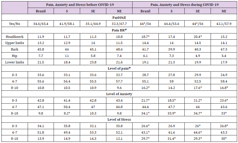
Table 2: Level of pain and mental health impact of the sample from the Brazilian general population (n = 1,848), aged from 18 years old and divided by the 3 mains Brazilian macro-regions, South (S) n=378, Southeast (SE) n=964 and Northeast (NE) n=431, before and during the COVID-19 pandemic. Values are presented as percentage (%)..

Note: PEx, Physical Exercise; BR, body regions; ᵆ percentages within the pain cases; *Chi-square statistical significance with Bonferroni post hoc correction, comparing before and after the COVID-19 pandemic.

Sociodemographic Characteristics and Sample Health Condition

The majority of the participants were young-adults (53.3%), followed by adults (23.4%), young (14.7%) and elderly (8.6%). Comparing Brazil with the mains macro-regions, the age distribution and sex was similar (P˃0.05). Between the respondents, 70% was female, 29.8% male and 0.2% did not want to declare. The mean±SD age was 39.5±13.3 years, body mass 71.4±26.3 kg, height 1.7±0.4 m and Body Mass Index (BMI) 25.4±7.8 kg.m-2. The S and NE regions presented less mean body mass and consequently less BMI compared to Brazil in general (24.5±4.3 and 24.8±4.1 kg.m- 2, respectively), (P˂0.05). Considering the marital status of the sample, 50.8% was married, 38.3% was single and 9.4% and 1.5% was, respectively divorced and widowed. The majority of the sample worked in the public (30.1%) or private sectors (28.2%). Students were 15.5%, retired 7% and unemployed 6.5%. The registered selfrelated current health condition showed that 56% of the sample had any disease, 14% respiratory diseases followed by musculoskeletal (10.7%), cardiac and related conditions (10.2%), mental health (8.4%) and 0.2% neurological diseases. Smoking habits occur in 4.1% of the total sample. Considering the sociodemographic and current health condition variables, no differences were found between Brazil and mains macro-regions (P˃0.05).

Social Isolation Status

Most of the individuals were in social isolation (~80%) at the time of the survey application and the ones that were not in social isolation (~15%) were not released from their jobs. Few respondents (1.5%) do not believe in the importance of social isolation.

Pain and Psychological Impact

The level of pain and psychological impact of the sample before and during the COVID-19 pandemic are presented on Table 2. Considering the pain before and during the COVID-19 pandemic for the Brazilian general population, the independent chi-square test showed that the COVID-19 pandemic influenced the pain perception [X2(1) = 33.2; P<0.0001]; the odds ratio for COVID-19 before/during is 1.477. When analyzing the different regions of Brazil, SE [X2(1) = 23.951; P<0.0001] e NE [X2(1)= 8.779; P<0.01] regions showed the same pattern, however, the S region accepted the null hypothesis [X2(1)= 1.384; P˃0.05]. To verify the body regions with pain before and during the COVID-19 pandemic for the Brazilian general population, the independent chi-square test showed that the COVID-19 pandemic influenced the body regions with pain [X2(4)= 15.215; P<0.01]; after the Bonferroni post hoc correction only the pain in head and neck regions were influenced by the pandemic, with increase in pain (P<0.001).

When analyzing the different regions of Brazil, only the SE [X2(4)= 11.956; P<0.001] region showed the same pattern for an increase in head and neck pain during the pandemic; however, the S and NE regions accepted the null hypothesis [X2(4)= 4.434; P˃0.05] and [X2(4)= 2.763; P˃0.05], respectively. The level of selfrelated pain analysis before and during the COVID-19 pandemic for the Brazilian general population, the independent chi-square test showed that the COVID-19 pandemic influenced the level of pain [X2(2)= 11.635; P<0.01]; after the Bonferroni post hoc correction the pain in the highest levels (8-10) were influenced by the pandemic, with increase in pain (p=0.001). When analyzing the different regions of Brazil, only the SE [X2(4)= 7.357; P<0.05] region showed the same pattern for the highest levels of pain during the pandemic after the post hoc (P<0.01); however, the S and NE regions accepted the null hypothesis [X2(2)= 1.823; P˃0.05] and [X2(2)= 5.146; P˃0.05], respectively.

The level of self-related anxiety before and during the COVID-19 pandemic for the Brazilian general population showed with the independent chi-square test that the COVID-19 pandemic influenced the level of anxiety [X2(2) = 376.904; P<0.0001]; after the Bonferroni post hoc correction the anxiety in the highest levels (8-10) were influenced by the pandemic, with increase in anxiety (P<0.01). When analyzing the different regions of Brazil, the S region [X2(2)= 92.292; P<0.0001] after the Bonferroni post hoc correction the anxiety in the smallest levels (0-3) decreased (P<0.0001) probably due to the increase of the highest levels of anxiety (8-10), (P<0.0001); the SE [X2(2)= 197.898; P<0.0001] and NE [X2(2)= 80.534; P<0.0001] regions showed the same pattern for the smallest and highest levels of anxiety during the pandemic after the post hoc (p<0.0001).

To verify the stress before and during the COVID-19 pandemic for the Brazilian general population, the independent chi-square test showed that the COVID-19 pandemic influenced the stress [X2(2)= 137.857; P<0.0001]; after the Bonferroni post hoc correction all self-related stress levels were influenced by the pandemic, with an decrease in the low (0-3) and moderate (4-7) levels and an increase in the highest stress levels (P<0.0001). When analyzing the different regions of Brazil, the S region [X2(2)= 29.617; P<0.0001] after the Bonferroni post hoc correction the anxiety in the highest levels of stress (8-10) increased during the pandemic (P<0.0001); the SE [X2(2)= 62.693;P<0.0001] after the post hoc for all stress levels (P<0.01); and NE [X2(2)= 41.933; P<0.0001] regions showed an increase for the highest levels of stress (P<0.0001) and a decrease for the smallest stress levels (P<0.01) during the pandemic.

Discussion

Introduction

The necessity of being confined at home as a consequence of the outbreak of COVID-19 caused great involvement and concerns worldwide, changing drastically people daily life and quality of life [1], the economy and the nations’ health systems, especially during the first wave of the pandemic. As a consequence of the Social Isolation (SI), a negative impact in the general health due to sedentary behaviors [2-6], pain [6,7] and lead to psychological conditions, such as anxiety and stress [6,8,9] would be possibly expected. The study of SI impact has been investigated on pain conditions and mental health. A longitudinal study about SI in people seeking treatment for pain management investigated the SI interference on pain and provided evidence that individuals with higher levels of SI status presented greater impact on pain levels and decreased physical function [10]. Another study investigated whether SI predicts the clinical outcomes of disability, anxiety, depression and pain in people with low back pain [11].

Emotional support, to participate in social activities, and satisfaction are inherent to human beings [10] and negative social experiences can induce profound effect on our emotional wellbeing [12]; loneliness on a daily basis, shows an up regulation of pro-inflammatory response genes, which may contribute to the risk of inflammatory diseases in individuals who experience chronically high levels of subjective SI [13]. The degree of SI can induce a higher or lower prevalence of physical or mental health problems [12]. Considering that the first wave of COVID-19 caused great public concern, because the population experienced the fear of the unknown, the fear of death and the amount of daily information about the pandemic “numbers”, the aim of this study was to use the validated questionnaire “Physical exercise level before and during social isolation” (PEF-COVID19) [14], to identify in Brazil and its mains macro-regions, the changes in the level of pain and psychological impact as stress and anxiety during the first wave of COVID-19 outbreak. Our hypothesis was that COVID-19 lockdown will result in major levels of self-related pain, anxiety and stress.

Materials And Methods

A self-administered questionnaire delivered through the e-mail and social media was conducted in this observational crosssectional study. The data collection happened in Brazil from 21st April until May 3rd 2020. Ethical approval was obtained from of the Hospital Universitário Pedro Ernesto (HUPE), Universidade do Estado do Rio de Janeiro (UERJ), Plataforma Brasil with the number CAAE 30649620.1.0000.5259.

Sample

Brazilian population over 18 years old was invited to participate of this study. The responders who agreed to participate, after an explanation of the consent form in the beginning of the survey, with ≥18 years old, were included. At any time, the responder could give up concluding the survey without any penalty or constraints. Brazilians were divided according to the macro-region where they lived during the survey application. Survey. The questionnaire PEFCOVID19 was created to assess the levels of physical exercise and psychological impact of general population before and during the social isolation due to the COVID-19 pandemic. The psychometric properties of this instrument were previously described [14], and it reports the validity indexes and the test-retest reliability and feasibility, being considered a valid and reliable instrument.

The PEF-COVID19 was divided into four sections,

(I) Subject’s characterization with demographic, anthropometric and health status questions;

(II) Physical exercise performed or not, pain, anxiety and stress before COVID-19;

(III) Confinement situation update;

(IV) Physical exercise performed or not, pain, anxiety and stress during COVID-19 [14].

Data Collection

The questionnaire was distributed using social and network media (Whatsapp, Facebook, Messenger, Linkedin), and email inviting people in general to participate and asking them to share with family and friends through the link: https://docs.google.com/ forms/d/e/1FAIpQLScgnHqfHH5NjLQHv6sLnbtz38eg1hBTu3U y3jxgIV0Ou7-_gw/viewform?usp=sf_link. Participant responses were secured where the data was recorded, scaled and scored in electronic sheets by custom Excel formulas for later statistical analysis.

Statistical Analysis

All data were exported to an Excel sheet. Nominal data were coded for statistical purposes. The statistical analyses were performed using IBM SPSS Statistics for Windows (version 21.0., IBM Corp., Armonk, NY, USA). Descriptive statistical analyses (n, %) were performed and the independent chi-square test. Post hoc analyses were performed on multiple category data with the Bonferroni correction. Comparisons between the groups (Brazil and mains Brazilian macro-regions) for the categorical variables of pain, stress, anxiety, sex, smoking habits, were made using chi squared analysis. Age distributions, mass, height, body mass index, years smoking, between Brazil and mains macro-regions, were compared using an independent t test.

Results

A total of 1,977 participants filled the survey. Prior to data analysis, 129 answers were excluded (6.5%) of the initial respondents because they were from other countries than Brazil or with age below 18 years-old, leaving a total of 1,848 questionnaires to be considered in the current study. Brazil has five different macroregions. Table 1 shows different Brazilian regions population, the number of respondents and the respondents’ percentage per millions of people from each region. South (S), Southeast (SE) and Northeast (NE) are the regions with the higher percentages of respondents per million of inhabitants. In general, the survey reached 8.8% of respondents per million of person considering the Brazilian population. As expected, Midwest and North regions, with the bigger territories and smaller population, had a little number of answers and for that reason these answers were analyzed in the Tables with the Brazilian population as a whole.

Table 1: Number of respondents per million from the different Brazilian regions.

Note: *DATASUS (http://tabnet.datasus.gov.br/cgi/tabcgi.exe?ibge/cnv/poptuf.def);

Table 2: Level of pain and mental health impact of the sample from the Brazilian general population (n = 1,848), aged from 18 years old and divided by the 3 mains Brazilian macro-regions, South (S) n=378, Southeast (SE) n=964 and Northeast (NE) n=431, before and during the COVID-19 pandemic. Values are presented as percentage (%)..

Note: PEx, Physical Exercise; BR, body regions; ᵆ percentages within the pain cases; *Chi-square statistical significance with Bonferroni post hoc correction, comparing before and after the COVID-19 pandemic.

Sociodemographic Characteristics and Sample Health Condition

The majority of the participants were young-adults (53.3%), followed by adults (23.4%), young (14.7%) and elderly (8.6%). Comparing Brazil with the mains macro-regions, the age distribution and sex was similar (P˃0.05). Between the respondents, 70% was female, 29.8% male and 0.2% did not want to declare. The mean±SD age was 39.5±13.3 years, body mass 71.4±26.3 kg, height 1.7±0.4 m and Body Mass Index (BMI) 25.4±7.8 kg.m-2. The S and NE regions presented less mean body mass and consequently less BMI compared to Brazil in general (24.5±4.3 and 24.8±4.1 kg.m- 2, respectively), (P˂0.05). Considering the marital status of the sample, 50.8% was married, 38.3% was single and 9.4% and 1.5% was, respectively divorced and widowed. The majority of the sample worked in the public (30.1%) or private sectors (28.2%). Students were 15.5%, retired 7% and unemployed 6.5%. The registered selfrelated current health condition showed that 56% of the sample had any disease, 14% respiratory diseases followed by musculoskeletal (10.7%), cardiac and related conditions (10.2%), mental health (8.4%) and 0.2% neurological diseases. Smoking habits occur in 4.1% of the total sample. Considering the sociodemographic and current health condition variables, no differences were found between Brazil and mains macro-regions (P˃0.05).

Social Isolation Status

Most of the individuals were in social isolation (~80%) at the time of the survey application and the ones that were not in social isolation (~15%) were not released from their jobs. Few respondents (1.5%) do not believe in the importance of social isolation.

Pain and Psychological Impact

The level of pain and psychological impact of the sample before and during the COVID-19 pandemic are presented on Table 2. Considering the pain before and during the COVID-19 pandemic for the Brazilian general population, the independent chi-square test showed that the COVID-19 pandemic influenced the pain perception [X2(1) = 33.2; P<0.0001]; the odds ratio for COVID-19 before/during is 1.477. When analyzing the different regions of Brazil, SE [X2(1) = 23.951; P<0.0001] e NE [X2(1)= 8.779; P<0.01] regions showed the same pattern, however, the S region accepted the null hypothesis [X2(1)= 1.384; P˃0.05]. To verify the body regions with pain before and during the COVID-19 pandemic for the Brazilian general population, the independent chi-square test showed that the COVID-19 pandemic influenced the body regions with pain [X2(4)= 15.215; P<0.01]; after the Bonferroni post hoc correction only the pain in head and neck regions were influenced by the pandemic, with increase in pain (P<0.001).

When analyzing the different regions of Brazil, only the SE [X2(4)= 11.956; P<0.001] region showed the same pattern for an increase in head and neck pain during the pandemic; however, the S and NE regions accepted the null hypothesis [X2(4)= 4.434; P˃0.05] and [X2(4)= 2.763; P˃0.05], respectively. The level of selfrelated pain analysis before and during the COVID-19 pandemic for the Brazilian general population, the independent chi-square test showed that the COVID-19 pandemic influenced the level of pain [X2(2)= 11.635; P<0.01]; after the Bonferroni post hoc correction the pain in the highest levels (8-10) were influenced by the pandemic, with increase in pain (p=0.001). When analyzing the different regions of Brazil, only the SE [X2(4)= 7.357; P<0.05] region showed the same pattern for the highest levels of pain during the pandemic after the post hoc (P<0.01); however, the S and NE regions accepted the null hypothesis [X2(2)= 1.823; P˃0.05] and [X2(2)= 5.146; P˃0.05], respectively.

The level of self-related anxiety before and during the COVID-19 pandemic for the Brazilian general population showed with the independent chi-square test that the COVID-19 pandemic influenced the level of anxiety [X2(2) = 376.904; P<0.0001]; after the Bonferroni post hoc correction the anxiety in the highest levels (8-10) were influenced by the pandemic, with increase in anxiety (P<0.01). When analyzing the different regions of Brazil, the S region [X2(2)= 92.292; P<0.0001] after the Bonferroni post hoc correction the anxiety in the smallest levels (0-3) decreased (P<0.0001) probably due to the increase of the highest levels of anxiety (8-10), (P<0.0001); the SE [X2(2)= 197.898; P<0.0001] and NE [X2(2)= 80.534; P<0.0001] regions showed the same pattern for the smallest and highest levels of anxiety during the pandemic after the post hoc (p<0.0001).

To verify the stress before and during the COVID-19 pandemic for the Brazilian general population, the independent chi-square test showed that the COVID-19 pandemic influenced the stress [X2(2)= 137.857; P<0.0001]; after the Bonferroni post hoc correction all self-related stress levels were influenced by the pandemic, with an decrease in the low (0-3) and moderate (4-7) levels and an increase in the highest stress levels (P<0.0001). When analyzing the different regions of Brazil, the S region [X2(2)= 29.617; P<0.0001] after the Bonferroni post hoc correction the anxiety in the highest levels of stress (8-10) increased during the pandemic (P<0.0001); the SE [X2(2)= 62.693;P<0.0001] after the post hoc for all stress levels (P<0.01); and NE [X2(2)= 41.933; P<0.0001] regions showed an increase for the highest levels of stress (P<0.0001) and a decrease for the smallest stress levels (P<0.01) during the pandemic.

Discussion

To the best of our knowledge, this is the first study to evaluate the level of pain and psychological impact as stress and anxiety together in Brazilian mains macro-regions during the COVID-19 outbreak. Our hypotheses were confirmed, and COVID-19 lockdown contributed negatively to increase the levels of selfrelated pain, anxiety and stress. It was observed that the majority of the respondents were following the social isolation during the first wave of the pandemic and government restrictions (about 80%) considering that in February 3rd the epidemic was declared a Public Health Emergency of National Concern [15] following the notifications of the disease spread and deaths in all continents [16]. The decrease of exercise and physical activity during the pandemic and SI can also generate mental health impacts, as stress or anxiety [17] that many will experience in the face of the feeling of fear [18] (fear of being sick or dying, possible loss of family and friends, economic impact) and isolation from common social life [19,20]. Moreover, this survey shows alarming results considering the level increase of anxiety and stress, which the highest levels (8- 10) in a scale with a maximum of 10 points increased significantly comparing before and during the pandemic for Brazil.

A recent study [21] investigated the fear of COVID-19, stress and anxiety in undergraduate students from Ecuador; they observed the complex relationships between fear, stress, and anxiety in the development of depression symptoms and how they can be taken into account in programs aimed to prevent and alleviate the mental health disorders. Two other studies presented also the effects of the pandemic on mental health for general Brazilian population [6,22] and in both studies anxiety, depression and stress scores were higher during the pandemic, and mainly the ones that were not physically active [22]. Also, the self-related pain level increased significantly mainly in the head and neck regions for Brazilians in general and SE macro-region inhabitants. Inappropriate posture watching television, using computers [23,24] and smartphones [25] or psychosocial stressors [20,26] might be the reason for the pain increase in such body regions. Furthermore, in a review [27], the authors verified strong consistent evidence for the relationship between both psychological distress, and time spent using screens for leisure and depressive symptomatology.

Moderate evidence supported the relationship between low self-esteem and screen use. Poorer mental health status was found among adolescents using screen time more than 2-3h per day, and gender differences exist. Corroborating with our findings, considering that SI can favor chronic stress and pain, Majumdar et al, 2020 [28] reported that the chronic stress of living through a pandemic led to a host of physical symptoms, like headaches, insomnia, digestive problems, hormonal imbalances, and fatigue. The southeastern region is the most populated and industrialized region of Brazil and probably suffered most an economic imbalance due to the pandemic. Since the outbreak of the novel coronavirus was first detected, the world has been challenged since the health systems were overloaded in large large-scale, economy income decreased and market volatility, unemployment, mental health issues and lifestyle changes with sedentary behavior increase, leading, perhaps, to an increase in chronic diseases.

Professions were also reinvented as populations never expected, mainly the education and heath area professions. Moreover, as it was pointed out by Zieff, et al. [29], targeting sedentary behavior is a simple strategy that may help counter physical and mental health concerns associated with COVID-19-related social restrictions. During the SI, Brazilians in general decreased the self-related levels of physical exercise and changed the exercise habits [5]. Of course, traditional strategies such as achieving optimal exercise and physical activity levels are also important and should be recommended. However, even under normal circumstances, the difficulty in promoting lasting exercise habits at multiple levels (e.g. individual, environment, policy) are well documented [4,5,29,30], and chances of maintaining or improving these factors is now even lower. Thus, relative to other lifestyle behaviors - sedentary behavior may be more amenable to change.

Moreover, reducing sedentary behavior may be less susceptible to influence from factors associated with health disparities such as age, race, and socio-economic status compared to exercise or physical activity. Sedentary behavior is a feasible health strategy that should be targeted during COVID-19. The strengths and the applications of the findings described in this study and considering the return to the normal life is still unexpected, the results from the current study can help to create measures to decrease mental health disturbances, facing the fear of the unknown. Moreover, as a perspective, it is expected that with the comparison of some parameters before and during the outbreak, the results can aid in the definition of policies to help the Brazilian population, in general, and in specific regions, due to the COVID-19 period post-pandemic. As a limitation, the number of respondents was different in the various regions that were investigated. Moreover, in two regions, North and Midwest, the number of respondents was reduced.

Due to the unknown home-quarantine in each region of the country, a specific adaptation of the questionnaire was not possible to be done. In general, the Brazilian regions have specific characteristics, as nutrition, political approaches, and local costumes. Also, self-reported instruments can induce bias. The findings obtained with the PEF-COVID19 showed, in general, that the Brazilian general population was affected by the pandemic considering the increased levels of self-related pain, stress and anxiety. SE macro-region is the economic center of Brazil presented a significant increase in the levels of self-related pain compared to the others mains macro-regions; the higher levels (8-10) of stress and anxiety increased significantly in all studied macroregions, with similar results. These results will be valuable to the establishment of actions to aid the population of Brazil to minimize, with different approaches, the undesirable commitment due the social distancing related to the COVID-19.

A recent study [21] investigated the fear of COVID-19, stress and anxiety in undergraduate students from Ecuador; they observed the complex relationships between fear, stress, and anxiety in the development of depression symptoms and how they can be taken into account in programs aimed to prevent and alleviate the mental health disorders. Two other studies presented also the effects of the pandemic on mental health for general Brazilian population [6,22] and in both studies anxiety, depression and stress scores were higher during the pandemic, and mainly the ones that were not physically active [22]. Also, the self-related pain level increased significantly mainly in the head and neck regions for Brazilians in general and SE macro-region inhabitants. Inappropriate posture watching television, using computers [23,24] and smartphones [25] or psychosocial stressors [20,26] might be the reason for the pain increase in such body regions. Furthermore, in a review [27], the authors verified strong consistent evidence for the relationship between both psychological distress, and time spent using screens for leisure and depressive symptomatology.

Moderate evidence supported the relationship between low self-esteem and screen use. Poorer mental health status was found among adolescents using screen time more than 2-3h per day, and gender differences exist. Corroborating with our findings, considering that SI can favor chronic stress and pain, Majumdar et al, 2020 [28] reported that the chronic stress of living through a pandemic led to a host of physical symptoms, like headaches, insomnia, digestive problems, hormonal imbalances, and fatigue. The southeastern region is the most populated and industrialized region of Brazil and probably suffered most an economic imbalance due to the pandemic. Since the outbreak of the novel coronavirus was first detected, the world has been challenged since the health systems were overloaded in large large-scale, economy income decreased and market volatility, unemployment, mental health issues and lifestyle changes with sedentary behavior increase, leading, perhaps, to an increase in chronic diseases.

Professions were also reinvented as populations never expected, mainly the education and heath area professions. Moreover, as it was pointed out by Zieff, et al. [29], targeting sedentary behavior is a simple strategy that may help counter physical and mental health concerns associated with COVID-19-related social restrictions. During the SI, Brazilians in general decreased the self-related levels of physical exercise and changed the exercise habits [5]. Of course, traditional strategies such as achieving optimal exercise and physical activity levels are also important and should be recommended. However, even under normal circumstances, the difficulty in promoting lasting exercise habits at multiple levels (e.g. individual, environment, policy) are well documented [4,5,29,30], and chances of maintaining or improving these factors is now even lower. Thus, relative to other lifestyle behaviors - sedentary behavior may be more amenable to change.

Moreover, reducing sedentary behavior may be less susceptible to influence from factors associated with health disparities such as age, race, and socio-economic status compared to exercise or physical activity. Sedentary behavior is a feasible health strategy that should be targeted during COVID-19. The strengths and the applications of the findings described in this study and considering the return to the normal life is still unexpected, the results from the current study can help to create measures to decrease mental health disturbances, facing the fear of the unknown. Moreover, as a perspective, it is expected that with the comparison of some parameters before and during the outbreak, the results can aid in the definition of policies to help the Brazilian population, in general, and in specific regions, due to the COVID-19 period post-pandemic. As a limitation, the number of respondents was different in the various regions that were investigated. Moreover, in two regions, North and Midwest, the number of respondents was reduced.

Due to the unknown home-quarantine in each region of the country, a specific adaptation of the questionnaire was not possible to be done. In general, the Brazilian regions have specific characteristics, as nutrition, political approaches, and local costumes. Also, self-reported instruments can induce bias. The findings obtained with the PEF-COVID19 showed, in general, that the Brazilian general population was affected by the pandemic considering the increased levels of self-related pain, stress and anxiety. SE macro-region is the economic center of Brazil presented a significant increase in the levels of self-related pain compared to the others mains macro-regions; the higher levels (8-10) of stress and anxiety increased significantly in all studied macroregions, with similar results. These results will be valuable to the establishment of actions to aid the population of Brazil to minimize, with different approaches, the undesirable commitment due the social distancing related to the COVID-19.

For more Articles on : https://biomedres01.blogspot.com/

Faba Bean Varieties Response to Phosphorus Application on Yield and Yield Components at Kulumsa Area of Tiyo District, Arsi Zone, South-Eastern Ethiopia

  Faba Bean Varieties Response to Phosphorus Application on Yield and Yield Components at Kulumsa Area of Tiyo District, Arsi Zone, South-Ea...