Showing posts with label Journals on Biomedical Engineering Journals on Biomedical Science Journal on Medical Genetics Journals on Cancer Medicine Journals on Medical Informatics. Show all posts

Thursday, December 1, 2022

A Tensional Network in the Knee


A Tensional Network in the Knee


The Medium of Haptic Perception

In contrast to the widespread assumption that the neural system generates impulses to the motor units in the skeletal muscle fibers, Turvey [1] place high importance on the fibrous collagenous connective tissues in the body; these are seen as providing a tensional network throughout the whole body, the biomechanical properties of which provide the framework for muscular force transmission as well as for the haptic sensory system. To identify the haptic system’s medium, Turvey focused on connective tissue and the conjunction of muscular, connective tissue net, and skeletal as the body’s proper characterization Turvey [1]. Myers has also posed the medium as a body-wide responsive physiological network—the myofascial meridian Myers [2]. Taking on ‘geometry’ first, cell biologist Donald Ingber placed one final piece of the puzzle: to view the body’s architecture in the light of ‘tensegrity’ geometry Ingber [3]. ‘Tensegrity’ was coined from the phrase’ tension integrity’ by the designer R. Buckminster Fuller (working from original structures developed by artist Kenneth Snelson Skelton [4]. As Snelson describes it Snelson [5], “The sculpture could be put into orbit in outer space, and it would maintain its form. Its forces are internally locked. These mechanical forces, compression, and tension or push and pull are invisible—just pure energy—in the same way that magnetic or electric fields are invisible.”
The tensegrity principle describes precisely the relationship between the connective tissues, the muscles, and the skeleton. For example, weight applied to shank/thigh bones would cause it to slide off its knee joint if it were not for the tensional balances that hold it in place and control its pivoting Swanson [6]. The invariant feature of tensegrity structures encompasses those that stabilize themselves through a phenomenon known as pre-stressing. Architects call this type of pre-stressed structural network, composed of opposing tension and compression elements that self-stabilize its shape by establishing a mechanical force balance, a tensegrity structure. Bio tensegrity is a term introduced by Dr. Stephen Levin and denotes the application of tensegrity’s principles to biological structures Hutson [7]. Tensional forces naturally transmit over the shortest distance between two points, so the elastic members of tensegrity structures are precisely positioned to withstand applied stress. For this reason, tensegrity structures offer a maximum amount of strength for any given amount of material Myers [2]. Furthermore, the invariant feature of a knee tensegrity system (specified by a given set of external forces such as the ground reaction force (GRF)) is a stable equilibrium if the structure returns to the initially given configuration after the application of arbitrarily small perturbations with respect to the functional knee axis (FKA) anywhere within the configuration Ingber [3].
Kim [8] showed how the knee tensegrity system manages the balance between tension and compression during locomotion by utilizing a unique combination of the FKA and GRF stimuli Kim [8]. When deformed by the shank to the ground via GRF, the strain is distributed over the whole structure, not locked into the joint itself by virtue of the assembly Phillips [9]. Thus, a reaction torque is zero on the knee joint if the GRF line of action intersects the joint axis. The configuration can exert a significant force on the ground without overloading the knee joint. This study aims to introduce the conception of the tensional network in muscles, i.e., the action lines that satisfy the given condition as the tensegrity’s structure is characterized by transmitting forces across the bones Kim [10]. Thus, we hypothesized that tensional forces in muscles are derivable from a scalar function, the work function.

N-lines as the Medium of Haptic Perception

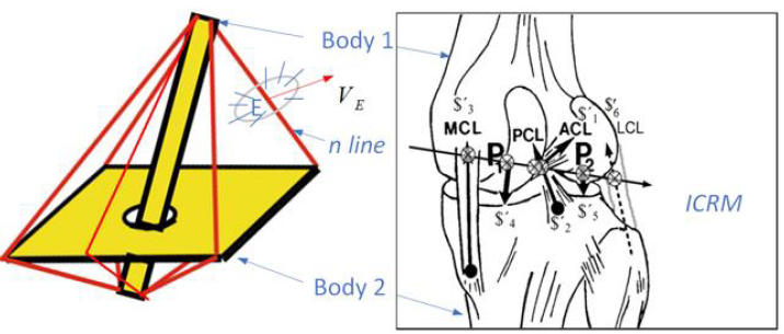
Neurophysiologist Nikolai Bernstein (1967) defined coordination as mastering the many degrees of freedom (DOF) of a particular movement by reducing the number of variables to be controlled Turvey [11]. Recently, a contemporary perspective on Bernstein’s concept of synergies has been proposed Profeta [12]. The muscle synergy is equivalent to the complexity of lines, a manifold approximated by individual fibers Kim, et al. [13]. Muscles are not functional units, even though this is a common misconception. Instead, most muscular movements are generated by many individual motor units distributed over some portions of one muscle, plus portions of other muscles. The tensional forces of these motor units are then transmitted to a complex network of fascia sheets, bags, and strings, which convert them into the final joint/body movement Myers [14]. We define an n-line in a body as a member of the tensional network by virtue of some constraint upon the body Phillips [9]. This will mean that, whereas all n-lines in a body will immediately become a tensional network as soon as the body begins to move, not all members in a tensional network in a moving body will have been n-lines before they began to move. The whole regulus of n-lines is a tensional network for the three constraints at the instant (Figure 1a). This single infinity of n-lines are the generators of the regulars of lines, a linear ruled surface, upon the hyperboloid. Given any three lines in a body (protectively independent) which are bespoken by virtue of some existing constraints to be n-lines in the body, the whole regulus of lines to which the three bespoken lines belong are also n-lines in the body.
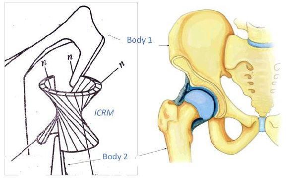
In (Figure 1), if any four forces along the n-lines are in equilibrium, their lines of action will reside in space as the lines of some regulus. What makes this remarkable is that, while any three of the lines of action are enough to define the hyperboloidal surface, the fourth line of action will be found, not to miss or to intersect that surface, but to lie automatically and exactly upon it. The object of the illustrated apparatus in (Figure 1) is to set up body 2 with 3 DOF with respect to body 1 in such a way that, within it, three n-lines do exist. The 3 DOF speaks about a body’s ICRM, its instantaneous capacity for relative motion with respect to some other body. It should next be clear that, given this infinity of n-lines in (Figure 1a), there is a corresponding infinity of hinges, which are the generators of the other regulus upon the same hyperboloid. The articulation between bones in (Figure 1b) is ball-and-socket type, which can take up the single infinity of hinges as the ICRM, which seem to be continuously registered by the joints; the changes of the angles seem to be episodically registered by their input to the nervous system Gibson [15]. The question to be answered is this: How does a perceiver feel what he is touching instead of the cutaneous impression and the bone posture as such Gibson [15]? Now to answer the question. In brief, The ICRM of the body (with its 3DOF) can be exactly reproduced by the substitution of (6DOF-3) direct points of contact between the body and its frame.

biomedres-openaccess-journal-bjstr

Figure 1:

(a) A regulus of n-lines represents the tensional network. It physically connects bodies 1 and 2. The whole regulus of hinges is an equivalent mechanical substitution for the three n-lines at the instant
(b) The articulation of bones is the ball-and-socket type.

Tensional Network

Skelton [16] defined a tensegrity configuration of rigid bodies as follows Skelton [16]. In the absence of external forces, let a set of rigid bodies in a specific configuration have torque less connections (e.g. via frictionless ball-joints). Then this configuration forms a tensegrity configuration if the given configuration can be stabilized by some set of internal tensile members, i.e. connected between the rigid bodies. The configuration is not a tensegrity configuration if no tensile members are required and/or no set of tensile members exist to stabilize the configuration. (p. 1) which are conjointly reciprocal to the ICRM as indicated by their intersections (at the ⊗ ’ s). A balance of forces happens when the virtual coefficient vanishes, being it the necessary and sufficient condition for knee equilibrium. The original anatomic schematics and lines of action were published previously Kim, et al. [17-19] and are used with the permission of Professor Michele Conconi. The video is available:
h t t p s : / / d r i v e . g o o g l e . c o m / f i l e / d / 1 8 _ Y t s z z T 3 _ IvNIken5uxObj4jmSd0Zs_/view?usp=sharing
Attached to body member 1, in (Figure 2a), by mean of five taut strings, there are five n-lines, exhibiting a body with 1 DOF. It follows that body 2 suffers constrained motion as the tensegrity system moves that the paths in body 1 of all pints in body 2 are predetermined. (Figure 2a) is a stable embodiment of the same tensegrity configuration, hence a tensegrity system, exhibiting a torque less connection between body 1 and body 2. The line n-E-n in (Figure 2a) is an n-line by this definition, for it is a tensional network by virtue of a constraint. A member in the tensional network in a moving body is any straight line that joins two points in the body whose linear velocities are perpendicular to the line. The instantaneous velocity at point E,, is not yet known without analysis. Still, we know that it will occur in one or another of the directions indicated by the planar pencil of possible directions vectors drawn upon the flat surface perpendicular to the n-line at the point there, E (Figure 2a). The intra-articular structures of the tensegrity system of the knee include the muscles, the anterior cruciate ligament (ACL), posterior cruciate ligament (PCL), medial collateral ligament (MCL), lateral collateral ligament (LCL), and articular contact in the medial (P1) and lateral (P2) compartments (Figure 2b) Kim [18]. We have shown that six n-lines or constraints are members of the “tensional network” and are spatially oriented in such ways that by imposing an internal tension or “pre-stress” to reduce the play in the system; this ensures immediate mechanical responsiveness (all others feel, i.e., that movement of one element) and reduces impact fatigue at the joint.

Please refer to the authors ‘ previous works towards a visualization of the linear complex set Kim [13]. Let us look at the case next of six forces in equilibrium. When six forces are in equilibrium, their lines of action in the n-lines in the tensional network will be members of the same linear complex. Line manifold contraction is a linear line complex Jessop [20] defined by screws ICRM (Figure 2b). Using the superposition principle of infinitesimal quantities of DOF Kim [21], we can apply these two types of displacement as a body twist around a screw called Instantaneous Screw Ball [22]. The lines remain within the complex in any screw motion along a line axis, forming a linear complex. Additional cognitive processes or internal representations are not needed to explain these phenomena, as perception and action are coupled. Perceptual systems are active sets of organs designed to reach equilibrium through synergies Smart [23]. Our previous research Kim, et al. [24] introduced the concept of measurable invariance of the knee perceptual organ. In such invariant, six constraints ($) are collectively reciprocal to the instantaneous knee screw (ICRM) indicated by ⊗ (Figure 2b). These metrics predicted the knee synergy model based on synergies Turvey [25]. Moreover, this perspective defines torque-free pure forces based on the tensegrity structure Kim, et al. [26,27,21,24].
It is important to note that this configuration is a tensegrity configuration, as the system is pre-stress able in the absence of external forces, such as ground reaction forces during actual locomotion Skelton [16]. It was shown the knee tensional network (KTN) has six constraints and that it can balance the forces between tension and compression in the joint such that no work results Huang [28]. The KTN can be pre-stressed to obtain the same configuration as if external loads were applied. The selected pre-stress may yield the same configuration in the swing phase (external forces are absent) as in the stance phase (external forces are present) Skelton [9]. Notably, preparedness is not only a reactive aspect of the movement apparatus, but it also relates to anticipatory adjustments that predispose a system to behave in a particular way Profeta [12].

biomedres-openaccess-journal-bjstr

Figure 2:

(a) A set of rigid bodies in a specific tensegrity configuration have torque less connections.
(b) The knee joint synergy is represented by six constraints ($' , 1,..,6 i i = ).

Ensembles of a Geometric Pattern

It has been shown that a special kind of line in a body, which is determined by the constraints, namely n-line, is a regularly occurring and probably useful kind of line in bio tensegrity. Moreover, an important theorem about the overall layout or tensional network of n-lines in a body has been presented: the beginnings of a geometric pattern that emerged; it has been found in some special cases only, that associated 1 DOF and 3 DOF of a body in tensegrity, there are ensembles of n-lines existing which are, respectively, linear complex and regulus. This study has shown that the appearance or otherwise of n-lines in a body is a mystery. Still, it can be said that throughout the knee tensional network run two forces in the two types of n-lines: the one is mechanically operated by direct contact between bodies Lanczos [29]; the other is the living force exerted by the neuromuscular mechanism. Sylvester has shown that when six n-lines in (Figure 2) are so situated that forces acting along them equilibrate when applied to a free rigid body, a certain determinant vanishes, and he speaks of the six lines so related as being in involution Ball [22]. We shall see in a later investigation that these ensembles of lines figure largely and continuously in the theory of freedom and constraint of the tensegrity.

For more Articles on: https://biomedres01.blogspot.com/

Tuesday, June 14, 2022

Fura Da Nono Drink and Its Implications in the Treatment of Malnutrition in Bauchi Metropolis of Bauchi State


Fura Da Nono Drink and Its Implications in the Treatment of Malnutrition in Bauchi Metropolis of Bauchi State


Introduction

In many cultures, especially the western world, humans continue to consume milk beyond infancy, using the animal milk especially from cattle, goats and sheep as food products. For many decades, cow milk has been processed into dairy products such as condensed milk, skimmed milk, ice cream, butter, yoghurt and the more durable cheese products to fight malnutrition [1,2]. Malnutrition, in all its forms, includes under nutrition (wasting, stunting, underweight), inadequate vitamins or minerals, overweight, obesity, and resulting diet-related no communicable diseases is a major health concern [3,4]. This has stimulated the science of nutritional biology to progress extensively over the last decade to develop food-based nutraceuticals as a form of highly personalized medicine or therapeutic agent. The believe that Fura danono has nutritional abilities is well known but the medicinal values not been adequately studied.Fura da Nono is a food drink with potentially tremendous but under-explored source of nutraceutical properties as compared to other regularly consumed food drinks [5]. Millet is an important staple for food and nutritional security in semi-arid regions of the world [6]. It is an important staple in several semi-arid and tropical regions of the world with excellent nutraceutical properties as well as ensuring food security in these areas even during harsh environment [7].

Millet, the Fura in Fura da nono is known for several health benefits and some of the health benefits are attributed to its polyphenol and dietary fibre contents [8,9]. When consumed according to appropriate national guidelines, milk and its derivatives contribute essential micro- and macronutrients to the diet, especially in infancy and childhood where bone mass growth is in a critical phase. Furthermore, preliminary evidence suggests potentially protective effects of milk against overweight, obesity, diabetes, and cardiovascular disease [10]. The incidence of malnutrition and diseases arising from it are increasing in an exponential manner globally and to combat them, a raise in demand for food containing rich food classes with higher levels of dietary fibre and health beneficial phytochemicals has to be done [11]. In recent years, progress in addressing all forms of malnutrition has seen a declining trend, alarmingly slow, with >150 million children still stunted (Global Nutrition Report, 2018), but the movement has increased awareness among people and hence increased health-conscious people demanding healthy food relatively more often. The growing public awareness of nutrition and health care research substantiates the benefits of millets and its phytochemicals potential such as polyphenols and dietary fibre on human health. Many phytochemicals with established or potential biological activity have been identified in plants. Plants, including many currently used as culinary spices and herbs, have been used as medicines, not certainly effectively, from ancient times. Polyphenols of many classes are widely spread in plants [12].

Plants having phytoestrogens, a type of polyphenols, have been administered for decades for gynecological conditions, such as fertility, menopausal, and menstrual problems; Nicotine, an alkaloid, from tobacco directly binds to nicotinic acetylcholine receptors in the body, accounting for its pharmacological effects [13]. Cardiac glycosides, digitoxin and a host of other phytochemicals have been found in plant-based foods to be essential for human health [14]. Fura danono is a food drink with potentially tremendous but under-explored source of nutraceutical properties as compared to other regularly consumed food drinks [6]. Millet is an important staple for food and nutritional security in semi-arid regions of the world (Salej et al. 2019). It is an important staple in several semiarid and tropical regions of the world with excellent nutraceutical properties as well as ensuring food security in these areas even during harsh environment [8]. Millet, the fura in Fura da nono is known for several health benefits and some of the health benefits are attributed to its polyphenol and dietary fibre contents [3,4].

Aim of Study

The general objective of this study was to evaluate the Fura da Nono drink, and its health implications in the treatment of malnutrition. Specifically, the study.

1) Evaluated the knowledge of the nutritional and medicinal benefits of Fura da Nono consumption in Bauchi.

2) Assessed the public’s awareness levels of the nutritional and medicinal benefits of Fura da Nono consumption.

3) Determined the impact of Fura da Nono drink in the Treatment of Malnutrition.

Materials and Method

Research Design

This research was a cross-sectional descriptive study, involving indigenes of Bauchi town and streets of Bauchi metropolis, in Bauchi Local Government Area of Bauchi State, Nigeria. A random sampling technique was used in selecting the volunteers for this study. A self-administered questionnaire was adopted in generating information on socio-demographic characteristics, knowledge, awareness and factors affecting perceived outcome of Fura da nono consumption. The research was conducted as face to face interview and administration of questionnaire to 220 volunteers. Questionnaires was pre-tested on volunteers from a community not selected for this study, a nearby community with same cultural heritage as the communities in this study. The questionnaire was structured into the following parts.

1) Demographic characteristics,

2) Knowledge of knowledge about the consumption of Fura danono

3) Awareness of the nutritional benefits of Fura da nono

4) Awareness of the medicinal benefits of Fura danono

5) Factors influencing the perception of the benefits of Fura da nono.

Respondents were allowed the freedom to participate on their own accord without any form of compulsion to do so after a comprehensive education on what the nature, benefits and procedure for the research.

Study Area

This study was conducted in Bauchi metropolis, Bauchi State, Nigeria. Bauchi is the headquarters of Bauchi State in the Northeastern Nigeria. Bauchi has an important town and is the commercial nerve centre of the state. The area is predominantly semi–Sahel Savanna with large expanses of dry forests and has a land area of approximately 3,890 square kilometers. It is also home to two different ethnic groups namely, the Hausa and Fulani’s. It has a population of 303,417 as at the 2006 census. It is home to Yankari Game Reserve in Nigeria. Agriculture is the main stay of the State’s economy. The major crops cultivated in the State are millet, maize, guinea corn and rice and vegetables (pepper, tomato, onions, cabbage and leaf vegetables among others). Households in the study area are predominantly involved in farming and rearing of animals, petty trading and civil service.

Study Population

The study population for this study was derived from the entire bauchi town, comprising mainly of those participating in nono processing and those who buy and consume Furada nono.

Sampling Procedures and Sample Size

A sample of 220 consumers of Fura da nono drinks within the streets of Bauchi metropolis were selected at random to participate in the research.

Data Collection

A structured questionnaire was used to collect primary data for the consumers of Fura da nono drinks. The Questionnaire was carefully structured and comprised of closed-ended questions for the opinions of respondents. Furthermore, an interview checklist for key informants (health extension workers) was also employed. This was because the questionnaire was an expensive way to gather data from a potentially large number of respondents. Often, they are the only feasible way to reach a number of respondents larger to allow good statistical analysis and outcome.

Primary Data Collection

Primary data was collected by administering the questionnaire to respondents. Socioeconomic characteristics of the household head such as sex, age and educational status. The data were also collected in collaboration with well-trained enumerators who had received prior skill on the objective of the study, the contents of the interview and interviewing techniques. Proper training of enumerators and supervision during the data collection process boosted the reliability of the data.

Limitations

Fura da Nono has a refreshing taste and can also be consumed as a weaning food for infants. The major limitation of this food drink was obtaining informed consent from volunteers that has to do with the effect of germs during the preparation of Fura da nono from the apparent unhygienic conditions under which it is prepared, its poor shelf-life stability, storage and processing systems.

Statistical Approach

The results were calculated using mean and standard error of means (SEM) respectively. The data from the obtained cardiopulmonary variables were analyzed using one way ANOVA test and for significant level of (p < 0.05) as taken below variables. All statistical analysis and data presentation were automated with the graph pad prism version 8.

Results

Adding milk and dairy products to your diet may prevent bone diseases like osteoporosis. Studies have linked milk and dairy to a lower risk of osteoporosis and fractures, especially in older adults. This finding shows that the consumers of fura da nono drink are fully aware of its nutritional value. 185(87.26%) of the respondent disagree that with the statement which said that fura da nono can help regulate body weight may be as a result of patty nature nono (cow milk) it increases body weight if consumes on regular basis. The respondents 124(58.49%) agree that fura da nono can help the child develop well in the womb while 12(5.66%) respondents strongly disagree with the statement. The respondents disagree that fura da nono relieves fatigue 76(35.85%) because lactose intolerance is a common digestive problem where the body is unable to digest lactose, a type of sugar mainly found in milk and dairy products and the respondents agree that fura da nonpromoted sleep 102(48.11%). Majority of the consumers 77(36.32%) agree that fura da nonincreasing immunity because Milk contains nutrients like probiotics, vitamin D and immunoglobulins that boost the immune system and in turn reduce the risk of allergies (Table 1).

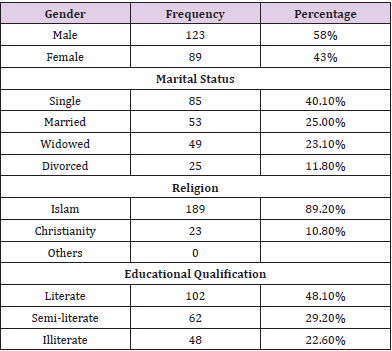
Table 1: Summary of Socio-Demographic Information of respondents.

Note: Demographic characteristics of the respondents are presented in Table I. A total of 212 persons responded to the questionnaire. Consumers of fura da nono were those who buy fura and either takes as meal or food supplement. The respondents were comparable in most demographic characteristics.The table shows that 123(56%) of the respondents are male while only 89(42%) of the respondents are female. The result indicates that males consume fura da nono more than females in Bauchi metropolis. The table further shows that majority of the respondents 40.1% of them are married while 25% of them are single and only 35% of the respondents where widowed and divorced. Majority of the respondents are Muslims (89.2%) while 10.8% of the respondents are Christians. This is because the residents in Bauchi LGA are predominantly a Muslims. None of the respondents are having any other religion apart from Islam and Christianity. table shows that 48% of the respondents are literate while 29.2% are semi-literate and only 22.6% are illiterate.

Discussion

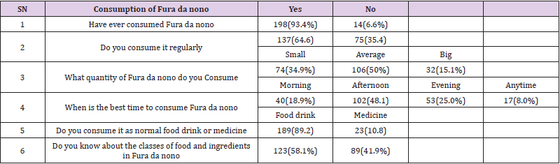
The findings in Table 2 shows responses of the respondents on the knowledge about the Consumption of Fura de nunu, about 93.4% of the respondents said that they have consumed Fura de nunu while only 6.6% of the respondents said that they have never consumed Fura de nunu also this finding shows that majority of the people living in Bauchi LGA are Fura de nunu consumers. Furthermore, the table indicates that majority of people in Bauchi LGA consume Fura de nunu regularly about 137(64.6%) while 106(50%) respondents consume Fura de nunu on average basis. Majority of the respondents said that the best time to consume Fura de nunu is afternoon which is the percentage occurrence. The information shows the reasons why respondents consumed Fura de nunu. The majority of the respondents indicated that the nutritional quality of Fura de nunu drinks.

Table 2: Knowledge on the Consumption of Fura Da Nono.

Note: Table II above shows responses of the respondents on the knowledge about the Consumption of Fura Da Nono. 93.4% of the respondents said that they have consumed fura da nono while only 6.6% of the respondents said that they have never consumed fura da nono. This finding shows that majority of the people living in Bauchi LGA are fura da nono consumers.The table indicates that majority of people in Bauchi LGA consume fura da nono regularly about 137(64.6%) while 106(50%) respondents consume fura da nono on average basis. Majority of the respondents said that the best time to consume Fura da nono is afternoon which is the percentage occurrence. The respondents 189(89.2%) said that they consume the drink as food drink not as no normal food while only 23 of them said they consumed as a regular food. Also majority 123(58.1%) of them are aware of the classes of food and ingredients in fura da nono. The information shows the reasons why respondents consumed fura da nono. The majority of the respondents indicated that the nutritional quality of fura da nono drinks.

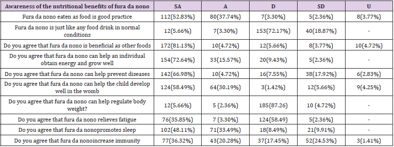
Also, it has been observed that a moderated consumption of Fura de nono is advocated such that the general public would take advantage of its antimicrobial effect in addition to its acclaimed physiological roles-lactation in nursing mothers, purgative effect and cure for flatulence as well as nutrient composition [15]. The peculiar sweet-tainted sour taste of Fura de nunu is noteworthy. The results obtained showed that Fura de nunu has antimicrobial properties and further supports the claim that the drink is medicinal. Again, the result of the findings was shown in Table 3 it clearly indicated that majority 80(37.74%) Agree that drinking Fura de nunu drink as food is good practice and majority 153 (72.17%) of them disagree statement that Fura de nunu is just like any food drink in normal conditions and strongly agree 172 (81.13%) that Fura de nunu is beneficial as other foods. Furthermore, the respondents 154 (72.64%) strongly agree that Fura de nunu can help an individual to obtain energy and grow well while 142(66.98%) of the respondents agree that Fura de nunu can help prevent diseases. This result was in line with the findings of Zhu K., Prince R.L. (2012) they observed adding milk and dairy products to your diet may prevent bone diseases like osteoporosis [16,17].

Table 3: Level of Awareness on the Nutritional Benefits of Fura Da Nono.

Note: Table III shows the Awareness of the Nutritional Benefits of Fura Da Nono, it also clearly indicated that majority 80(37.74%) Agree that eaten Fura Da Nono as food is good practice and majority 153(72.17%) of them disagree statement that Fura Da Nono is just like any food drink in normal conditions and strongly agree 172(81.13%) that fura da nono is beneficial as other foods. Furthermore, the respondents 154(72.64%) strongly agree that fura da nono can help an individual to obtain energy and grow well while 142(66.98%) of the respondents agree that fura da nono can help prevent diseases.

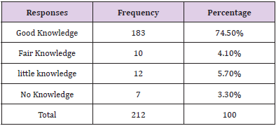
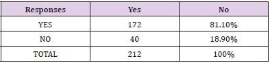
Studies have linked milk and dairy to a lower risk of osteoporosis and fractures, especially in older adults. This finding shows that the consumers of Fura de nunu drink are fully aware of its nutritional value. Most of the consumers are fully aware of the nutritional value of the fura da nono drinks because millet from which fura is made can help reduce the effects of migraines and heart attacks due to the presence of magnesium. Although oats have been widely publicized for their heart-protective properties, millet is a grain that should also be included on your list of heart-healthy choices because of its status as a good source of magnesium. Magnesium has been shown in studies to reduce the severity of asthma and to reduce the frequency of migraine attacks. Magnesium has also been shown to lower high blood pressure and reduce the risk of heart attack, especially in people with atherosclerosis or diabetic heart disease. From Table 4. Majority of the respondents (74.5%) have good knowledge on the use of fura da nono in tackling malnutrition. While (5.7%) of them have little knowledge. Research indicates that Cow’s milk protein is a key ingredient in products used for treatment of severe acute malnutrition (SAM), such as F-100 and ready-to-use therapeutic foods (RUTFs). Also, the respondents 3.3% of them have no knowledge on nutritional value of Fura de nunu. Also, Table 5 indicates that majority of the respondents (81.1%) use to mixed Fura de nunu with herbs to cure malnutrition.

Table 4: Percentage Awareness Levels of Fura da nono as a cure for malnutrition.

Table 5: Practice of Fura mixture with other herbs for malnutrition.

A research confirmed that cow milk alternately with sugarmaize- soybean vegetable oil preparation is an acceptable alternative in case of stock-outs in conventional therapeutic milk. Fura da nono” (fermented milk-cereal mix) is a highly nutritious beverage which is a two-in-one product, consisting of a cereal, ‘Fura’, made from millet and ‘nono’ a fermented milk product similar to yoghurt. Fura da nono is sold from calabash converted with mat using scopes made from calabash. In the market, Fura is mixed with nono in a bowl for customers. Usually, one bowl is used in mixing for all the customers, without cleaning. Depending on the consistency, the product is used as food, refreshing drink and a weaning food for infants. More so, in table VI shows the opinions and the perceptions of the respondents. Majority 142(66.98%) strongly agree that Fura de nunu decreases your dependence on medication. Also, the table further shows that 112(52.83%) respondents strongly agree that Fura de nunu is medicinal. In Table 4&6 it has been observed that majority (58.1%) of the respondents are not taken Fura de nunu as medicine for malnutrition and for other reasons and also majority (52.83%) of the respondents strongly agree that they take Fura de nunu for malnutrition, they achieved the purpose. This shows that consumers of Fura de nunu drink are knowledgeable of the food they take. This finding is similar with the research of WHO (2019) that cow milk mixture with millet is loaded with numerous essential nutrients and is widely considered as a healing food it is rich in fats, calcium, phosphorus, potassium which help in regulating and maintaining elevated blood pressure. Furthermore, the respondent’s majority 153(72.17%) strongly disagree that Fura da nono is a specially recommended in malnutrition.

Table 6: Practice of Fura mixture with other herbs for malnutrition.

Note: Table VI shows the opinions and the perceptions of the respondents. 142(66.98%) strongly agree that fura da nono decreases your dependence on medication while 38(17.92%) respondents strongly disagree with the statement. Also, the table further shows that 112(52.83%) strongly agree that fura da nono is a medicinal drink while 5(2.36%) strongly disagree that fura da nono is a medicinal drink. Furthermore, the respondents’ majority 153(72.17%) strongly disagree that fura da nono is a specially recommended in malnutrition. This shows that respondents are very attached to fura da nono not only for medicinal, hunger but also for pleasure.

Conclusion

Results from this study found out that Nono drink, which is basically a milk drink is a good source of protein while the calcium in milk helps in building strong bones. It also helps your breast and uterine tissue to grow during pregnancy, and it plays a role in your increasing blood supply. If you do not consume enough calcium to sustain the needs of your developing baby, your body will take calcium from your bones, decreasing your bone mass and putting you at risk for osteoporosis.

For more Articles on : https://biomedres01.blogspot.com/ 


Thursday, April 28, 2022

Immunomodulatory Effect of Lactoferrin on Mucosal Immunity of Uterus in Pregnant Rat


Immunomodulatory Effect of Lactoferrin on Mucosal Immunity of Uterus in Pregnant Rat


Introduction

Disturbances in early Pregnancy and endometrial receptivity are the major causes of subfertility and also impact placental improvement and fetal growth [1], which has implications for the phenotype of the offspring and capacity to withstand health challenges in later life [2]. Key regulators of the conception and implantation events are maternal tract cytokines and immune cells within the female reproductive tract, while the receptivity of the endometrium is dependent on immune cells present at the implantation site [3]. In mice and rats, implantation was between days 4 and 5 of Pregnancy, considering the first day of Pregnancy as the day on which a vaginal plug or spermatozoa are present in the vagina [4]. Lactoferrin (LF) was a very important part of the human body’s natural defense system [5]. Lactoferrin is a cellsecreted mediator that links innate and adaptive immune function in mammals. It is a pleiotropic molecule that directly supports the influence of presenting cells for the development of T-helper cell polarization [6].

In vitro, Lactoferrin stimulates the growth of lymphocytes [7], natural killer activity [8], and the release of interleukin-8 (IL- 8) from neutrophils. Further, Lactoferrin stimulates the release of IL 1, IL 2, and tumor necrosis factor (TNF) from leukocytes or complement activation [9]. LF and its derivatives have pleiotropic functions, including broad-spectrum antimicrobial activity, regulation of cell growth and differentiation, and intonation of inflammatory as well as humoral and cellular immune responses [10]. During Pregnancy, the peripheral-specific immune response is shifted away from a type 1 cellular immune response towards a type 2 humoral immune response [11]. During rat pregnancy, both monocyte and granulocytes increased in number in circulation [12]. There is an absolute monocities during Pregnancy, especially in the first trimester, but decreases as gestation advances. Monocytes help in preventing fetal allograft rejection by infiltrating the decidual tissue from seven to twenty weeks of gestation, possibly through prostaglandin E2 mediated immunosuppression [13].

In fact, the uteroplacental tissue produces an array of antiinflammatory cytokines as interleukin 10 that are expressed in the uterus, oviducts, and ovaries of cycling mice and during Pregnancy in the uterine myometrium and cervix [14-20]. Thus, the balance between pro-inflammatory cytokines as tumor necrosis factor (TNF) and interleukin 1a and anti-inflammatory cytokines is essential to a successful pregnancy outcome [21]. IL1A is one of the major cytokines that participate in the local regulation of many reproductive affairs. IL1A also plays a role in regulating ovulation [22]. TNF-α has been shown to influence hormone synthesis, placental architecture, embryonic and follicle development, steroidogenesis, uterine cyclicity, placental differentiation, and parturition [23,24]. [25-30] hypothesized that Pregnancy is a stressful condition as it leads to increased levels of oxidants and therefore reduces Total Antioxidant Capacity (TAC). C- Reactive Protein (CRP) is a sensitive marker of systemic inflammation and is primarily synthesized in hepatocytes in response to infection and tissue injury. Production of CRP is stimulated by the release of proinflammatory cytokines, including interleukin-1, interleukin-6, and tumor necrosis factor-alpha. Although sometimes referred to as an acute-phase reactant, CRP accompanies both acute and chronic inflammatory disorders [31].

[32] showed that progesterone reduced macrophage migration into the murine uterus, while [33-36] showed that estrogen, progesterone, and human chronic gonadotrophin did not affect migration of macrophage cells in guinea pigs. [37-45] showed that progesterone(p4) withdrawal is associated with the initiation of labor. The effect of P4 on immunity has been revealed, mainly at pregnancy concentrations. These effects are primarily mediated via the intracellular P4 receptors (PR), PR-A, and PR-B, which act as transcription factors [46]. Upon Pregnancy, estrogen is shown to promote uterine blood flow, myometrial growth, stimulate breast growth, and at term, promote cervical softening and expression of myometrial receptors. Also, estrogen was suggested to affect different immune cell populations in their number and function and thereby contribute to fetal tolerance [47]. Leukocytic infiltration during early Pregnancy indicated that decidual natural killer cells makeup around 30% of the uterine stromal compartment, mainly at the implantation site where they are found scattered throughout the stroma and thickly clustered around glands and spiral arteries in the uterus [48]. So, the present study aimed to evaluate the immunological effect of Lactoferrin on some cytokines, immune cells, and mucosal immunity of uterine tissue in the rat during pregnancy.

Materials and Methods

All procedures of laboratory work carried out under guidelines for biosecurity and infection control according to Guidelines for Veterinary personal biosecurity (2013) & Australian Veterinary Infection control manual (2014).

Experimental Animals

The study was conducted on twenty-four mature female Sprague Dawley rats and six mature male rats weighing 250 g. B. wt, divided into two groups, twelve female rats in each group. All animals were purchased from a laboratory animal house in, Helwan – Egypt. They were housed in separate cages in the physiology department, Faculty of veterinary medicine, Mansoura University. They were kept in a controlled environment for two weeks before the experiment for accommodation; maintained under normal condition, and rats were fed a basal control diet; water and diet were given ad-libitum throughout the experimental period. All females were examined for the detection of the stage of the estrus cycle by taking vaginal smear and examine it under the microscope to determine which female in the estrous phase to determine the best time of insemination according to the methods of [49].

Diet and Additives

The basal diet was prepared by the Faculty of Agriculture, Mansoura University, according to standard levels of NRC. The diet was produced in the form of pellets and was admitted to all groups of the study.

Lactoferrin

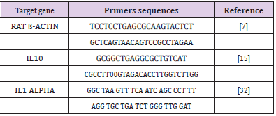
Bovine Lactoferrin (BLF), an immune-modulatory substance, was purchased from Sigma company; the commercial name is L9507 in the form of a powder that was dissolved by distilled water and was prepared to be given to twelve mature female rats (lactoferrin group) in a dose of 50microgram/kg orally by stomach tube every day before mating by one week and persisted after mating till one week according to (Yamauchi et al., 2000). Oligonucleotide primers used in SYBR Green real-time PCR: They are shown in Table 1.

Table 1: Oligonucleotide primers and probes used in SYBR Green real-time PCR.

Blood and Tissue Samples

At different time points (5hrs,1 day,3 days, and 7 days) after mating, blood samples were collected from retro-orbital venous plexus and separated into two parts, one for hematological assay and the other for serum separation that stored frozen until hormonal and biochemical assay. Uterine tissue samples were collected after anesthesia and slaughter of three rats of two groups at each time point and stored frozen for PCR assay and other tissue parts stored in 10% neutral buffer formaldehyde for histopathological examination.

PCR

RNA Extraction, according to (Yuan, et al. [50]): RNA extraction from tissue samples was applied using QIAamp RNeasy Mini kit (Qiagen, Germany, GmbH) when 30 mg of the tissue sample was added to 600 μl RLT buffer containing 10 μl β-mercaptothion per 1 ml. For the homogenization of samples, tubes were placed into the adaptor sets, which are fixed into the clamps of the Qiagen tissue Lyser. Disruption was performed in 2 minutes high-speed (30 Hz) shaking step. One volume of 70% ethanol was added to the cleared lysate, and the steps were completed according to the Purification of Total RNA from Animal Tissues protocol of the QIAamp Rneasy Mini kit (Qiagen, Germany, GmbH). N.B. On column DNase, digestion was done to remove residual DNA.

Preparation of PCR Master Mix according to Quantitect SYBR:

a) Oligonucleotide Primers: Primers used were supplied from Metabion (Germany) and are listed in Table 1.

b) cDNA Synthesis: RNA was reversely transcribed into cDNA using Thermo Scientific Revert Aid Reverse Transcriptase (Thermo Scientific). Where 20- μl reaction containing 4 μl of the 5X Reaction Buffer, 1 μl of Revert Aid Reverse Transcriptase (200 U/ μL), 0.5 μl of (20 U) Thermo Scientific™ RiboLock Rnase Inhibitor (#EO0381), 0.5 μl (20 pmol) Random hexamer (#SO142), 2 μl of dNTP Mix [10 mM each (#R0191)] 10 μl of water, and 2 μl of RNA template. The reaction was performed in an Applied biosystem 2720 thermal cycler. The mixture was incubated for 10 min at 25°C, followed by 60 min at 42°C.

c) SYBR Green rt-PCR: Primers were utilized in a 25- μl reaction containing 12.5 μl of the 2x QuantiTect SYBR Green PCR Master Mix (Qiagen, Germany, GmbH), 0.5 μl of each primer of 20 pmol concentration, 10.5 μl of water, and 1 μl of cDNA template. The reaction was performed in a Stratagene MX3005P real-time PCR machine. A primary denaturation step was done at 94OC for 15 min, followed by 40 cycles of 94°C for 15 sec., 60°C for 30 sec. and 72°C for 30 sec. A Dissociation curve was performed for one cycle, including 94OC for 1 min., 60°C for 1min., and 94°C for 1 min.

d) Analysis of the SYBR Green rt-PCR Results: Amplification curves and ct values were determined by the Stratagene MX3005P software. To estimate the variation of gene expression on the RNA of the different samples, the CT of each sample was compared with that of the positive control group according to the “ΔΔCt” method stated by [51].

Biochemical Assay

1-Estrogen: Estrogen was diagnostic by using IMMULITE and IMMULITE 1000 Analyzer for the quantitative measurement of Estradiol in serum, as an aid in the differential diagnosis of amenorrhea, and monitoring8 of ovulation induction with and without stimulating in Assisted Reproductive Technology (ART) according to [52].

2-progesterone: Progesterone was diagnostic by using IMMULITE and IMMULITE 1000 Analyzer for the quantitative measurement of progesterone in serum, as an aid in the differential diagnosis of amenorrhea, and monitoring of ovulation induction with and without stimulating in Assisted Reproductive Technology (ART) according to [53].

3- TAC (Total Antioxidant Capacity): Total antioxidant capacity is measured by the colorimetric determination method by ready-made diagnostic kits provided by Bio-diagnostic, according to [54].

4- TNFα (Tumor Necrosis Factor): TNFα was estimated by using ready-made Rat Tumor Necrosis Factor-α (TNFα). ELISA Kit presented by Quantikine Company according to [55].

5- CRP (C- Reactive Protein): The rapid latex agglutination test is for the qualitative screening and quantitative determination of C-Reactive Protein (CRP) in serum presented by ATLAS MEDICAL LATEX Kit, according to [56].

Tissue Sampling

Both uterine horns were excised from all pregnant rats and one sample from each horn was taken and fixed immediately in 10% neutral buffered formalin solution. All specimens were routinely processed in ascending grades of ethyl alcohol (80%, 90%, 100%), cleared in xylene, and embedded in paraffin wax to prepare 5μm thick paraffin sections. Sections of 5 μm thickness were cut and picked up on uncoated slides, dried, deparaffinized with xylene (3x3minutes) and rehydrated with graded alcohol (5x2minutes), washed and stained with H&E according to [57], and stained with Giemsa stain to determine eosinophil cell count in uterine tissue sections. Uterine sections from day 7 of gestation were additionally stained with Masson’s trichrome. The stained sections were dehydrated in absolute ethyl alcohol, cleared in xylene, and mounted with Mount-Quick liquid cover glass medium to the surface of the slides. Histological changes were examined by light microscopy (binocular, Olympus). Histological pictures were picked up using a Digital camera (Canon 5 megapixels, 3.2x optical zoom).

Histopathological Examination and Statistical Analysis

The data of histopathological examination of morphometric measurements of epithelial height and diameter of uterine glands were subjected to student T-test to differentiate between control and lactoferrin treated group to compare the difference between means. Data were expressed as means ± standard errors. The difference between means was considered significant when (P < 0.05). The remaining data obtained for rats of the two experiments were expressed as means ± SD. Statistical Analysis of data was carried out by the computer package program (SPSS,1994) version 16, using the two-way Analysis of variance M-ANOVA between variables at group level and time level followed by Duncans Multiple Range Test (DMRT) for testing the significant differences between variables [58].

Results

Effect of Lactoferrin on Total Leukocytic Count (WBCS×103 /μl)

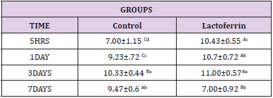
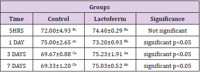
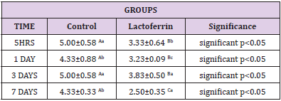
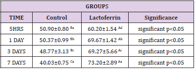
As shown in Table 2 results indicated an increase in Wbcs at 5hrs, 1day, and 3 days after mating in the lactoferrin group compared with the control group at the same time.

Table 2: Effect of Lactoferrin on total leukocytic count (WBCS×103 /μl).

Effect of Lactoferrin on Neutrophil%

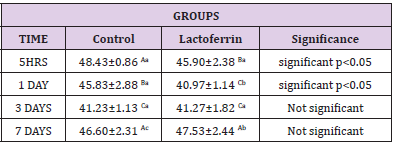
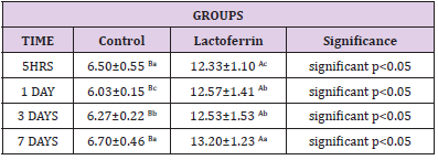
As shown in Table 3, results indicated an increase in neutrophil percent in the lactoferrin group at 5hrs, 3 days, and 7 days after mating compared with the control group.

Table 3: Effect of Lactoferrin on neutrophil%.

Effect of Lactoferrin on Lymphocyte%

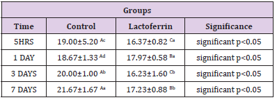
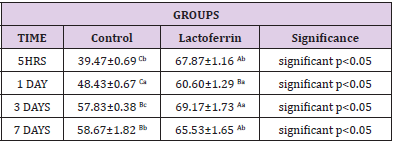
As shown in Table 4, results demonstrated a decrease in lymphocyte percent in the lactoferrin group at 5hrs, 1day, 3 days, and7 days after mating.

Table 4: Effect of Lactoferrin on lymphocyte%.

Effect of Lactoferrin on Eosinophil%

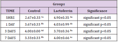
As shown in Table 5, results indicated that the eosinophil percent increase in Lactoferrin at 5hrs, 1day, and 7 days.

Table 5: Effect of Lactoferrin on eosinophil%.

Effect of Lactoferrin on Basophil%

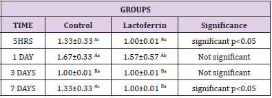
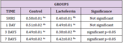
As shown in Table 6, results indicated that basophil decrease in the lactoferrin group at 5hrs, 1 day, and 7 days.

Table 6: Effect of Lactoferrin on basophil%.

Effect of Lactoferrin on Monocyte%

As shown in Table 7, results indicated that the monocyte percent decrease in the lactoferrin group at 5hrs, 1day, 3days, and 7 days.

Table 7: Effect of Lactoferrin on monocyte%.

Effect of Lactoferrin on Estrogen(ng/ml)

As shown in Table 8, results indicated that estrogen decrease in the lactoferrin group at 5hrs and 1 day when compared to the control group.

Table 8: Effect of lactoferrin on estrogen(ng/ml).

Effect of Lactoferrin on Progesterone (ng/ml)

As shown in Table 9, results indicated an increase in progesterone hormone in the lactoferrin group at 5hrs, 1 day, 3 days, and 7 days after mating when compared to the control group.

Table 9: Effect of Lactoferrin on progesterone (ng/ml).

Effect of Lactoferrin on Total Antioxidant Capacity (TAC) (μ mol/L):

As shown in Table 10, results indicated a decrease in the lactoferrin group at 5hrs, 1day, 3days, and 7 days when compared with the control group.

Table 10: Effect of Lactoferrin on total antioxidant capacity (TAC) (μ mol/L).

Effect of Lactoferrin on Tumor Necrosis Factor (TNF) (pg/ml)

As shown in Table 11, results indicated an increase in TNF at 5hrs, 1 day, 3 days, and 7 days in the lactoferrin group when compared to the control group.

Table 11: Effect of Lactoferrin on tumor necrosis factor (TNF) (pg/ml).

Effect of Lactoferrin on C-Reactive Protein (mg/L)

As shown in Table 12, results indicated an increase in C reactive protein in the lactoferrin group at 5hrs, 1day, 3days, and 7 days when compared to the control group.

Table 12: Effect of Lactoferrin on C-reactive protein (mg/L).

Effect of Lactoferrin on IL1a and IL10 by Real-Time PCR:

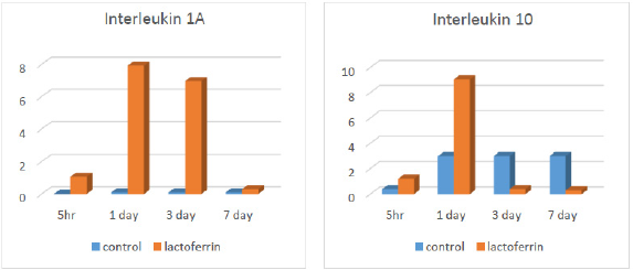
As illustrated from Figure 1, results indicated an increase in interleukin 1A in the lactoferrin group at 5hrs, 1 day, and 3 days after mating when compared to the control group. At the same time, results indicated an increase in interleukin 10 in the lactoferrin group at 5hrs and 1 day after mating but decreased at 3 days and 7 days when compared with the control group.

Figure 1: Effect of Lactoferrin on IL1a and IL10 by real-time PCR.

Histopathological Analysis for Rat Uterus

Rat Uterus After 5 Hours of Gestation

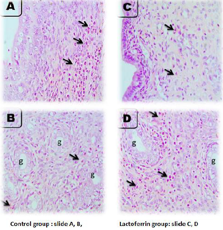
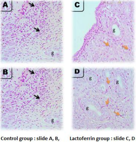
As illustrated from Figure 2, the microscopic picture shows rat uterus after 5 hours of gestation shows heavy eosinophils infiltration in superficial endometrium (arrows) (A, B) and in between uterine glands (g) (arrows). Few stromal cell density and very few eosinophils’ infiltrations are seen in superficial endometrium (arrows), and heavy eosinophils infiltrations are seen in between uterine glands (g) (arrows) in Lactoferrin treated rats I. Control (slide A and B), Lactoferrin group (C and D).

Figure 2: Microscopic picture shows rat uterus after 5 hours of gestation.

Rat Uterus at 1 Day of Gestation

As illustrated from Figure 3, the microscopic picture shows rat uterus at 1 day of gestation shows focal PMNs infiltration in superficial endometrium (black arrows) (A) and fewer eosinophils between the uterine glands (g) than after 5 hours (orange arrows) (B) in control rats. Very few eosinophils’ infiltrations are seen in superficial endometrium (orange arrows) (C), and between uterine glands (g) (orange arrows) (D) in Lactoferrin treated rats. H&E, X: 400.Note: density of uterine glands increased in Lactoferrin treated rats (D) when compared with (B). H&E. The Control group presented in (slide A and B) and Lactoferrin group presented in slide (C and D).

Figure 3: Microscopic picture shows rat uterus at 1 day of gestation.

Rat Uterus at 3 Days of Gestation

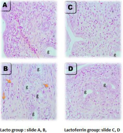
As illustrated from Figure 4, the microscopic picture shows rat uterus at 3 days of gestation shows minimal leukocytic cells infiltration in superficial endometrium (A) and between the uterine glands (orange arrows) (g) (B) in control rats. Few eosinophils’ infiltrations are seen in superficial endometrium (orange arrows) (C), and between uterine glands (g) (orange arrows) (D) in Lactoferrin treated rats. H and E. Microscopic picture shows rat uterus at 3 days of gestation shows eosinophils infiltration in the myometrium (arrows) (A) in control rats and Lactoferrin treated rats (C). Uterine glands in control (B) and Lactoferrin treated rats(D).

Figure 4: Microscopic picture shows rat uterus at 3 days of gestation.

Rat Uterus at 7 Days of Gestation

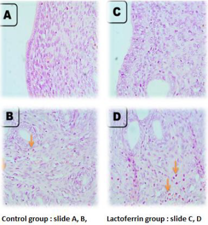
As illustrated from Figure 5, the microscopic picture shows rat uterus at 7 days of gestation shows superficial endometrium (A) and uterine glands (g) with few eosinophils’ infiltration (orange arrows) (B) in control rats. Few eosinophils’ infiltrations are not seen in superficial endometrium (C) but seen between uterine glands (g) (orange arrows) (D) in Lactoferrin treated rats. Note: density and diameter of uterine glands (g) increased in (D) in Lactoferrin treated rats, H&E. Control (slide A and B), Lactoferrin group (C and D).

Figure 5: Microscopic picture shows rat uterus at 7 days of gestation.

Effects of Lactoferrin Administration to Pregnant Rat on Eosinophil Count in Uterine Tissue at a Different Time (5hrs, 1day, 3days, and 7 days) of Pregnancy

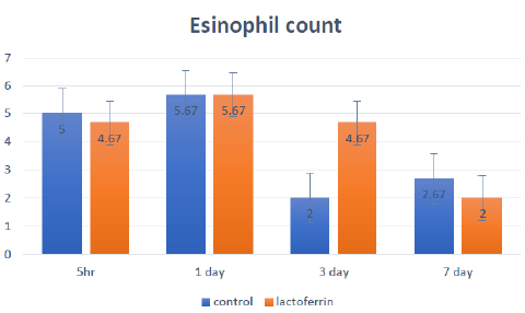
The effects of lactoferrin administration to pregnant rats on eosinophil count in uterine tissue at a different time (5hrs, 1day, 3days, and 7 days) of Pregnancy is shown and summarized in Figure 6.

Figure 6: Effects of lactoferrin administration to pregnant rat on eosinophil count in uterine tissue at different time (5hrs, 1day, 3days and 7 days) of Pregnancy

Discussion

From the results, we should remember that we are dealing mainly with concentrations of a number of immunity markers either wbcs in plasma or immunity markers , hormones and cytokines in serum to detect effect of LF on immunity mainly during early period of pregnancy. Results of the present investigation were recorded in (Tables 2-7) showed that white blood cells number increased at 5hrs and 3 days after insemination and decreased at 7 days after insemination in the lactoferrin group when compared with the control group. Lf was a natural compound of mammalian secretions which have a protective effects range from antimicrobial activities against variety of pathogens, including bacteria, viruses, fungi and parasites, to anti-inflammatory effect. Its functions rely not only on the capacity of Lf to bind iron but also on its immunomodulatory effect by its cellular and molecular mechanisms with both host and pathogen. Lf can interact with antigen presenting cells, reduce excessive inflammation and stimulate host immune responses, as well as identifying cell targets and receptors and this was important in the maintenance of immune system homeostasis [59]. Neutrophils increased at 5hrs, 3days, and 7 days after insemination, while basophil and eosinophil increased at 5hrs, 1 day, and 7 days after insemination in the lactoferrin group.

While, Lymphocyte and monocyte showed a significant decrease in the lactoferrin group at 5hrs, 1 day, 3 days, and 7 days after insemination. This result agreed with [60], who showed that Lactoferrin had a role in regulating innate and adaptive immune response, regulating the secretion of cytokines and regulated growth, differentiation, and activation of different leukocyte cells and so increased the number of circulated leukocytes. This agrees with [61], who showed that both monocytes and granulocytes increased and immune systems activated during Pregnancy in the rat. [62] reported that Lactoferrin regulated the immune system by stimulating endometrial and stromal cell proliferation and stimulating immune cells against antigen as well as [63] reported that Lactoferrin increased recruitment of neutrophil in mice circulation. Besides, [64] recorded that Lactoferrin increased the number of natural killer cells and modulated myelopoiesis with increased phagocytosis against a specific antigen. In the same respect, [65] showed that WBCs increased during rat pregnancy, and this was inconsistent with the significant increase in the number of white blood cells as presented in (Table 2).

Estrogen increased in the lactoferrin group, particularly at 7 days after insemination, and showed decreased at 5hrs and 1day after insemination. Estrogen was considered an immune modulator substance that could improve the immune response of the female reproductive tract in rodents, as shown by [66]. This agreed with (Hamid et al. 2012) showed that rats implantation period was initiated on day 5 and completed by day 7 of the pregnancy and during this implantation period, the dominant hormone modulator to morohological and functional changes were estrogen and progesterone and this may explain why estrogen increased at our study at 7 days taking into consideration the difference between species as difference between rat and human in time of endometrium changes and hormones secretion as shown [67] showed that estrogen played a pivotal role in early Pregnancy in mice as, during the first two days after insemination, the preovulatory estrogenstimulated proliferation of the luminal and glandular epithelial cells in addition to potentiated with progesterone hormone that secreted from corpus luteum and stimulated stromal cell proliferation on 4 days which was the day of the implantation process. Furthermore, [68] showed that there was a close relationship between estrogen and Lactoferrin as an estrogen-regulated expression of Lactoferrin in the endometrial epithelium of rat, particularly along all stages of the estrus cycle as well as lactoferrin levels changed with the change in estrogen level and so this could explain the increase in estrogen hormone in lactoferrin group after insemination.

Progesterone increased at 5hrs, 3 days, and 7 days in the lactoferrin group compared to the control group, as showed in Table 10. [69] reported that progesterone hormone inhibited estrogen hormone and decreased lactoferrin gene expression in uterine epithelial and stromal cells by controlling progesterone receptor and affecting estrogen receptors in vitro using uterine rat tissue. Also, [70] reported that progesterone was secreted in hamsters at the preovulatory stage from an extra luteal tissue source for allowed characteristics appearance of estrus behavior, and this could explain the increase in progesterone hormone in the lactoferrin group. Total Antioxidant Capacity(TAC) increased in the lactoferrin group at 1 day after insemination when compared to the control group as showed in Table 10 and this agrees with [71] who showed that Lactoferrin (LF) was believed to be safer than the drugs currently used for hypertension treatment due to its antioxidant effect. It has been reported that two weeks of lactoferrin supplementation has been able to increase the hydrophilic antioxidant capacity in healthy humans [72]. Besides, [73] suggested that Lactoferrin contributed to oxidoreductive reactions at the cell membrane, and LF had an antioxidant effect on red blood cells through inhibition of lipid peroxidation and hemolysis. In the same respect, LF was an important specialized iron scavenger, and its antioxidant activity was most likely related to its ability to bind ferrous and ferric ions. Thus, LF may inhibit the iron-catalyzed formation of hydroxyl radicals.

Tumor necrosis factor (TNF) decreased at 3 days and then increased at 5hrs, 1 day, and 7 days in the lactoferrin group as presented in Table 11. This increase may be due to secretion of TNF at early gestation as that different cytokine profiles might be beneficial or harmful at different stages of Pregnancy as TNF-a which was proinflammatory cytokines seem to be crucial during the implantation process, whereas high levels of these cytokines may be harmful later in Pregnancy. Also, showed that tumor necrosis factor was multi potent cytokines and had an essential role during early Pregnancy by the stimulated embryo and placental growth. In contrast, revealed that TNF had an adverse impact on Pregnancy and inhibited blastocyst growth in the rat. This conflicted impact was shown by reported that the timing of secretion, the concentration of TNF, and stimulatory signals directed TNF to be either useful or harmful toward Pregnancy. Nakashima 2012 suggested that stimulation of Th 1 dominance directed TNF to stimulate lymphokine-activated killer cells that destructed trophoblast cells and caused pregnancy loss. Moreover, Clark 1998 suggested that TNF stimulated maternal immune rejection to the embryo through the affected blood supply by applying vasculitis. This data could explain the significant increase in TNF in the lactoferrin group, particularly at 5hrs, 1 day, 3 days, and 7 days of Pregnancy. C-Reactive Protein (CRP) increased at 5hrs, 1 day, 3 days, and 7 days of Pregnancy in the lactoferrin group when compared to the control group, as shown in Table 12. This in agreement who found a slight rise in the CRP values throughout Pregnancy, and who detected maximal concentrations of CRP at 24-48 hours after the inducing stimulus or infection and so may be increased due to presence of sperm and then the presence of embryo as immune response. Moreover, CRP increased in inflammation and applied a proinflammatory function and stimulated secretion of cytokines and granulocyte-macrophage colony-stimulating factor as well as had anti-inflammatory functions as prevented migration and adhesion of neutrophil to endothelial cells to provide immune tolerance and aided in implantation and this meant that presence of embryo or sperm caused inflammatory response and stimulated secretion of CRP. Interleukin 1a (IL1A) increased at 5hrs, 1day, and 3 days in the lactoferrin group, as showed in Figure 1. This in agreement with, who showed that the interleukin1(IL1) family as IL1alpha and IL1 b secreted by early embryonic signals and aided in the preparation of a receptive maternal endometrium and may play an important role in embryo implantation.

Interleukin 10 increased in the lactoferrin group, particularly at 5hrs and 1 day of Pregnancy as shown in Figure 1. This in agreement with recorded that an up-regulation of anti-inflammatory IL-10 was found after lactoferrin (LF) administration in rats with colitis. Moreover, reported that bovine LF regulated cytokines production by splenocytes of obstructive jaundice rats. Also, LF enhanced the secretion of the anti-inflammatory cytokines IL-10 and IL-4 and reduced colitis in rats. Interleukin 10 entered in the initial formation of the fetus and supported implantation during the early period of Pregnancy as proved by Tewari 2009 that IL-10 role in Pregnancy was a potent protector against vascular dysfunction that was associated with hypertension and inflammation during Pregnancy as contributing to the regulation of maternal immune tolerance during Pregnancy and helped in the extra villous trophoblast and endothelial cell formation. Besides, illustrated that interleukin 10 in mice reached high levels in the first and second trimester of Pregnancy for improving placental growth and supporting trophoblast invasion. Histopathological examination of uterine tissue specimens presented in Figures 2-5 showed that the rat uterus was formed of three layers; endometrium, myometrium, and perimetrium. The endometrial glandular epithelium was a tall simple columnar with rounded basal nuclei. Few Polymorphonuclear Cells (PMNs) were seen in the lumen of the uterus and uterine glands.

The stroma contained spindle-shaped stromal cells and blood vessels. The myometrium was formed of inner circular and outer longitudinal smooth muscle layers with numerous blood capillaries in the perimetrium. Histopathological examination after administration of Lactoferrin to female rats revealed that there was an increase in eosinophilic and neutrophil infiltration in endometrium and myometrium, increased stromal cells, and increased in epithelial cell thickness and folding at 5hrs, 1day, 3 days, and 7 days after insemination. This agreed showed that there was a dramatic increase in cellular proliferation, endometrial tissues proliferation neovascularization, and blood flow during early rat pregnancy. Also, Guerra-Infante 1999 showed that neutrophil cells increased in the count but decreased in phagocytic activity during Pregnancy. Moreover, there was an increase in a blood vessel with decreased congestion and an increase in the number and diameter of the uterine gland when compared with the control group. This may provide nutrition to the embryo at early Pregnancy as the obtained results are declared and presented in Figures 2-5. This result inconsistency with Filant and Spencer, 2013 showed that uterine glands and blood vessels supported embryo with essential substances like proteins and sugars during early Pregnancy for providing the fetus with adequate nutrition. This could explain the increase of uterine gland number in early Pregnancy, particularly in the lactoferrin group, as shown in Figures 2-5.

In early pregnant women and rats, demonstrated that decidual cells differentiated and clumped together to form masses around cells to be in direct contact allowing the transportation of food between mother and fetus. The increase in stromal cells and epithelial cell proliferation in the lactoferrin group agreed who showed that Lactoferrin had a stimulatory and inhibitory effect on cell division and proliferation as it stimulated growth and division of somebody cells and inhibited others as mammary epithelial cells, fibroblasts, and tumor cells that may be according to the type of cells with unknown mechanism. In the same respect, demonstrated that estrogen and progesterone regulated expression of Lactoferrin in rat uterine tissue and so during early pregnancy, estrogen enhanced the uterine epithelium for the secretion of Lactoferrin and so increased epithelial and stromal cell proliferation. Nichols and McKee 1990 showed that Lactoferrin’s role in the proliferation of epithelial cells was due to its ability for iron transportation inside the cells. Moreover, Hagiwara 1995 reported that the role of Lactoferrin in stimulating cell proliferation was due to stimulation of growth factor as one of its modulatory actions. Eosinophil count increased in the lactoferrin group as showed in Figure 6 in uterine tissue, particularly at 3 days after insemination when compared with the control group. These data get inconsistent with the results of added that eosinophil was decreased in uterine tissue with the advancement of Pregnancy. However, Robertson 2000 reported that eosinophil cells were present in rodent uterine tissue, particularly at early Pregnancy.

Their numbers increased after exposure to semen after mating within the uterine stroma, particularly at the area adjacent to the luminal and glandular epithelium. The migration of eosinophil and leukocyte to uterine tissue maybe had a role in implantation and attracted to granulocyte-macrophage colony-stimulating factor, interleukin 4 and interleukin 5 molecules. This could explain that Lactoferrin increased eosinophil particularly at 5hrs and 1 day after insemination in uterine tissues, and this may explain that eosinophil had a role in the implantation of the embryo and it may secrete chemical mediators or cytokines as Rantes, Eotaxin, MIP, and IL5 that aided in early embryo acceptance by immunity and may have a role in early implantation of the embryo.

Conclusion

Lactoferrin increases the number of leukocytes, especially lymphocytes, eosinophil, and basophil, and increases TNF and C-reactive protein in serum. In addition to that, increase the concentration of interleukin 1 A and interleukin 10, especially with the advancement of pregnancy at 3 and 7 days of gestation. So Lactoferrin has a positive effect on stimulating the general and mucosal immune system during pregnancy.

For more Articles on : https://biomedres01.blogspot.com/
 

Effect of Nutritional Intervention on the Temporal Dynamics of Muscle Strength Adaptation

  Effect of Nutritional Intervention on the Temporal Dynamics of Muscle Strength Adaptation Introduction Muscular adaptation is determined b...