Friday, April 30, 2021

Garlic Polyphenols: A Diet Based Therapy

Garlic Polyphenols: A Diet Based Therapy

Introduction

Garlic (Allium sativum L.) belongs to the family Alliaceae and has been derived from the Celtic word ‘all’ meaning pungent and nature’s boon to mankind. More than 5000 years, garlic has been used as curative and food based bioactive agent for physiological disorders. Garlic plant is mentioned in the Ancient Israel (The Talmud), Egypt (Codex Ebers), Bible and India (Vedas and Purans, Charak Sanghita). Chinese people believe that consumption of garlic as diet based therapy is most effective against human metabolic syndromes such as aging, oxidative stress, cancer, wound healing, diabetes, heart diseases and obesity [1]. Hippocrates observed that garlic was inhibited the cancer cells lines and also prevented from the intestinal and extra-intestinal diseases. Prophet Hazrat Mohammad (PBUH) recommended it for treating scorpion stings and cured health’s related disorders. In Historia Naturalis by Pliny, it was more effective against dog & snake bites as well as gastro-intestinal disorders. Allicin is sulfur containing compound formed through the action of alliinase enzyme on alliin and show the anti-platelet and hypolipidemic activity on human health. There are multiple evidences which confirm the uses of garlic as nature tonic to cure diarrhea in ancient times. In Nigeria, it is used to treat diarrhea, abdominal discomfort, respiratory tract, and otitis media infections.

In Europe, India and Pakistan, consumption of garlic was found to cure asthma, hay fever, and common colds, respectively [2,3]. Garlic is a promising source of bioactive compounds such as allicin, disulfides, alliin, allyl sulfides, cycloalliin, ajoene, allyl disulfides, methionine, cysteine, cystine, diallyl sulfides, glutathione, pseudoscordinine, dimethyl sulfides, sulfones, allyl trisulfides, thiosulfates, scordinine, methyl sulfides, tetra thiol, trisulfides cysteine, and sulfoxides [4]. It is prominent source of calcium, phosphorous, iron, nicotinic acid, thiamine, riboflavin, and vitamin C, respectively. Garlic comprises of citral, linalool, geraniol, α-phellandrene, valeraldehyde, and propionic aldehyde components [5]. They play a significant role against cancer, cataract, cardiovascular disease (CVD), hypercholesterolemia, obesity and hypertension. Maceration of garlic produces oil that good source of 3-vinyl-[4H]-1,2-dithiin, 2-vinyl-[4H]-1,3-dithiin, ajoene and allyl sulfides, whilst steam distillation of garlic oil (SDGO) gives diallyltrisulfide and diallyl disulfide [6] (Tables 1 & 2).

biomedres-openaccess-journal-bjstr

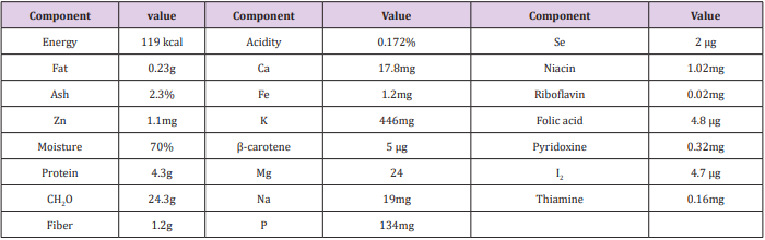
Table 1: Nutritional profile of raw garlic/100g.

biomedres-openaccess-journal-bjstr

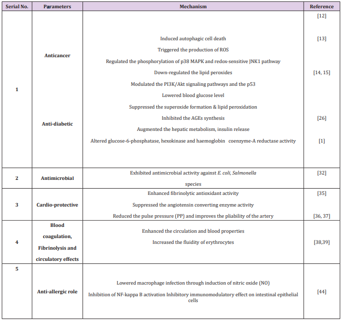
Table 2: Health perspectives of garlic.

Antioxidant Potential of Garlic

Nencini et al. [7] determined the antioxidant potential of 15% hydroethanolic extracts of aged diverse parts (bulblets, bulbs, flowers, leaves, and flower bulblets) of Allium subhirsutum L, Allium roseum L. Allium neapolitanum for 20 months. The aged garlic leaves extracts show the higher antioxidant potential followed by flowers and bulbs, whilst bulblets and flower bulblets exhibit no antioxidant potential. Nencini et al. [8] also determined the effect of garlic (bulbs or leaves, 250mg/kg) daily for 5 days in liver of ethanol induced male albino rats. They determined that garlic enhances the GSH and lowers the MDA levels, GR, CAT and SOD activities in liver injury through ethanol. Park et al. [9] evaluated the antioxidant potential garlic extracts through providing different diverse processing conditions. Aged-garlic extract (AGE) yields total phenolic contents (562.6 ± 1.92 mg/100 g GAE) as compared to heated garlic extract (HGE) or raw garlic extract (RGE). In H2O2 induced DNA damage of rats, the ED50 (19.3μg/mL) and inhibition activity (70.8%) of aged garlic extract were reported, whereas ED50 value (38.6μg/mL) of raw garlic extract for 4-hydroxynonenal induced DNA damage followed by aged garlic extract>heated garlic extract was observed. Raw and aged garlic comprises of high quantity of phenolic contents and exhibit high anti-genotoxic activity. They showed chemopreventive role against deleterious effects of H2O2 against DNA and 4-hydroxy-2,3-trans-nonenal (HNE) [9].

Health Perspectives

Anticancer Role: Garlic and garlic oil have been found to provide significant protection against N-nitrosodiethylamine-induced hepatocarcinogenesis. Garlic contains fat and water soluble sulfur compounds that exhibits the anti-cancer mechanisms through the ameliorating oxidative stress, improving immune function, and inhibiting metabolic carcinogenic activation [10]. A group of peers, Choi and Park [11] determined that garlic oil has anticancer role by inducing apoptosis, inhibiting differentiation & tumor angiogenesis and reversing multidrug resistance. Garlic is a potential phytochemical candidate against colon cancer cells, glioblastoma cells and hepato carcinoma cells. It induces apoptosis mechanism in different cancer cells. Allicin, which is the most abundant component in garlic, could induce autophagic cell death in Hep G2 cells [12]. Garlic consists of diallyl disulfide (DADS), diallyl sulfide (DAS), and diallyltrisulfide (DATS) which protect from the carcinogenesis. The supplementation of garlic triggers the production of ROS in glioblastoma cells and induces apoptosis through regulating phosphorylation of p38 MAPK and redox-sensitive JNK1 pathway.

JNK (c-Jun N-terminal kinases) is a potent inhibitor and dominantly lower the cell death by enhancing intracellular free [Ca (2+)], activating caspase-4, calreticulin, calpain, caspase-3 and caspase-9, mitochondrial release of cytochrome c and Smac into the cytosol, down-regulating Bcl-2, and overexpression of Bax [13]. Garlic dietary phytochemicals inhibit, delay or reverse skin carcinogenesis due to presence of organosulfur compounds i.e. ajoene, allicin, S-llyl cysteine (SAC) and allyl sulfides. The oral administration of garlic prevents the mice from chemical carcinogen induced skin papillomagenesis [14]. It up-regulates the carcinogenesis progression and down-regulates the lipid peroxides, as well as also modulates PI3K/Akt signaling pathways and the p53 in the skin papilloma cells [15]. Similarly, in another study conducted by Cherng et al. [16], they investigated that supplementation of DAS significantly decreased the expression of apoptotic sunburn cells, proliferative cell nuclear antigen (PCNA), UVB-induced thymine dimer-positive cells, and terminal deoxynucleotidyl transferasemediated dUTP nick end labeling whilst enhanced the p21/Cip1- positive cell population and p53 in the epidermis. Moreover, suppression of COX-2, prostaglandin E2 (PGE2), NF-κB, and NO level were reported after DAS administration, respectively [16].

There are different methods like digital rectal examination (DRE) and prostate specific antigen (PSA) use in blood sera and enhance the prostate cancer diagnosis among male population at early stages [17]. The use of androgen drugs suppresses the hormone stimulation of the prostate; on the other side, show deleterious effects on body [18]. Garlic bioactive components inhibit the proliferation of various cancer cells through the histone deacetylase (HDAC) inhibitory activity, down-regulation of the Akt and up-regulation of apoptosis of prostate cancer cells [19,20]. The earlier findings of Kim et al. [10] reported that administration of garlic compounds prevented the in vitro and in vivo rats from the prostate cancer. These compounds induced repression of the androgen receptor (AR), apoptosis, and cell cycle arrest in prostate cancer cells. Besides this, they also enhanced the histone acetylation and apoptosis in prostate cancer cells of mice [21, 22]. The administration of S-allylcysteine at the rate of 1-100 mmol/L in A2780 cells in concentration- and time-dependent fashions momentously suppressed the proliferation of cells. It also induced apoptosis, accompanying by lowering the expression of pro-caspase-3, Parp-1, and Bcl-2, and enhancing active caspase-3 and Bax. Similarly, it also reduces the migration of A2780 cells, and significantly decreases the protein expression of Wnt5a, p-AKT and c-Jun [23].

Antidiabetic Role: Diabetes is a metabolic disorder and produces the abnormal levels of glucose in the bloodstream. It is growing emerging public health problem and estimated to be 333 million people by 2025. Herbal plants are being consumed as food by the patient as well as healthy person. Synthetic oral antihypoglycemic agents are more effective to lower the hyperglycaemia level but on other side, they produce the side effects [24]. Allicin is a bioactive component and prevent from the diabetes through lowering the blood glucose level. The oral administration of allicin enhances the free amino acid content and hepatic glycogen, lowers the triglyceride and glucose in serum sucrose-fed rabbits. Similarly, the administration of garlic extracts to streptozotocin-diabetic rats reduced the sugar level, inhibited superoxide formation and lipid per-oxidation. S-allyl cysteine inhibits the AGEs synthesis [25]. DATS, AM, and DAS show the hypoglycemic action that augmented the hepatic metabolism, and insulin release [26]. Allicin controls the blood glucose level in serum and alters the activities of liver hexokinase glucose-6-phosphatase and haemoglobin coenzyme-A reductase in rabbits. It significantly increases the free amino acids and liver glycogen that lower the liver serum proteins, triglycerides level (aorta and liver) and fasting blood sugar (FBS) as compared to high sucrose-fed diet rabbits [1].

The supplementation of diallylthiosulfinate and S-methyl cysteine sulfoxide (SMCS) to alloxan diabetic rats that activates 3-hydroxy- 3-methyl-glutaryl (HMG) Co-A reductase, hexokinase, glucose-6-phosphatase, and lecithin- cholesterol acyltransferase (LCAT) enzymes [27]. Padiya et al. [28] also determined the hypoglycaemic effect of garlic that enhanced the insulin sensitivity and lowered the body weight gain in fructose fed rats. The previous findings of Osman et al. [29], they reported that daily intraperitoneal administration of allicin at the rate of 8 mg/kg diabetic experimental volunteers for 30 days. It effectively reduced the antiislet cell antibodies ICA level through following mechanism (1) the lowered pan B cell marker (CD19), lowered pan T cell marker (CD90), decreased pan innate cells marker (CD11b), prevented from the damage of langerghans islet cell.The hypoglycaemic action of garlic polyphenols could possibly due to release of bound insulin and high pancreatic secretion of insulin from β-cells. These polyphenols lower the blood glucose level, TG, LDL, VLDL and enhance the HDL level in streptozotocin and alloxan-induced diabetes mellitus rats. S- allyl cysteine restores its function in streptozotocin-diabetic rats by preventing free radicals production through modulating the NADPH oxidase. DAT lowered the fasting blood glucose (FBG), LDL and increased the HDL level in diabetic patients for 12 weeks [30].

Antimicrobial Activity: Antimicrobial drugs have failed to provide response to micro-organisms due to their expensive prices, side effects and greater risk of death [31]. The garlic containing extracts show antimicrobial activity against different types of bacteria such as gram positive (A streptococcus, s. pneumonia group, s. aureus and bacillus anthrax) and gram negative (salmonella species, citrobacter enterobacter, kilabsella and E. coli) [32]. Hindi [33] reported that thiosulfinates in garlic are responsible for the antimicrobial activity in rats. Allicin also exhibits the antibacterial action against some fish pathogenic bacteria. The 10% v/v solution of allicin in dimethyl sulfoxide (DMSO) was also showed anti-bacterial activity against aeromonas veronii, aeromonassobria, aeromonascaviae, aeromonashydrophila and streptococcus iniae. The allicin (MICs; 125 µg/ ml) concentration for aeromonashydrophila and 250-500µg/ml for aeromonas veronii, aeromonas caviae and aeromonas sobria in dimethyl sulfoxide was found to be effective [34].

Cardiovascular Role: Allicin, DATS, DADS and AM polyphenols perform their antioxidant mechanism and prevent from cardiovascular disease by enhancing fibrinolytic antioxidant activity, inhibiting platelet aggregation, reducting serum lipids and blood pressure levels [35]. The previous findings of Thomson et al. [36] reported that single intravenous dose of garlic extract (10- 100mg/kg) dose-dependently suppressed the blood thromboxane B2 concentration in rats. Garlic also up-regulates the cellular glutathione levels in vascular endothelial cells and prevents endothelial dysfunction. The prophylactic supplementation of aged garlic extract prior to ischemia reperfusion suppresses the production of free radicals and prevent from the depletion of glutathione antioxidant enzyme. Similarly, they also suppress the fibrosis development in kidney and liver organs by inhibiting the neutrophil migration. Allicin and DATS suppress the angiotensin converting enzyme activity that lowered the blood pressure and cardio-protective effect in kidney and liver. The aged garlic extract (AGE) reduces the pulse pressure (PP) and improves the pliability of the artery. Additionally, they lower the deleterious effects that produce the papilloma in the forestomach, lower erythrocytes and enhance reticulocytes [37].

Blood Coagulation, Fibrinolysis and Circulatory Effects: In oxidized erythrocytes of rats, the supplementation of aged garlic extract has been found to enhance the blood circulation via preventing hemolysis and lipid peroxidation. Allicin, AM improve the erythrocytes fluidity, whereas unable to protect the erythrocytes from t-butyl hydroperoxide-induced hemolysis [38]. They also modulate the production and function of both constricting factors (endothelin-1) and endothelium-derived relaxing factor (NO) in pulmonary arteries of mice [39]. Garlic juice and g-glutamylcysteines have been proved to show the beneficial effects on heart rate and may lower the blood pressure through inhibiting angiotensin-converting enzyme in vitro [40].

Immunomodulatory Activity: Aged garlic extract, and their bioactive components exhibit the immunomodulatory properties due to presence of organosulfur compounds. The concentration of garlic extract is dominantly effective on IL-2 and INF-γ gene expression of stimulated lymphocytes. These compounds also reduce the macrophage infection through the induction of NO production in vitro [41]. Allicin prevents from the immunemediated liver damage of T cells of mice and as well as also inhibit the NF-kappa B activation. A group of researchers [42], they investigated that allicin supplementation prevented from the intestinal inflammation though inhibitory effect on peripheral blood, and intestinal epithelial cells [42].

Antiallergic Role: Allergic responses are the activation of FceRI receptor on the plasma membrane of mast and basophilic cells is known to liberate the b-hexosaminidase, histamine, cytokines, prostaglandins and leukotrienes allergic mediators. Likewise, arachidonic acid metabolites such as prostaglandins and leukotrienes cause acute and chronic allergic inflammatory reactions [43]. Supplementation of allicin and AM can inhibit the passive cutaneous anaphylaxis (PCA) and immunoglobulin E-mediated allergic response in RBL-2H3 cells. Similarly, ethyl acetate extract of garlic polyphenols inhibits the release of TNF-a and b-hexosaminidase. They suppress the cPLA2, Syk, COX- 2 and 5-LO factors in rats [44].

Conclusion

Garlic is mainly used as commodity worldwide for its health promoting perspectives in human such as prevention from cancer insurgence and diabetes, lowering the cardiovascular complications, allergy responses, and aging. Bioactive compounds from garlic exert anti-carcinogenic potential target multiple pathways, inclusive of the cell cycle, apoptotic cell death and angiogenic pathway. The active form of alliinase enzyme can be stabilized into biocompatible materials such as alginate beads and layered double hydroxides. This review encompasses the multiple health effects of garlic and its bio-active constituents with references to health perspectives.

More BJSTR Articles : https://biomedres01.blogspot.com/

A Case of Huge Follicular Adenomatoid Odontogenic Tumor Treated by Fenestration and Delayed Enucleation

A Case of Huge Follicular Adenomatoid Odontogenic Tumor Treated by Fenestration and Delayed Enucleation

Introduction

Adenomatoid odontogenic tumor (AOT), first described by Stafne in 1948 [1], is a rare and benign epithelial tumor that most commonly occurs in the anterior maxillary region (especially in the canine region) of younger women [2]. There are three types of AOT: follicular, which has a central lesion associated with an impacted tooth; extrafollicular, which has no relation with an impacted tooth; and peripheral [3]. The follicular type is often associated with the crown and part of the root of an impacted tooth, typically affecting the maxillary canines but rarely affecting the permanent molars [4]. Radiographically, a cystic lesion containing teeth and a calcified body may be present, while histologically, the tumor cells appear as duct- or corolla-like structures with calcified material [5]. In this report, we describe a rare case of a huge follicular-type AOT that extended into the maxillary sinus of a young woman.

Case Report

A 26-year-old woman presented to our department with swelling of the left cheek for several months. There was no history of trauma, discharge, or any altered sensation. Intraorally, she complained of pain from the left upper central incisor to the left upper first premolar, and examination revealed that the buccal and palatal gingiva overlying this area was swollen. However, the swelling was non-tender to palpation and had a firm consistency. Also, the left upper canine was not present. Extra-orally, the swelling was diffuse and extended on the left cheek. Panoramic X-ray examination revealed a radiolucent area with an impacted left upper lateral incisor extending into the left maxillary sinus (Figure 1A). The roots of the left upper median incisor and canine were separate, but the roots of left upper premolars and first molar had been absorbed. Subsequent contrast-enhanced computed tomography (Figures 1B & 1C) revealed the presence of a lesion measuring 40 × 40 × 36 mm. Many radiopacities were found within the radiolucent lesion, including calcified material and impacted tooth. The lesion extended to the nasal cavity and maxillary sinus, the buccal cortical bone was thinning, and the palatine bone had been absorbed.

biomedres-openaccess-journal-bjstr

Figure 1: Panoramic X-ray and CT at first visit and 6 months after fenestration.

The size of lesion on Panoramic radiograph (A) and CT (B,C) at first visit was approximately 40×40×36 mm and Many radiopacities were found within the radiolucent lesion, including calcified material and impacted tooth. Changes in the lesion were monitored by follow-up CT(D,E), and a reduction in the tumor size with accompanying bone renewal were confirmed about 6 months after the fenestration procedure.

Biopsy was performed under local anesthesia in an outpatient clinic. Serous liquid, calcified material, and a thick fibrous capsule were encountered after perforating the labial cortical plate, and a jelly-like specimen was collected that had a cystic appearance. At this time, we therefore performed a fenestration to reduce the tumor size, stimulate bone renewal, and improve the chance of a favorable cosmetic outcome. A histologic diagnosis of folliculartype AOT was made from the biopsy sample. Changes in the lesion were monitored by follow-up CT, and a reduction in the tumor size with accompanying bone renewal were confirmed about 6 months after the fenestration procedure (Figures 1D & 1E). Therefore, we performed enucleation of the maxillary tumor and apicoectomy of the left upper canine under general anesthesia (Figure 2A). The tumor was approached intraorally via a reflected mucoperiosteal flap extending from the right upper lateral incisor to the left upper first molar. To remove bone from around the fenestration, the firm mass was separated from the adjoining bone along its margin and removed with the involved tooth (left upper lateral incisor).

Apicoectomy was also performed for the involved canine, but no other teeth were associated with the lesion. Hemostasis was achieved, and primary closure was completed with absorbable sutures. The total size of the enucleated tissue, including the impacted tooth, was about 35 mm and had a thick cyst-like coating (Figures 2A & 2B). As at biopsy, the contents of the enucleated tumor had a jelly-like consistency. The histopathological report of the enucleated lesion confirmed the diagnosis of follicular-type AOT. Ova-l and polygon-shaped cells were present with ductal and corolla-like structures (Figures 2C & 2D). A mass of calcifications was also present. At 1 year after the final surgery, the left cheek swelling, and the facial asymmetry had greatly improved (Figure 3). Bone renewal was also confirmed and the deformity of the bone of the front wall of the maxillary sinus and the alveolar bone of the buccal side had improved. Of note, there had been no surgical site infection or clinical recurrence.

biomedres-openaccess-journal-bjstr

Figure 2: Intraoperative views, enucleated tumor and histological image.

We performed enucleation of the maxillary tumor and apicoectomy of the left upper canine under general anesthesia (A). The total size of the enucleated tissue, including the impacted tooth, was about 35 mm and had a thick cyst-like coating (B). The histopathological report of the enucleated lesion confirmed the diagnosis of follicular-type AOT. Ova-l and polygon-shaped cells were present with ductal and corolla-like structures (C,D).

biomedres-openaccess-journal-bjstr

Figure 3: CT findings at 1 years after surgery.

At 1 year after the final surgery, the left cheek swelling, and the facial asymmetry had greatly improved. Bone renewal was also confirmed and the deformity of the bone of the front wall of the maxillary sinus and the alveolar bone of the buccal side had improved.

Discussion

AOT was first described in 1905 by Steensland. In 1907, Dreibladt called this lesion a pseudo-adenoameloblastoma, while in 1915, Harbitz proposed the term adamantoma; however, it was not until 1948 that Stafne [1] considered the lesion to be a distinct entity. Although it has since been thought of as a subtype of ameloblastoma, in 1969 Philipsen et al. coined the term “adenomatoid odontogenic tumor” that was adopted by the World Health Organization (WHO) classification for odontogenic tumors in 1971 [6]. It has been reported that AOTs account for 2%–7% of all odontogenic tumors [4-8]. They are more common in the maxilla than in the mandible (a ratio of 2:1), and they also occur more frequently in females (64%–65%) and between the ages of 10 and 29 years (74%–81%) compared with other groups [7,8]. Philipsen [2,4] also reported that the male to female ratio was 6:5, nearly all cases occurred during the second decade of life (age 8 to 39 years), and most cases occurred in patients of Asian origin.

The typical presentation is with a maxillary swelling that can be either painless or painful. AOT is a slow-growing benign tumor that is not prone to recurrence. It has intraosseous and peripheral forms, with the former being differentiated into follicular and extrafollicular types based on radiographic findings [9]. Philipsen reported that the follicular, extrafollicular, and peripheral types occurred in 70.9%, 26.7%, and 2.3% of cases, respectively [2,4]. AOTs usually measure 10–30 mm, but they may grow to as large as 70 mm [10]. Horino [9] reported that the average size was 27.6 mm, but that the average of those that developed into the maxillary sinus was only 41.6 mm. Our maxillary case was consistent with this average. As mentioned, AOTs are usually solid, but may occasionally be cystic; indeed, only 11 cases have been described that arise in association with a dentigerous cyst [4]. That said, AOT has previously been described as an adenomatoid odontogenic cyst based on its form and histology [2]. Our case is therefore consistent with these alternative reports.

AOT often occurs in anterior areas, and about 70% of all cases are associated with an impacted tooth. The maxillary canines account for 41.7% of these cases, with all four canines involved in 60.1% of cases [4-10]. Therefore, it is perhaps unsurprising that 77% of follicular-type AOTs are initially diagnosed as dentigerous cysts [7]. Although the first premolar, third molar, second molar, and lateral incisor have been reported to be involved, the maxillary canine is the most often involved tooth. Interestingly, Philipsen reported that only one casehad a well-defined radiopaque mass, finding that all other lesions appeared as circumscribed unilocular radiolucent lesions around an unerupted tooth. Our patient was a young Asian woman with an AOT associated with an unerupted maxillary lateral incisor that was displacing the maxillary canine. Philipsen et al. [2] postulated that follicular-type AOT develops from cell nests within the dental lamina that surround the tooth. Furthermore, they suggest that the extrafollicular variants form from the epithelial nests situated at the borders of the conduit of tooth eruption. Consequently, the tooth would not be embedded by the developing AOT during eruption. As noted in the macroscopic observations, the tumor in the current case had a tissue tag that was attached to the epithelial lining in the bony depression.

Microscopic examination revealed that the tissue tag belonged to the epithelial lining of the dentigerous cyst. This raised three possibilities. First, the dentigerous cyst with the unerupted lateral incisor may have developed initially, and through a stimulus that is yet to be determined, the AOT arose from the epithelial nests in the dental lamina within the odontogenic cystic lining. Second, the findings reflect two different pathologies that were occurring concurrently in the lesion. Third, this may have been an entirely new variant. However, AOT has been reported to derive from a calcifying epithelial odontogenic cyst [2]. AOT is usually encapsulated and has few recurrences, allowing definitive treatment by surgical enucleation and curettage at presentation. However, our case was relatively large, and the maxillary deformity was a concern for the enucleation surgery. At biopsy, the tumor had a cyst-like structure with liquid or jelly-like material, so we initially performed fenestration to reduce the tumor size and improve the facial deformity. As a result, we could perform the enucleation with minimal surgical invasion, and satisfactory clinical results could be obtained without leaving unsatisfactory facial deformity. In cases such as ours, where the AOT has a cystic component and the patient has facial deformity, we advocate performing enucleation surgery after reducing the tumor size by fenestration surgery.

Preventive Medicine

More BJSTR Articles : https://biomedres01.blogspot.com/

Promoting an All-Inclusive Epidemiological Studies of Parkinson Disease (PD) in Ethnic Minorities of African Descent

Promoting an All-Inclusive Epidemiological Studies of Parkinson Disease (PD) in Ethnic Minorities of African Descent

Introduction

Parkinson Disease (PD) is a neurodegenerative disease characterized by the loss of the Substantia Nigra (SN) accommodating dopaminergic neurons responsible for the production of dopamine (DA), an excitatory neurotransmitter, in the mid brain [1,2]. Because DA is involved in the relay of messages to the part of the brain that controls movement and coordination (Motor Cortex) via the Basal Ganglia and the Thalamus, its loss results in the complete or partial loss of movement [3]. PD symptoms can be categorized into four groups based on the individual and severity. These include tremor of the hands, arms, legs, jaw and face; bradykinesia or slowness of movement; rigidity or stiffness of the limbs and trunk; and postural instability or impaired balance and coordination [4]. It is termed a progressive neurological disorder because it worsens with time [4]. The cause of PD is not fully understood. So far, no cure has been found for PD, but certain treatments are recommended for patients based on the severity of the symptoms. Because PD’s pathology is linked to loss of dopaminergic neurons that produces DA, most drugs are designed and produced as synthetic or semisynthetic DA to replenish insufficient endogenous DA in order to reverse the symptoms. The most prominent drug in this category is the levodopa, a semi-synthetic form of DA [5]. The final conversion of levodopa to DA is facilitated in the brain [6]. Although this drug and other variants have been shown to mitigate the symptoms of PD, they have not been able to reverse them completely. Apart from medications, surgical therapy, lifestyle modifications, physical therapy and change of diets are other alternative therapies that may be recommended on an individual basis [4].

Etiology and Pathophysiology

Most PD development can be traced to the loss of the SN in the mid brain (National Parkinson Foundation). The brain SN accommodates dopaminergic neurons that produce DA, an excitatory neurotransmitter that plays a critical role in movement and coordination [1,2]. Thus, the decreased production of DA results in movement disorder, characterized by tremors in people with PD [4]. Apart from the damaged or loss of SN, genetic mutations of some certain genes have been implicated in PD development. Some of these genes include PARK2 (parkin), PARK7, PINK1 (PARK6), LRRK2 (PARK8) and SNCA (PARK1), PARK 7(DJ-1) and PARK2 (ATP13A2) [7]. In most cases, mutations of these genes have been linked to the impairment of the SN [8], although the mechanisms are yet to be clearly defined in many cases. It is also possible that PD may be caused by mutations in some other genes that are yet to be discovered. Scientists and researchers have also discovered a correlation between PD and protein aggregates of alpha-synuclein, the major component of Lewy bodies, and a signature and marker for dementia and other neurodegenerative, neurological and cardiovascular diseases [9,10]. Drugs that can dissolve this protein aggregates have been developed and in preclinical testing stages [11]. While the pathology and pathophysiology of most PD development can be accounted for, about 15% are idiopathic [12].

Prevalence of PD in the United States

Because of the lack of adequate statistics, especially in the sub-Sahara (SS), it is very difficult to state categorically how many people are living with this disease worldwide. However, according to the Parkinson’s Disease Foundation, about one million people are living with this disease in the United States (US). Also, about sixty thousand new cases are detected each year minus those that go undetected. Studies have also shown that PD is about one and a half time as common in men as in women and people with a family history are 10% more likely to have it than people with no family history [4,13]. Although the basis for the prevalence of PD in men compared to women has not been fully understood, studies have shown that it may not be unconnected with exposure of men to toxic chemicals or head injuries, both of which have been characterized as possible PD risk factors [13]. It is also believed that estrogen may provide additional protection for females’ nervous system [13]. The elucidation of the underlying mechanism of the gender-based discrimination against PD development will definitely shed more light on the pathophysiology of PD. Apart from gender, other factors that may influence PD prevalence include age, genetics, social life and family history.

Prevalence of PD in Ethnic Minorities in the United States

Most data available on the prevalence and incidence of PD are based on studies done in the US, Europe and some parts of Asia. Data are lacking on the prevalence and incidence of this disease in the Sub-Saharan (SS). Although available data of studies done in the US, with the second largest population of black people after Nigeria and Brazil, showed that black people may have a low incidence of PD compared to Caucasians [14], this does not necessarily translate to the overall low prevalence of PD among black people. Many of these studies are really vulnerable to statistical biases and their outcomes have been termed unacceptable. It is necessary and important to conduct more comprehensives studies to really ascertain the prevalence of PD in minorities, especially of African descent. In Africa, the historical ancestral homeland of black minorities in the US, reliable and substantial data are lacking on the prevalence and incidence of PD. Except for South Africa, Nigeria, Ghana and Zambia, research studies both at the molecular and epidemiological levels on PD are essentially nonexistent. The unreliable epidemiological data on PD in American minorities of African descent and the lack of substantial data in Africa makes it difficult to make any substantial conclusion on the prevalence and incidence of PD between Caucasian and Blacks vis-à-vis sex, age, demography etc.

Why Epidemiological Studies of PD of Minorities is Essential

The importance of a comprehensive report on the incidence and prevalence of PD in African American, and Black people in general, cannot be overemphasized. It will definitely provide a platform to organize a comprehensive treatment scheme to manage the disease among this ethnic minority. Also, reliable epidemiological data on this disease on blacks could reveal new novel information about the genetic and environmental basis of the development and evolution of this disease. This is predicated on the fact that epidemiological studies of some diseases have revealed novel pattern of inheritance and polymorphisms, providing more information on genetic variations and diversities. Africa has been termed a dark continent when it comes to the knowledge of most neurological diseases, including PD [15]. It is generally believed PD prevalence and incidence in black people are actually more than reported in few countries where they are reported. Many factors are responsible for this development. Firstly, there is a paucity of qualified trained staff and personnel that can diagnose PD. It is projected that there will be a significant shortage of neurologists that can manage PD in the US by 2025 if the present rate of turnover is not increased [16]. Secondly, the reluctance of people to participate in clinical trials and epidemiological studies. Cultural beliefs, religious sentiments and prejudice, especially in Africa, may also discourage people with PD from coming out to declare their status.

Correcting PD Management Disparities in African Americans

Comprehensive Epidemiological Studies

The first step in correcting the disparities of the management of PD in African American minorities is to conduct comprehensive epidemiological studies on this group of people. While comprehensive epidemiological studies of this disease are also recommended in the sub-Saharan to provide a comprehensive conclusion on the prevalence and incidence of this disease in black people as a whole, the financial and demographic burdens may be quite challenging. Therefore, for a start, the studies can be limited to Africans in the United States and the research extended to the subSaharan in the future. The epidemiological studies must emphasize the frequencies and pattern of PD distribution. The frequencies studies must emphasize the prevalence and incidence with respect to time, space, age, gender and social status.

Clinical and Genetic studies (Genome Wide Association Studies)

African Americans should be encouraged to participate in large scale screenings to identify potential gene mutations which could results in PD development. These genes include PARK2 (parkin), PARK7, PINK1 (PARK6), LRRK2 (PARK8) and SNCA (PARK1), PARK 7(DJ-1) and PARK2 (ATP13A2). More genetic studies are recommended for African Americans in order to identify novel genes mutations that can also cause PD. Genetics studies is a powerful tool that can reveal a lot of information about the vulnerability and susceptibility to a disease vis-à-vis sex, age, race and demography. For instance, genetic studies have shown that female ethnic minorities of African descent are vulnerable to having and dying from certain types of breast cancer compared to their Caucasian counterpart [17]. In addition, novel race-specific polymorphisms, including single nuclear polymorphisms and copy number polymorphisms can be identified and patients can be advised in advance of their chances of having PD.

Periodic Screening

Periodic screenings should be encouraged among minorities to access and examine their health conditions and their propensity to having PD. This screening may reveal past family histories of PD. People with family histories of PD have about ten chances of having PD compared to people with no family histories.

Affordable Health Care Provision

Although the government is doing a lot to make health care affordable to patients, there is still need for improvement especially with regard to minorities. This is predicated on the facts that most minorities, the majority of whom live below the middle class, cannot afford the minimum health insurance plans. Also, as PD requires a medical specialist attention, copayment should be waived by the Insurance companies. Many patients may be forced to hide their family histories of PD to avoid being classified as preexisting by the insurance companies since most preexisting classified patients are rejected by insurance companies or in the worst scenario case accepted with a high premium insurance rate which may be unaffordable to the patients. The implications of hiding a family history of a disease, including PD, is that it prevents early intervention measures. This should not only be discouraged but should be discontinued.

Individual Focused Interventions

There is a general misconception that health care disparities among ethnic groups can be eradicated by simply improving the quality of health care of the general population. Past studies have shown that this may not be true after all. A 15-year progress analysis of carried out to investigate black-white health disparities in the United States vis-à-vis improved general quality of health in Chicago area from 1990 to 2005 showed that rather than closing the health disparities gap, it was further widened, although the qualities of health care for the two groups were significantly improved [18]. A recent study showed that individual-focused interventions may be very effective in managing chronic diseases. This is based on the premise that it provides a combination of prevention interventions that consider individual lifestyle, genetics and environment rather than general characteristics [19]. Individually focused intervention should also be employed in the management of PD.

Sensitization of Local Communities

Also, efforts should be directed at increasing the number of health care providers, especially medical specialists in areas that are densely populated by minorities. Studies have shown that minorities with terminal diseases, including PD, are less likely to be accepted by specialists due to prejudice about their ability to pay. To prevent this situation, government and private agencies should encouraged private care providers to assist and educate minorities, especially those with low income on the need to enroll in the Medicare program of the government. They should be made to understand that been healthy at all time is a personal responsibility and no effort should be spared in getting access to adequate treatment, even when the patients cannot afford it.

Clinical Trials

Minorities should be encouraged to participate in clinical trials of potential new drugs for PD treatment. Because PD affects a relatively small number of people, pre-clinical trials to identify new drugs for its treatment has proven relatively difficult. Although studies have shown that a lot of minorities are skeptical about clinical studies and their outcomes, these sentiments and prejudice can be eradicated through proper education and sensitization about the importance of clinical trials in the management of diseases, including PD [20]. The idea of monetary compensation to encourage participation should also be encouraged.

More BJSTR Articles : https://biomedres01.blogspot.com/

Thursday, April 29, 2021

The Effect of Visual-Sport Specific Exercise on Shooting Skill Performance in Football Players

The Effect of Visual-Sport Specific Exercise on Shooting Skill Performance in Football Players

Introduction

One of the key issues with effective design of learning and practice situations is the information resources used to control motor skills; For example, recognizing the sources of information used by skilled people to control movements can help guide the attention of athletes to these resources. Vision is one of the main sensory systems involved in the implementation of many sports skills. This system has a direct connection with brain sensory centers [1]. These centers are responsible for controlling the position of the body in space, which is especially important during physical activity and exercise; however, despite its fundamental role, during the design of training programs, its importance is less important for educators and athletes [2]. This may be due to the limited time available for training or the lack of research to demonstrate the benefits of visual training [3]. Although visual exercises in sports are not new concepts; but these exercises have done more in laboratories and clinical environments that cost a lot to the athlete [2]. On the other hand, the results of laboratory research are most widely used in clinical conditions and their application in sporting environments has not yet reached the stage of implementation [4]. In general, research shows that athletes carry out specific exercises to enhance their visual skills and increase their decision-making skills, which they have a much better performance than beginners [5].

Therefore, identifying the mechanisms involved in this better performance can help researchers to provide more accurate and applied planning for the purpose of talent detection and educating athletes [6]. Generally, it has been accepted that skilled performers do not have vision skills, rather, the cognitive foundations of the advances that enable them to receive perceptual information more efficiently than their counterparts who are less skilled [7]. Wood and Abernethy (2001) showed that some visual skills (which he called “hardware skills”) were more intrinsic and did not respond to visual training exercises. While software skills are more adventitious and affected by visual exercises [3]. Ludeke & Ferreira (2003) declare that visual hardware skills include perspicuity, matching, and profound perception that are more intrinsic and software or cognitive components include vision perception, vision focus, visual response time, peripheral awareness and visualization [8]. These skills are more influenced by practice and experience and can be improved and upgraded. Regardless of whether visual skills are practicable or not, research suggests that visual exercises can develop brain-related visual areas such as visual memory, Background shape perception, and directional.

This helps the athlete to be able to use them well in their sport field [1]. In many skills, if athletes do not pay attention to important symptoms at the start of their work, their chances of success in the performance will be greatly reduced. Skills such as determining the place of Sending pass in football or deciding what kind of move a defender should do in basketball or football depends entirely on the player’s attention to the visual symptoms of action [9]. To participate in a competitive sport, such as cricket and football, one of the important aspects that every coachman must always remember is to achieve the best possible performance of the entire body, including the visual system. The influence of the player’s ability to act quickly and correctly depends on how effective the visual system can process the information. Effective visual skills are one of the most important assets that a player can have. When we consider the selected sport that the athlete is participating in, the odds are those visual skills that play a very important role in that particular activity [10]. Several researchers such as Rio, Maki and Fuber (1986) have stated that when there are vision, individuals rely on it as the main source of information, even if the information is not accurate enough to lead to successful performance.

To retrieve information from the external environment, we strongly rely on our vision system. There is no doubt that athletic performance is related to perceptual cognitive skills as well as motor and physical abilities, over the past years, perceptual skills have received significant research interest in sports, one of the main differences between good and elite athletes is that elite athletes move their eyes more quickly and more adequately [11]. Although evidence of the effectiveness of visual exercises is available on sports performances [12]; But there are some contradictory findings in this regard; For example, Wood and Abernethy studies (2001, 1997) showed that the utility of these programs when exercising with physical exercises is not more than physical exercise [3,13]. In this regard, fallen (1396) the effect of visual and skill training on visual and performance skills, retention and transmission of badminton short backhand service [14]; Farsi, Abdoli and Basiri (1393) The Effect of Vision and Skills Training on forhand learning in tennis on the Table [15]; Azimzadeh, Ghasemi and Gholami (1394) The Effect of a Selected vision Exercise Program and Two Tennis Skills on the Table [1]; Hopwood, Mann, Farrow and Nielsen (2011) The Effect of Perceived Vision Exercise on the Performance of Skilled Cricket Players [16]; Veale, Pearce, Koehn & Carlson (2008) The effect of vision program on visual skills of professional football players [17], Abernethy and Wood (2001) examined the effect of generalized vision training on the performance of two groups of athletes in rocket science [3].

biomedres-openaccess-journal-bjstr

Table 1: Description of researcher-made visual-sport exercises.

Regarding the backgrounds, most research on rocket sports and Adult male professional samples has been carried out and fewer research has been done in the field of football in the country, due to the importance of visual information on performance and specifically on the athlete’s female society. Because football is a popular sport and a political, cultural, social and sporting event and today, the success of football teams is a measure of the development of a country, Therefore, research in its various domains, in particular psychological and psychoanalysis, can contribute to the development of this field and its strategies should be used by players, coaches, etc. Doing research in this regard could be the reason for future research. Therefore, in order to study the effectiveness of visual and physical exercises on the performance of the sport, especially the women’s football field that has recently gained great honors, it seems necessary to provide valuable information, In this regard, a research titled “Effect of sport-vision exercises on the performance of shooting skill of soccer girls in Karaj” Was performed.

Methodology

Research Method

The method of this research was semi experimental with the applied nature and random sampling of the subjects in the experimental and control groups, along with pre-test and post-test, and the data was collected by field.

Society and Statistical Sample

The statistical population of this study was all girls of football school in Alborz province in 1397. From the present society, 45 female footballers with an average age of 19 years were selected from club teams in Alborz province (Karaj) with less than 1 month of training as an accessible and purposeful sample and randomly divided into 3 groups (2 experimental groups and 1 group Control). The criteria for participation in this study included physical and mental health (according to health records), family, activity level, physical characteristics, parental satisfaction, and willingness and volunteering of the participants themselves.

Research Tool

In the present study, the standard futsal hall, ball, goalkeeper, standard stopwatch, More- Christian Shot test, Revien & Gabor vision exercises (1981), researcher-made sports vision exercises (colored goalkeepers, colored caissons) were used as a tool.

Researcher-Made Sports Vision Exercises

The Exercises sports vision consisted of four colored goalkeeper, each randomly lit for 3 seconds, shot at the futsal goal, according to colored caissons, for visual acuity and shoot. These exercises were considered for 12 minutes. In order to avoid differences in instructional guidelines and feedback, an AFC official football coach was used in experimental groups. The formal validity of this exercise was confirmed by the 3 AFC official football coaches and three faculty members in the field of physical education and sports science (Table 1).

Revien & Gabor Exercises

A visual instruction is used to perform visual training exercises that include:

A. Exercise for stimulation of light: In this exercise, a manual flashlight turns on and off to allow the central cells of the retina to enhance the light and darkness, and ultimately improve the sharpness.

B. Snail rotation workout: In this exercise, the person looks for a rotating spiral for a long time. This creates an illusion of increasing size in seeing objects. In this way, the perceptual system of the athlete becomes familiar with this illusion, and this illusion is not temporary in sporting movements that require head rotation. In this way, the perceptual system of the athlete becomes familiar with this illusion, and this illusion is not temporary in sporting movements that require head rotation.

C. Twine attached to the ball workout: In this exercise, you must move your kenning from the ball on the twine to the other ball (at approximately 3 meters) and vice versa. This exercise improves adaptive ease and convergence of eyes skills.

D. Practice with swing balls: Participants should follow a fluctuating ball in the condition of their heads are immobile. This exercise improves the Sakadian fluent motion of the eyes.

E. Chasing a ball with a finger: In this exercise, you should follow the swing ball with your fingertip. This exercise can improve the coordination of the athlete’s eyes and hands.

F. Practice rotating colors: In this exercise, there is a round plate with different black spots (in terms of shape and size) on it, rotating at different speeds. The athlete should follow the points on the screen. This exercise improves environmental awareness.

G. Practice the ball in the carton: In this exercise, there is a cardboard box with 6 to 12 colored orbs. In the middle of the cardboard there is a black spot that the participants must, while setting their eyes on it, move the orbs in the card with their fingers on specific paths.

H. Back and forth workouts: In this exercise, which is used to improve visual memory, there are between 50 and 100 cards, in the middle of each one a black circle and on two sides of the circle, two multi-digit number are spaced from the same distance, but their spacing varies in different cards. As the cards move quickly to the examiner’s hands, participants must read the numbers on the cards. Revien and Gabor (1981) say that this exercise improves the speed of assessment and peripheral vision.

I. Practice pulling the rope: An 8-meter rope is connected to two sides of the wall at a distance of 4 meters from the participant so that the participant can capture both ends of the head with two hands. The purpose of this exercise is to insert colored orbs in each of the ropes, so that they are spaced equal. This practice improves perception of the depth of athletes. Before and after the visual training, the participants performed warm-up and chill exercises in accordance with the Revien and Gabor Practice Guide (1981).

More Christian’s Shoot Skill

This standard test is characterized by a distance of 120 centimeters away from the goalkeepers. Then they are divided into two circles of 120 centimeters in diameter, and in total there are 4 identical circles on each side of the gate. From a distance of 16 meters in four stages, each stage, four shoots are shot. For points to score, for example, if a player wants to shoot on the right and the top of the goal and the shoot to hit the same place, 10 points and if the shoot is lowered to the bottom of the goal, 4 points will be awarded. The balls placed on the ground will not receive points. The final score was 16 times. So far no specific norm has been proposed for it, and in Iran this test has been used repeatedly in various researches [18].

Data Collection Method

First, after receiving the reference, coordination with the managers and coaches of the selected football clubs of Alborz province (Karaj) was carried out. Then, 45 female soccer players (1 month from the date of registration and familiar with the basic skills) with a mean age of 19 years were volunteered to participate in the study and randomly divided into 3 groups (2 experimental and 1 control group). The generalities of the research process and the purpose were fully explained to the participants, and they were assured that all information would be kept confidential and would not be available to anyone. Then all the participants completed the consent form of the participation in the research and were present with full satisfaction in the research. Researchers have provided the best possible environmental conditions for participation in the research to the extent possible and for the participants to meet all the ethical and value aspects of the research. The participants were also given the opportunity to withdraw at any stage of the study for any reason. This research was conducted in three stages: pre-test, intervention, post-test.

Pre-Test Stage: The participants performed a 3rd attempt block as a pre-test from the first penalty point of the Futsal Ground before they started their training efforts. This was done while they did not do any visual training. At this stage, the scores of each participant were recorded.

Acquisition Stage: The acquisition stage consisted of eight sessions of practice, in which participants participated in each session according to the training table under the supervision of the trained coach. The visual training group received visual training sessions per week for 3 sessions (2 weeks and 12 minutes each session). Visual – Sport exercise group, 2 sessions per week (2 weeks each session and 12 minutes each) in each session, and 50 exercises made a shoot at the goal.

Post-Test Stage: 48 hours after the acquisition stage, the participants in the transfer test, which shoot 10 meters (second penalty in the futsal) from the goal and the points marked the goalkeeper were smaller. Each participant performed eight shoots (10 meters) with a superior leg. In this test, the accuracy of the shoot was examined. It should be noted that the control group did not perform any other physical and mental exercises, except for its usual exercises. The intervention involved 6 sessions, including 1 session of the pre-test and 8 sessions of the acquisition phase and 1 session of the post-test. It should be noted that to increase the accuracy of the results after the end of each week of training, samples were tested.

In this research, descriptive statistics were used. Inferential statistics, Shapiro-Wilk test was used to verify the validity of data, and covariance test and Bonferroni’s post hoc test to test hypotheses at 95% confidence level using spss version 22 software.

Findings: Table 2, Mean and Performance Deviation Shooting skill of soccer girls show a group of visual-sports exercises, visual training and control in pre and post-test.

biomedres-openaccess-journal-bjstr

Table 2: Describes the variable performance of soccer shoot skill.

The results showed: There is a significant difference between the average adjusted performance of shooters of soccer girls in the group of visual training, visual-sport and control exercises (F(2,40)= 9.36, P = 0.000, r2 = 0.31). The results of Bonferroni’s post hoc test to determine the source of the differences showed that there is no significant difference between the averages corrected shoot performance of footballer girls in the visual training and control group (P = 0.300, BCa 95% Cl[-6.05, 1.08]) which means that visual exercises had no significant effect on the performance of soccer girls shots.

a) There is a significant difference between the average adjusted performance of shooters of soccer girls in visualsport and control exercises (P = 0.002, BCa 95% Cl [8.2, 3.9]) which means that visual-sport exercises have improved the performance of soccer girls shots (Table 3).

biomedres-openaccess-journal-bjstr

Table 3: Shapiro-Wilk test results.

b) There was a significant difference between the average adjusted performance of girls’ soccer shootings in the group of visual training and visual-sport exercises (P = 0.005, BCa 95% Cl[20.5, 3.9]) which means that sports-visual exercises compared with visual exercises resulted in a significant improvement in the skill of shooting soccer girls.

Discussion and Conclusion

The purpose of this study was to compare the effect of visualsport exercises on the performance of shoot skills on girl’s football player in Karaj. Results of data analysis showed that visual exercises had no significant effect on the performance of soccer girl’s shoot skill. But visual-sport exercises have improved the performance of the soccer girls shoot skill; also, visual-sport exercises compared to visual exercises led to a significant improvement in the soccer girls shoot skill. The results of this study were compared with Fallen (1396), Mahmoudi and Badami (1396), Farsi, Abdoli and Basiri (1393), Azimzadeh, Ghasemi and Gholami (1394), Shoja (1394), Hopwood, Mann, Farrow and Nielsen (2011) is the same [1,14- 16,19,20]; It is also consistent with the findings of Sherman (1980) and Balasaheb (2008) [12,21]. Sherman saw the effect of visualsport exercises on static visual acuity, dynamic visual acuity (depth perception), 3D vision, accurate mental imagery, eye movement and speed, environmental perception, and visualization in his research, He stated that all of these visual abilities are taught and improved with visual training [21]. In the study of the effect of visual training exercises on the performance of cricket throwing skills, found that six weeks of visual training affects deep athletic perception, as well as improving visual skills, including deep perception.

Balasaheb (2008) showed that if the visual exercises are designed in accordance with the requirements of the task, it can significantly increase the average impact on the ball in the cricket field, which results in this study is consistent with the present study [12]. Wilson & Falchel (2009) stated that recovery from visual training exercises in eye movements, focusing skills, visual perceptual environments, and visual perceptual skills will be transferred to playground; therefore, it does help athletes to achieve optimal performance and reach the next level, regardless of how rigorously they were at the given level. They believe that in order to engage in a competitive sport such as cricket and football, one of the important aspects that every coachman must always remember is to achieve the best possible performance of the entire body, including the visual system. The effectiveness of the athlete’s ability to act quickly and correctly depends on how effective the visual system can process the information. Effective visual skills are one of the most important tools an athlete can have. When we consider the selected sport that the athlete is taking on, chances are those visual skills that play a very important role in that particular activity [4]. Ayoubi, Mirinezhad and Fouladian (1396) state that skilled athletes are in better position than ordinary athletes in terms of depth perception. Vision is superior to perceptual-motor responses, which is superior to other sensory qualities. Effective motion behavior is entirely dependent on perception.

Perception of vision is also a problem that scientists have been focusing on for centuries. In order to effectively carry out many of the tasks in motion, accurate judgments about moving objects in space are necessary. On the other hand, this ability depends on vision perception. On the other hand, the ability to recognize the distance to have an effective performance plays a decisive role. In fact, deep-seated perception is a determining factor in all dynamic responsive exercises, which require the evaluation of appropriate visual factors that are important for understanding depth. In addition to speed assessment, reappraisal is also important in all of these sports because athletes need to process fast information to determine the best stimulated response. Therefore, according to the results, we can conclude that the performance of this study is similar to that of Ayoubi, Mirinezhad and Fouladian, due to both hereditary factors and their related exercises or their sport [10]. The results of this study showed that visual exercises had no significant effect on the performance of soccer girls shoot. It can be said that in order to enhance the visual skills of a sporting skill, the practice of that skill alone is not enough. Rather, exercising with visual training can bring the best results.

In this case, the perceived motor skills of the desired skill will be improved through specialized sporting exercises, and also visual training can be used as an auxiliary factor to develop the skill needs of the vision [1]. One of the reasons for lack of skill development in this group is probably a short training period, perhaps a 2-week practice, like the 4-week practice of Abernethy and Wood, could not significantly improve athletic and visual skills [3]. Therefore, we conclude that, in order to improve the performance of sports, especially the skill of football girls, to use visual exercises along with physical exercises, we should use it in combination and use it according to the results of this study, recommended to coaches, athletes, sports teachers, etc. It is suggested that in the future researches, the title of the present study should be more accurate and by measuring the visual, physical, perceptual factors related to performance, on women and men footballers of all levels (beginner, semi-professional and non-professional) in all age groups and, if possible, comparisons between disciplines The ball is made according to the same nature.

Acknowledgment

Special thanks to the sincere cooperation of the Alborz Provincial Football Association, the directors, supervisors and coaches of the selected football clubs in Karaj, and especially the football girls participating in this study.

More BJSTR Articles : https://biomedres01.blogspot.com/

Metabolic Syndrome and Berberine: A Framework of Situation

  Metabolic Syndrome and Berberine: A Framework of Situation Introduction The metabolic syndrome is a cluster of risk factors associated wit...