Showing posts with label #Scientific Research Articles on Biomedical #Journal of Biomedical Research and Review #Biomedical Research Articles #Journal on medical science #Open access medical journal. Show all posts

Saturday, October 3, 2020

Effect of Hyperlipidemia on Cell Mediated Immunity; Could it be as Predisposing Factor of Cancer Risk?

Effect of Hyperlipidemia on Cell Mediated Immunity; Could it be as Predisposing Factor of Cancer Risk?

Introduction

Hyperlipidemia, increased serum cholesterol and triglycerides, as a metabolic disease results from impaired lipid metabolism by excessive intake of cholesterol or genetic deficiency. High blood cholesterol levels are caused by kidney diseases, diabetes, medication side effects, hypothyroidism, metabolic syndrome, Polycystic Ovary Syndrome (PCOS) and Cushing’s syndrome [1,2]. Hyperlipidemia not only causes the progress of atherosclerosis primarily as a result of increased serum cholesterol levels, but also leads to systemic inflammation that can be effective on disease development [3-5]. A variety of studies reported the effects of dietary fats on diseases such as age-related memory loss [6], infertility and endometriosis [7,8], multiple sclerosis [9], breast cancer [10,11], prostate cancer [12], colon cancer [13] and non-Hodgkin’s lymphoma [14]. The immune system is a complex array of organs, tissues and specialized cells that protects us from outside invaders and tumor initiation in body. Studies to determine which metabolic disorder or combination of disorders in obese people increases their cancer risk have been inconclusive [15]. High level serum lipids lead to altered rates of cholesterol in the cell membrane and cytoplasm of macrophages. Therefore, hyperlipidemia might inhibit innate immune responses, cytokines release and inflammatory response and impair proper immune response to bacterial challenge [16] and increase risk of cancer [17,18].

On the other hand, in vitro studies show an increase in the number of IL-4 producing Th2 cells [19] and a decrease in Th1 response and cell immunity in ApoE-/- mice [20-22]. By the measurement of cholesterol levels in patients with superficial esophageal cancer, Akihiro Sako et al. suggests that high levels of serum lipid might provide suitable conditions for the progress of lymph node metastasis in the early stage of esophageal cancer [23]. In contrast, some previous studies demonstrate low serum levels of cholesterol and Triglycerides (TG) could lead to cancer morbidity [24,25]. Although all the studies about the effects of hyperlipidemia on immune response were dispersed, its relation to cancer is still controversial. We hypothesized that hyperlipidemia could reduce Th1 related cytokines and increased risk of cancer in human. For considering this purpose we decided to investigate the effect of high level of cholesterol and LDL on cell mediated immunity and expression of Th1 related cytokines in human.

Materials and Methods

Study Population

Forty untreated hypercholesterolemia patients in the clinic department of Hazrat Rasoul hospital and forty healthy individuals joined this study. The diagnosis of hypercholesterolemia was based upon an appropriate clinical history and measurement of LDL rate per weight. They all gave written informed consent and the local ethics committee approved the study protocol.

Inclusion and Exclusion Criteria

The patients who smoked or had other current inflammatory, infectious diseases or neurological disorder, cardiovascular, diabetes and allergic diseases were excluded from the study. The control group were healthy individuals and they had no other hyperlipidemia diseases (Table 1).

biomedres-openaccess-journal-bjstr

Table 1: Demographic and clinical characteristics of samples.

PBMC Isolation

We collected 4 ml of peripheral blood samples from the cubital vein of two study groups of normal controls and patients in EDTA-containing tubes. Then, Peripheral Blood Mononuclear Cells (PBMCs) were separated from total blood samples based on gradient density centrifugation technique by Ficoll hypaque (Pharmacia, Uppsula, Sweden).

RNA Isolation and cDNA Synthesis

The total cellular RNA was extracted from PBMC by High Pure RNA Isolation Kit (Roche, Germany) according to the manufacturer’s instructions. To normalize RNA concentration, we measured its absorbance by using the Nano Drop 2000 instrument (Wilmington, USA) at 260 nm. The RNA (400 ng/μl) from each sample was used to synthesize the first-strand cDNA by the cDNA synthesis kit (Fermentase, Germany). Similarly, cDNA synthesis was carried out based on the manufacture’s protocols. Ultimately, the cDNA samples were kept at -70oC until we used them for PCR.

PCR and Real-Time PCR Analyses

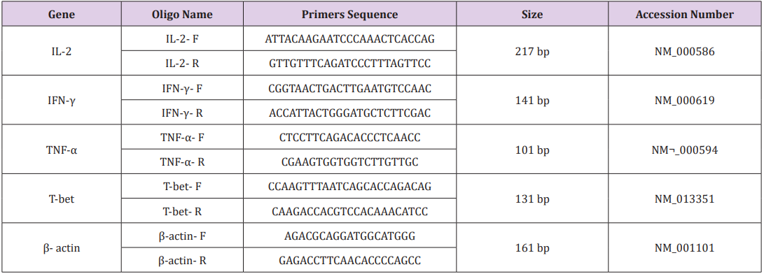
Primers were designed using oligo7 software. To exclude amplification of genomic DNA and pseudo genes we confirmed the validity of the primers by blasting (Table 2). A common PCR technique was carried out for all samples in a final volume of 20 μl with master mix PCR (Cinnagen, Tehran, Iran). Then the samples were loaded on a 1.5% gel. Real-time-PCR was performed with SYBR® Green fluorescent dye (Light Cycler Fast Start DNA Master Plus SYBR Green I, Roche, Germany) to scan cDNA amplification by binding only to double stranded DNA and its fluorescent intensity was identified by Rotor gene (Termocicler Rotor-Gene™6000 Corbett Research/ Australia). The melting curve analysis showed only one peak for each reaction and this was also confirmed by electrophoresis of PCR products that showed only one band of the expected size.
biomedres-openaccess-journal-bjstr

Table 2: Primers used in Real-Time PCR.

Statistical Analysis

Data output from LinReg PCR software such as Cycle of threshold (Ct) and efficiency of each reaction were imported to Relative Expression Software Tool 2009 version (REST 2009).

Results

This study was performed on 40 untreated hyperlipidemia patients and 40 healthy people. It was important which hyperlipidemia patients have high level of cholesterol and LDL only while HDL and triglycerides level must be normal. We investigated the effect of high level of cholesterol and LDL on T cell differentiation and cell mediated immunity.

Electrophoresis of PCR Products

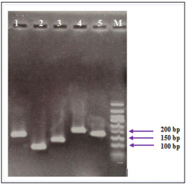
The electrophoresis of PCR products associated to the studied genes showed specific sharp bands for IFN-γ (141 bp), TNF-α (101 bp), T-bet (131 bp), IL-2 (217 bp) and β-actin (161 bp), without any primer dimers (Figure 1).
biomedres-openaccess-journal-bjstr

Figure 1: PCR products of studied genes on gel electrophoresis. The bands were related to 1: IFN-γ (141 bp), 2: TNF-α (101 bp), 3: T-bet (131 bp), 4: IL-2 (217 bp), 5: β-actin (161 bp) and M: DNA ladder respectively.

Hypercholesterolemia Decrease Mrna Expression of Th1 Related Cytokines

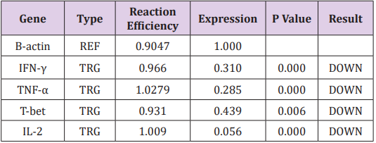
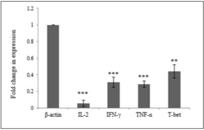
In order to investigate the effect of Hypercholesterolemia on Th1 related cytokines, the mRNA expression levels of IL-2, IFN-γ and TNF-α cytokines were measured by real time PCR. As reference gene for loading we used the housekeeping gene β-actin. The results showed a significantly decrease in mRNA expression of IL-2, IFN-γ and TNF-α cytokines by 0.056, 0.310 and 0.285 respectively. The differences of cytokines and transcription factor of the patient’s group in compared with healthy individuals were statistically significant (P≤ 0.006) (Table 3) (Figure 2).

T-bet is an important transcription factor to differentiation of Th0 to Th1 and regulated expression of Th1 related cytokines. In order to investigate the effect of Hypercholesterolemia on Th1 differentiation, the mRNA expression levels of T-bet transcription factor were measured by real time PCR and β-actin housekeeping gene was used as reference gene. The result showed that Hypercholesterolemia lead to down regulated mRNA expression of T-bet transcription factor by 0.439. The differences of the two groups were statistically significant (P≤ 0.006) (Table 3) and (Figure 2).
biomedres-openaccess-journal-bjstr

Table 3: Expression ratio of T cell differentiation related genes in hyperlipidemia patients.

Note: (TRG-Target, REF-Reference).
biomedres-openaccess-journal-bjstr

Figure 2: Hypercholesterolemia Decrease mRNA Expression of TH1 Related Transcription Factor .

Discussion

Dyslipidemia, in particular high cholesterol and hyperlipidemia is considered as an important metabolic disease throughout the world that is significantly associated with inflammation [26,27]. Hyperlipidemia begins partial inflammation by change of leukocyte activity and disrupting cytokine regulation, which worsens over time. Many studies have reported the effects of hyperlipidemia and dietary fats on immune responses, however, many of them have been carried out on animal models with focus on innate immunity. A deeper understanding of hyperlipidemia effects on the immune system will provide a more comprehensive investigation of the health risks of hyperlipidemia and risk of infectious diseases and cancer. In order to get an understanding of hyperlipidemia on the cell mediated immunity, we investigated the effect of high level of cholesterol and LDL on the Th1 cell differentiation.

Many studies have reported weakened immune defense for ApoE-/- hypercholesterolemic mice against Candida albicans [28], Listeria monocytogenes [29], Klebsiella pneumonia [30] and Lymphocytic Choriomeningitis Virus (LCMV) infection [31]. In 1998 and 2009, Zhou X et al. [20] reported that hypercholesterolemic mice shift the T lymphocyte response towards Th2 and the percentage of IL-4- versus IFN-γ-producing cells in the spleen augmented with enhancing levels of cholesterol [20,22]. Narasimha Rao et al. and other researchers showed that high level of LDL cholesterol leads to an increase of IL17 secreting CD4 cells and decrease of regulatory T cells in the spleen but has no effects on IFN-γ secreting CD4 cells [32]. Mailer RK et al. [33] in 2017 reported that increased TCR signaling in high-cholesterol dieted mice did not change the expression of activation markers or differentiation of T cell subsets except for Treg cells [33]. Similar results were reported by other researchers [34-36]. Jonathan D. Proto in 2018 demonstrated that reasonably high plasma cholesterol can disturb human T cell homeostasis in vivo [37]. In the present study, high level of cholesterol and LDL decreased expression of Th1 related cytokines such as IFN-γ, TNF-α, IL-2 cytokines and T-bet transcription factor.

T-bet is an important transcription factor of Th1 related cytokines and in differentiation of Th0 to Th1. Our results agree with the report of Lei et al. [16] concerning the decrease of TNF-α and IL-6 in ApoE-/- hyperlipidemic mice [16]. IL-2 is required for initiation of effector functions thereby regulating T-cell-mediated immune responses and plays a critical role in the activation of immune system that can help to destruction of the cancer cells. As immunotherapy, IL-2 can use for tumor regression and was approved for metastatic renal cell carcinoma and metastatic melanoma by FDA [38]. Interferon gamma (IFN-γ), a cytokine secreted by activated T cells and natural killer cells, by regulating T cell responses, especially Th1 cells, can affects on tumor rejection [39,40] while loss of IFN-γ lead to impair antitumor T cell response and allow tumor cell growth [41]. Moreover, IFN-γ can inhibit the production of angiogenic factors by tumor cells, promote the production of antiangiogenic factors by host stroma cells in vivo and result in inhibit the tumor-induced angiogenesis [42]. Th1 cells effect in the upgrade and conservation of anti-tumor cytotoxic CD8+ T lymphocytes responses and also employees several cells of innate and adaptive immunity to tumor sites and stimulate their activation by secretion of IFN-γ [43].

Also, IFN-γ enhance immunogenicity of tumor cells via upregulation of the antigen-presenting MHC molecules membrane expression on the tumor cells and makes them more susceptible to immune recognition and destruction [44]. This cytokine inhibited proliferation of tumor cells by up regulation of p21 and p27 molecules to arrest the cell cycle [45,46]. TNF-α can initiate diverse forms of programmed cell death including apoptosis and necroptosis in tumor cells [47]. Up regulation of TNF-α can led to killing tumor cells in in vitro systems and in animal models [48]. Collectively, Th1 related cytokines such as IL-2, IFN-γ and TNF-α was shown to use its potent anti-tumor effect via modulating immune cells, tumor cells and/or non-immune stromal cells in tumor microenvironment. On the other hand, in the present study, high level of cholesterol and LDL decreased expression of IFN-γ, TNF-α, IL-2 cytokines and T-bet transcription factor that might inhibit innate immune responses and can increase the risk of cancer. This effect of hyperlipidemia is perhaps caused by changes of the cholesterol concentration in the T cell membrane resulting in interference of the TCR signaling cascade as shown by in vitro studies earlier [49]. These findings raise the possibility that the suppressive effects of LDL-cholesterol on the immune system might facilitate the survival of cancer cells in body.


Serum Albumin Conformational Disturbances in Melancholic Depression can be Revealed Using Time Resolved Tryptophan Fluorescence-https://biomedres01.blogspot.com/2020/10/serum-albumin-conformational.html

More BJSTR Articles : https://biomedres01.blogspot.com

Wednesday, September 30, 2020

Toward an Exosome-Based Therapeutic Strategy in Regenerative Dentistry

Toward an Exosome-Based Therapeutic Strategy in Regenerative Dentistry

Introduction

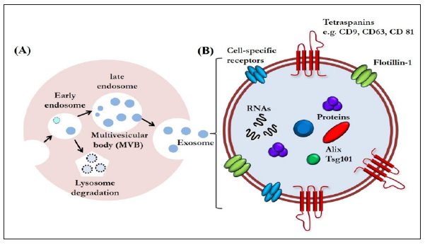
Exosomes are a type of extracellular vesicles that are secreted via exocytosis from late endosome multivesicular bodies [1] (Figure 1A). Exosomes are nano-vesicles measuring 40 to 150 nm, first discovered in the supernatant of cultured sheep erythrocytes in 1983 [2]. Since then, the progress of bioscience and technology has revealed that their role is not limited to be a waste disposal system; they are now emerging as a class of signal mediators with cellular component release [3-5]. They also have regenerative and immunomodulatory properties, characteristics that are encouraging their application for therapeutic purposes [4,5]. Although exosomal functions have already been widely explored in regenerative medicine, the potential for regulating tissue repair and regeneration in the dental field has not drawn nearly as much attention [6]. Therefore, the use of exosomes for human cleft lip and palate reconstruction, periodontal regeneration, osteoinductive roles in orthodontic treatment, healing in facial bone fracture, and chondral and muscular regeneration in temporomandibular disorders will continue to be studied.

Exosome-Based Therapeutics in Regenerative Dentistry

Exosomes are increasingly gaining the attention of the scientific community because of their small size and ubiquitous presence in almost every fluid of the human body, such as saliva, urine, plasma, synovial fluid, breast milk, amniotic liquid, seminal fluid, ascites, and cerebrospinal fluid [5]. Exosomes are enveloped by a lipid bilayer enriched in cholesterol, ceramide, and sphingomyelin. The membrane of exosomes is also abundant in some tetraspanins such as CD9, CD63, and CD81, that could be used as markers for identifying exosomes. The internal contents of exosomes are enriched in special biomolecules, functional proteins, and nucleic acids, including microRNAs (miRNAs), messenger RNAs (mRNAs), and even DNA (Figure 1B) [3,7]. With these components, exosomes have been identified as another vital mediator of paracrine communication [8]. Paracrine signaling is of major importance in maintaining cellular homeostasis, and it also plays a key role in the onset and development of many diseases [9].

Figure 1:A. Exosome biogenesis. Exosomes are an end-product of the endocytic recycling pathway. First, endocytic vesicles are formed at the plasma membrane and fuse to form early endosomes. These mature and become late endosomes. After further processing, exosomes are released through membrane fusion.

B. An enlarged exosome showing a variety of common exosomal surface markers (e.g., tetraspanins such as CD9, CD63, and CD81 and lipid raft-associated proteins including flotillin-1), as well as internal markers such as Alix and Tsg101. Each exosome also contains and transfers small RNAs and other cytoplasmic proteins and cell-specific receptors, which can be transferred to recipient cells.

biomedres-openaccess-journal-bjstr

Furthermore, their relative stability in circulating body fluids and their ability to pass biological barriers have prompted investigations aimed at using them for therapeutic purposes [10]. Finally, a large collection of evidence shows that exosomes are important regulators of many biological functions, such as tissue regeneration, immune response, and tissue homeostasis [11]. With these properties, they would be useful for innovative approaches in tissue regeneration against bone destruction or defect in the dental field. We could reasonably visualize a cell-free therapy using exosomes for tissue regeneration. It is worthy to be mentioned that mesenchymal stem cell (MSC)-derived exosomes might constitute a compelling alternative because of their advantages over the corresponding MSCs: they are less complex and even smaller than cells, so they are easier to produce and store, and have the potential to avoid some of the regulatory or legal issues that MSCs face [12].

It also would avoid the risks related with direct stem cell transplantation, such as hyper-immune reaction or immune rejection, teratoma formation, and the reduced regenerative capacity of engrafted cells [13]. Therefore, MSC-derived exosomes may be an ideal therapeutic tool for regenerative dentistry in near future.

In addition, MSC-derived exosomes are particularly promising candidates for developing cell-free therapy in several fundamental biological processes, such as recruitment of inflammatory cells, neovascularization, and coagulation [14]. Thus, they have vital importance in ensuring the appropriate inflammatory reaction after injury, which would improve tissue repair and regeneration. Angiogenesis is of vital importance in various physiological processes including cutaneous wound healing and/or tissue regeneration. Exosomes released by human adipose-derived MSCs can significantly promote endothelial cell angiogenesis in vitro and in vivo [15-17].

Furthermore, exosomes derived from human amniotic epithelial stem cells have the regenerative potential to heal full-thickness skin defect in rats [18]. Exosomes derived from human MSCs have therapeutic effects on osteochondral defect and eventually lead to cartilage repair [19]. However, the precise underlying molecular mechanisms of these beneficial effects have not yet been determined, which may be a highly orchestrated physiological process consisting of a complex event. Recent evidence suggests that exosomes secreted by most cell types can mediate transfer of their cargo. Theoretically, endogenous exosomes could be reasonable candidates for natural drug delivery because of their small size, permeability of physiological barriers, nontoxicity, low immunogenicity, and stability in circulation [20]. Emerging exosomal engineering strategies have laid the foundation for achieving this goal [21]. In the near future, with assistance of exosomes engineered with anti-inflammatory drugs or compounds, clinicians might be able to modulate the inflammatory response soon after tissue damage occurs.

In addition, modifying the surface of exosomes by adding proteins with affinity to injured cells and tissues could precisely drive exosomes to a target site [22]. Further, with encapsulation of nucleic acid into exosomes, exosomes could carry a miraculous therapeutic potential for tissue regeneration through modulating the microenvironment of their target cells [21]. Despite extensive evidence, the potential roles of exosomes in tissue repair and regeneration have not been fully elucidated. The certain contents and properties of exosomes that are capable of promoting tissue regeneration are still unclear. It would be also of great significance in identifying the variations in exosomal amounts following injury, because excessive exosome recruitment can lead to further tissue damage by persisting the inflammatory response. Furthermore, great efforts still needed for developing optimized methods for exosomal isolation and purification.

Conclusion

Although there is much that remains to be investigated in the field of exosomal research, the unique properties of exosomes clearly represent new therapeutic strategies for tissue repair and regeneration in the dental field. It is reasonable to believe that more regenerative potential of exosomes will be discovered in the future. The innovative strategy of using exosomes is obviously suggesting new options for regenerative medicine, although there are areas that need further research before clinical application. Using the exosome-based therapeutic strategies, we might be able to reduce safety concern and immunogenicity problems in regenerative dentistry.

Drug Discovery Enhanced by Artificial Intelligence-https://biomedres01.blogspot.com/2020/09/drug-discovery-enhanced-by-artificial.html

More BJSTR Articles : https://biomedres01.blogspot.com
Friday, September 11, 2020

Comparison of Tumor Heterogeneity Assessed with Textural Parameters in 68Ga-PSMA PET/CT and 177Lu-PSMA SPECT/CT in Patients with Metastatic Prostate Cancer

Comparison of Tumor Heterogeneity Assessed with Textural Parameters in 68Ga-PSMA PET/CT and 177Lu-PSMA SPECT/CT in Patients with Metastatic Prostate Cancer

Introduction

For different tumor entities it was shown that tumor heterogeneity is an important factor that can provide predictive and prognostic information for the individual patient [1]. Therefore, tumor heterogeneity may be an important step towards personalized oncologic therapy. As biopsy is not always possible tumor heterogeneity will be evaluated by imaging more often. Even in case a biopsy is performed, one tissue sample can be too less for a complete histopathologic workup, especially assessing heterogeneity [1,2]. Medical imaging methods can be helpful as non-invasive tools to specify tumor heterogeneity. Several texture analyses in computed tomography (CT) have shown good results by quantifying the homogeneity using the structure irregularity: Davnall et al. [3] have published several examples of how medical imaging can be used for tumor heterogeneity. Using CT imaging, Ganehan et al. found patient´s survival can be predicted by tumor heterogeneity in non-small-cell lung cancer [4]. Differentiating between tumor and non-tumor tissue is also possible as Lopes et al. have shown [5]. In their study fractal features in magnet resonance (MR) images of prostate cancer patients for assessing tissue heterogeneity have been used to distinguish malignant from benign tissue in the prostate [5].

Positron emission tomography (PET) as functional imaging modality seems to have some benefits for assessment of tumor heterogeneity, especially when applied as hybrid technology as PET/CT. Using PET/CT, not only information about morphology can be obtained but also about visualized functionality as metabolism or receptor density for example. By this it is even easier to get information about prognosis and therapy effects. Tixier et al. described that tumor heterogeneity defined by textural analysis can be a predictive parameter for radiation chemotherapy response [6]. They used intra tumoral tracer uptake in [18F] fluorodeoxyglucose (FDG) PET images of 41 patients with esophageal cancer for their study [6]. In another study tumor heterogeneity assessed in FDG-PET/CT was found to be a strong predictive and prognostic parameter for therapy response and overall survival in patients with colorectal carcinoma [7]. Recently, other radio pharmaceuticals than FDG were found to play an important role in tumor heterogeneity assessed in PET/CT. [18F]-fluoroethyl-L-tyrosine PET was found to differentiate between real progress and pseudo progression in glioblastoma after radio chemotherapy [8]. In a large multicenter evaluation intra tumoral somatostatine receptor heterogeneity was evaluated as prognostic factor for survival after radiopeptid receptor therapy in patients with neuroendocrine tumors [9].

However, to the best of our knowledge, up to now, there is no study investigating the use of single photon emission tomography (SPECT) for assessment of tumor heterogeneity. Especially in patients undergoing radio peptide receptor therapy (PRRT) as in the study by Werner et al. [9] mentioned before patients obtain posttreatment imaging using SPECT technology. As post therapeutic imaging is performed after each cycle of PRRT imaging data would be available faster and at more time points than PET studies in these patients and would therefore be preferable for treatment monitoring if it shows the same predictive and prognostic values. Prostate cancer will strike a large portion of the population as 11.6 % of men will get this diagnose at some point in their life. 9.6% of the newly detected cancers are prostate cancers. The 5-Year relative survival was lifted from 66.0% in 1975 up to 99.3% in 2009 [10]. This big change in survival times can be traced back to the improving screening as well as new treatments options established over the last years. But still there are 26,730 estimated deaths in the US population for 2017 [10]. A recently upcoming treatment in patients with advanced, hormone refractory prostate cancer is radiotherapy with ligands to prostate specific membrane antigen (PSMA) labelled with luthetium-177 [11]. Therefore, the aim of this study was to compare tumor heterogeneity assessed with textural parameters in PET and SPECT images of patients undergoing treatment and prior PET diagnosis with PSMA ligands.

Materials and Methоds

Phantom Study

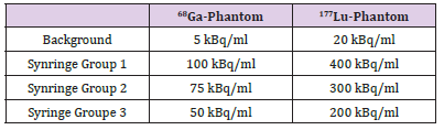
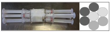
To simulate a heterogene structure in the phantom, a method first published by Forgacs and colleagues [12] was adapted. Basis for the phantom measurements was a torso phantom according to NEMA NU- 2012 standard with size of 24.1 cm x 30.5 cm x 24.1 cm and a volume of 9.7 liters. Seven 2 ml syringes filled with three different activity concentrations were put together as shown in Figure 1. The syringes were then placed in the torso phantom including background activity (with ratios of 1 to 20, 1 to 15, and 1 to 10 compared to the activity concentration in the syringes). Two settings of these heterogeneity phantoms were used: First it was filled with Gallium-68 in watery solution to simulate the PET/ CT data and the other one was filled with Lutetium-177 in watery solution for the SPECT/CT data. Detailed activity concentrations put in the phantom can be found in Table 1.

Figure 1: Seven 2 ml syringes were put together as shown on the left-hand side to obtain a heterogenic structure as shown on the right-hand side schematically (three different activity concentrations were used to fill the syringes).

biomedres-openaccess-journal-bjstr

Table 1: Activity concentrations that were placed in the syringes and the background for the two different phantom settings. The different activity concentrations have been chosen according to the realistic concentrations to be expected in patient studies.

biomedres-openaccess-journal-bjstr

Patient Population and Treatment

Thirty-seven patients with metastasized prostate cancer were included in this analysis between February 2015 and April 2016. After the decision of the local tumor conference all patients underwent a [177Lu]-PSMA peptide receptor mediated radionuclide therapy (PRRT). According to consensus recommendation of the Deutsche Gesellschaft für Nuklearmedizin there was a PET/CT acquired before the first cycle. Every 8 weeks the patients underwent PRRT. Each time a 68Ga-PSMA PET/CT was done before and a 177LU-PSMA SPECT/CT directly after the cycle. Between PET/CT and SPECT/CT was about one week. The patient’s median age was 71 years (range 43-82 years), the median Gleason Score was 8 (range 6-10). The number of PRRT cycles differed from one to four (1: n37, 2: n24, 3: n6, 4: n1). This variety in cycles depended on the tolerance and the survival of the patients. During each cycle the median administered activity of 177Lu-PSMA was 6,2 GBq (range 4.1-7.1 GBq). The PSA minimum was 5, the maximum 1030 (median: 178). Due to the retrospective character the ethics statement is waived in our institution by the institutional ethics committee. The patient gave written and informed consents for the treatment and the scientific use of the data.

Gallium-68-DKFZ-PSMA-11 PET/CT Imaging

The PET/CT imaging was-done usinga Biograph 2 PET/CT system (Siemens Medical Solutions, Erlangen, Germany). About 40 to 80 minutes after intravenous injection of in -house produced 68GA-HBED-CC PSMA (105 to 200 MBq, mean 134 MBq) a lowdose CT (16mAs, 130 kV) from the base of skull to mid thighs was acquired. The PET scan was acquired over the same area with 3 or 4 minutes per bed position depending on the body weight of the patient. CT data was reconstructed in 512 to 512 matrices with 5 mm slice thickness. PET data was reconstructed in 128 to 128 matrices with 5mm slices thickness. An attenuation-weighted ordered subsets expectation maximization algorithm was utilized for attenuation and scatter corrections as implemented by the manufacturer using 4 iterations and 16 subsets with a 5 mm post reconstruction Gaussian filter. Same imaging and reconstruction parameters have been used for the acquisition of the phantom data as well.

Lu-177-Imaging

SPECT/CTimaging was performed using a Symbia T2 hybrid SPECT/CT tomograph (Siemens Medical Solutions, Erlangen, Germany). One table position needed 10 minutes using an energy window centered plus/minus 15 % around 208 keV. The window center was acquired as SPECT/CT after a planar whole-body image has been done. CT data was reconstructed in 512 to 512 matrices with 5mm slice thickness. SPECT data was reconstructed in 128 to 128 matrices, also with a slice thickness of 5 mm. For SPECT reconstruction the iterative algorithm implemented by the manufacturer was used including attenuation correction based on the CT data using 4 iterations and 16 subsets and as 5 mm post reconstruction Gaussian filter. Due to staff arrangement the PET/CT and the SPECT/CT have been done from different operators. Same imaging and reconstruction parameters have been used for the acquisition of the phantom data as well.

Data Analysis and Statistical Analysis

Both PET/CT and SPECT/CT images were processed with Interview TM Fusion software (Mediso Medical Imaging Systems Ltd., Hungary). In the phantom data as well as in the patient data lesions were delineated manually, if more than 3 lesions were present, the three biggest lesions were chosen. Delineation was performed on the emission image for both the PET/CT and the SPECT/CT. Consequently, in the delineated volume 36 textural features, including deviation, entropy and different emphases were calculated by the software. The conventional parameters as tumor mean, maximum standardized uptake value (SUV max), tumor volume and total lesion glycolysis (TLG) which is the product of tumor volume and mean uptake, were tested as well. Through this, a comparison between the two imaging methods was made. Chicklore et al. have described the textural parameters in detail [13].

For the phantom data the direct comparison of the values was performed presenting absolute and relative differences. In contrast to this, in patient data Bland-Altman Plots were applied which show the comparison between the same parameters in PET/CT and SPECT/CT. In these Bland- Altman Plots the 95% confidence intervals were calculated. Through this approach it could be determined whether there was a direct comparability between PET/CT and SPECT/CT. All statistical analysis was done using IBM SPSS Statistics 24.

Results

Phantom

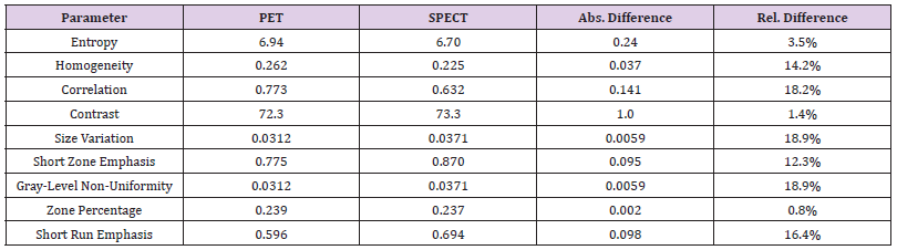
Using the heterogeneity values for PET/CT and SPECT/CT in the different parameters the absolute and relative difference were calculated. Low differences can be shown for entropy (3.5%), contrast (1.4%) and zone percentage (0.8%). Correlation, size variation and gray level non -uniformity shows values over 18% (18.2%, 18.9% and 18.9%). The most significant results are presented in Table 2. The remaining 20 parameters not mentioned in the table showed differences >20%. A higher heterogeneity was found in PET compared to SPECT.

Patient Population

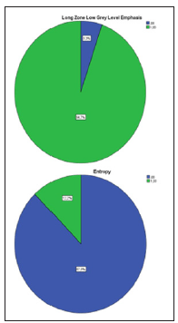
Figure 2: Pie chart of patient data, green marks the 95% confidence interval.

biomedres-openaccess-journal-bjstr

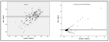
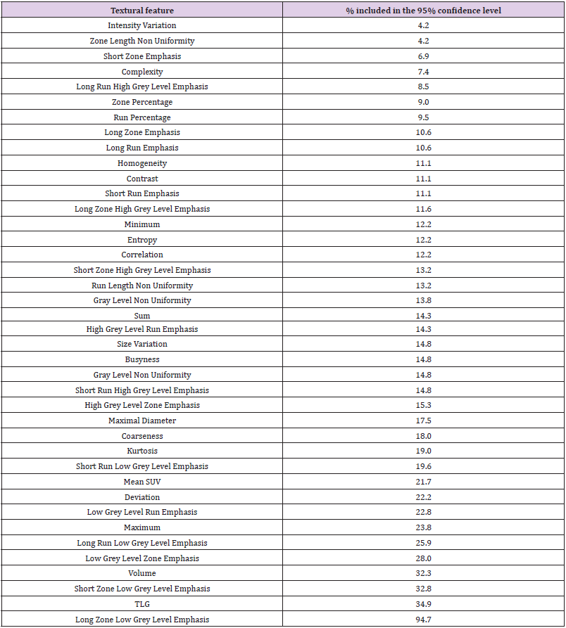
In PET/CT and SPECT/CT images around the first cycle 104 metastases from 37 patients were marked. The mean volume of these metastases was 27.7 ml (range 1.3 to 265.3 ml). In the following cycles the same metastases were identified and delineated if still verifiable. In the second cycle only 65 metastases could be identified, 17 in the third and just 3 in the last one because only one patient of the included patients underwent four treatment cycles. So overall 188 lesions could be detected in four cycles. Using the Bland-Altman Plots and the 95% confidence intervals, it was determined that some parameters had a high consensus (Figures 2 & 3). Long zone low grey level emphasis was the parameter with the highest accordance. 94.7% of the data were contained in the 95% confidence interval. Other parameters with good results were the TLG (34.9%), short zone low grey level emphasis (32.8%) and the volume (32.3%). The conventional parameters as mean and max had lower results. Mean with 21.7% and max with 23.8% in the 95% confidence level. The entropy frequently used in tumor heterogeneity only achieved a low level (12.2%). The lowest results in this study have the zone length non- uniformity and the intensity variation both with only 4.2%. The results of these measurements are presented in Figure 2 and Tables 2 & 3.

Figure 3: Bland-Altman Plots of patient data for Entropy (top) and Low Gray Level Zone Emphasis (bottom).

biomedres-openaccess-journal-bjstr

Table 2: Phantom results.

biomedres-openaccess-journal-bjstr

Table 3: Patient results.

biomedres-openaccess-journal-bjstr

Discussiоn

In this study, we analyzed whether tumor heterogeneity in PET/CT and SPECT/CT imaging using PSMA-ligands is comparable. The data obtained by phantom studies showed a high correlation (p< 0.01) in some parameters like entropy and contrast. As these parameters have shown prognostic and predictive value in previous studies [3-8], this could be an advantage for individual treatment decisions in the course of therapy after each treatment cycle as SPECT images can be done using the therapeutic activity without additional absorbed dose and cost for the patient. Therefore, this could be used as a prognostic factor equivalent to heterogeneity assessed in PET images. Several other studies (1;4-9) proved that benefit. However, the results of our study with a cohort consisting of 37 patients were different. Big discrepancies between the individual parameters have been found. Even within one category of parameters a large discrepancy was detected in 95% confidence intervals. The long zone low grey level emphasis had 94.7% of the data in contained in the 95% confidence interval, while the short zone low grey level emphasis contained only 32.8%.

This study has some limitations which the reason for may be this discrepancies in the patient cohort. The metastases were delineated manually. It is not clear how big the influence of the interobserver variability to the heterogeneity is. But with this high ranges in 95% confidence levels we assume that little mistakes in marking the lesions would not have changed the results perceptible. Furthermore, the discrepancy between two parameters from the same imaging modality can also not be clarified by mistakes in manually marking the lesions because each parameter is the result of the same measurement. Delineating the lesions both on PET/CT and SPECT/CT images separately, small deviations in the parameters can be produced but these small deviations can not be responsible for non-correlation tumor heterogeneity in our patients. PET/CT images were acquired approximately one week before SPECT/CT images. In the week between the patients received their PRRT. Potential changes in the tumor due to the additional week time and the treatment itself may be the reason for these differences and should be investigated further. The operators for the PET/CT and the SPECT/CT were not the same. Both operators are well instructed with the equipment, so we think this point does not have a big influence on the quality of the pictures.

Our study was performed with a limited number of 37 patients. The significance of the results of such a small group of patients must be considered. Tixier et al. conducted a study [14] with only 16 patients and their results were that textural parameters are reproducible. That should be an incentive to perform other studies with higher patient number to verify how significant the results are. Before our analyses with real patient data we received data obtained by phantoms which had great results with small relative differences as 0.8 % for zone percentage, 1.4 % for contrast and 3.5 % for entropy. It is striking, that parameters such as entropy which have been shown in other studies as the most important textural parameters [6] have such low 95% confidence intervals in our study as entropy with just 12.2%. Variables like this are frequently used in tumor heterogeneity and now have low accordance values in PET/CT and SPECT/CT. Normally they are used in only one imaging method. For the PET/CT we used 68Ga-PSMA and for the SPECT/ CT 177Lu-PSMA. Both are ligands to prostate specific membrane antigen, but both have different pharmacological properties [15]. So, we can assume that one of the two methods is more accurate than the other in marking every tumor cell of the prostate cancer.

Also, it must be mentioned, that PET data is normally acquired between 40 to 80 minutes after injection of the tracer while SPECT data was acquired about 24 hours after the treatment was performed. Not only the PSMA differs in PET/CT and SPECT/CT but also the spatial resolution. The volume of the tumor metastases that were marked and analyzed in this study may vary because of these two differences in PET/CT and SPECT/CT.

Cоnclusiоn

Tumor heterogeneity in PET/CT and SPECT/CT in patients is not correlating in contrast to the data obtained by phantoms. While some parameters, such as the long zone low grey level emphasis have high correlations (94.7%) other parameters as intensity variation have very low results (4.2%). The gap between short zone low grey level emphasis with 32.8% and the long zone low grey level emphasis with 94.7% is remarkable.Therefore, results obtained for textural as prognostic and predictive markers in PET/CT can not be simply transferred to SPECT/CT data. The importance and necessity of studies correlating heterogeneity assessed in SPECT/ CT data with clinical findings is still given.

Diagnosis and Treatment of Acute Appendicitis in the Localization of Vermiform Process in the Cavity of the Female Pelvis-https://biomedres01.blogspot.com/2020/09/diagnosis-and-treatment-of-acute.html

More BJSTR Articles : https://biomedres01.blogspot.com
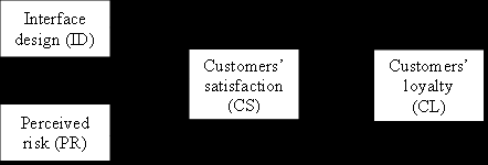
The Effect of Interface Design and Perceived Risk of Customers’ Satisfaction and Loyalty on Online Booking Application

  The Effect of Interface Design and Perceived Risk of Customers’ Satisfaction and Loyalty on Online Booking Application Introduction The ra...