Wednesday, September 30, 2020

Malignant Transformation of Exophytic Sinonasal Papilloma: Case Report

Malignant Transformation of Exophytic Sinonasal Papilloma: Case Report

Introduction

Schneiderian papillomas (SPs) of the nose (also known as sinonasal papillomas) were described by Ward in 1854. In 1991, the World Health Organization classified sinonasal papillomas into three distinct histopathological subtypes: exophytic, inverted, and oncocytic. The incidence of sinonasal papillomas is approximately 2.3 cases per 100,000 population per year. The inverted and the exophytic types are more common than the oncocytic. Malignant transformation of SP is estimated in the literature from 2 to 27 %and appears mostly in inverted papillomas [1]. Malignant transformation of exophytic SP is exceptionally rare.

Case Report

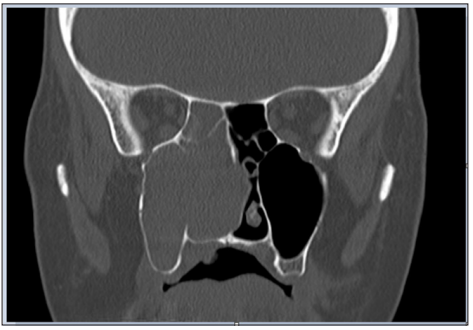
A 48-year-old woman presented to the hospital with nasal obstruction. She had noticed this problem first time 27 years before. During the last months a tissue mass appeared in the nasal entrance, which deformed the right alar part of the nose. Nasal endoscopy and CT scan of paranasal sinuses showed a polyplike soft tissue mass, that filled the right sinus maxillaris, destructed its medial wall and the right ethmoid cells (Figure 1). This tissue enlargement posteriorly reached the epipharynx and anteriorly bulged into the left common nasal cavity through a septal defect. Endoscopic surgery was performed in general anesthesia. The lesion was removed in multiple parts. The tissue samples from the right nasal cavity and from the right sinus maxillaris have shown the structure of a typical exophytic Schneiderian papilloma with a part of invasive high grade planocellular carcinoma (Figure 2). Histologically exophytic proliferations with a fibrovascular core covered by transitional type epithelium could be seen, in some locations squamosus metaplasia and koilocytosis were present. The malignant transformation represented a proportion of 10- 15% of the sample. At the margins of the malignant transformed area extensive features of dysplasia in the whole thickness of the epithelium was found, which represented the in situ component of the tumor.

In the invasive area there were nests of dysplastic squamosus cells, without any sign of keratinization. The tumor did not show desmoplastic reaction, but this area showed a rich inflammatory infiltrate. In the normal SP area Ki67 proliferating ratio was low, while at the base of the epithelium, in the in situ and the invasive areas the proliferation activity was much more elevated in the full thickness of the epithelial component. The tumor and the dysplastic area was p16 positive while the non-malignant epithelium was negative. We subjected the removed tissue sample to HPV DNA analysis using PCR. Although koilocytes were present on histological examination, no HPV DNA was found in this case. Gynecological examination has found no papilloma-like lesion and HPV detection from cervical excretion was also negative. The classification of the tumor in the TNM system was T1N0M0. The postoperative CT scan showed mucosal thickness inside the right maxillary sinus without bone destruction. The radicality of the removal could not be defined from the tissue specimen because of its fragmentation, therefore postoperative radiotherapy was given to the patient.

Figure 1: CT scan of paranasal sinuses shows a soft tissue mass, that fills the right sinus maxillaris, destructs its medial wall and the right ethmoid cells.
biomedres-openaccess-journal-bjstr

On the postoperative MRI scans mucosal thickness could be observed in the right sinus maxillaris. The thickened mucosa from the right sinus maxillaris was removed in multiple parts. Histological examination found SP fragments in the polypoid mucosa without any sign of malignancy. The patient underwent follow-up examination monthly. Due to the missing part of the medial right sinus maxillaris wall we could check endoscopically the whole right sinus maxillaris and operating field. To date we have not found any sign of recurrent papilloma or carcinoma

Discussion

Nasal obstruction is the most common symptom of SP, followed by epistaxis, pain and asymptomaticy[2]. Exophytic papillomas almost exclusively arise from the nasal septum with rare cases arising from the lateral nasal wall [3]. The most common localisation is the transitional zone between the squamosus epithelium and the respiratory type epithelium [4]. Glatre et al. described the unifocal and the multifocal form of exophitic SPs [4]. He suggested that the multifocal form, named also florid nasal papillomatosis, has a higher recurrence than the unifocal form. These lesions are usually unilateral, the bilateral location is uncommon. In larger series, papillomas are bilateral in about 8% of cases [5]. In our case the extremely enlarged laesion was unifocal, with its origin from nearby the natural orificium of the right maxillary sinus. Patients with the exophytic subtype are usually younger, presenting in their fourth decade of life and the remaining two subtypes were more likely to occur in older patients [3]. Male predominance can be observed by exophytic and inverted papillomas, whereas the oncocytic SP has shown no sex predilection [2,3,6].

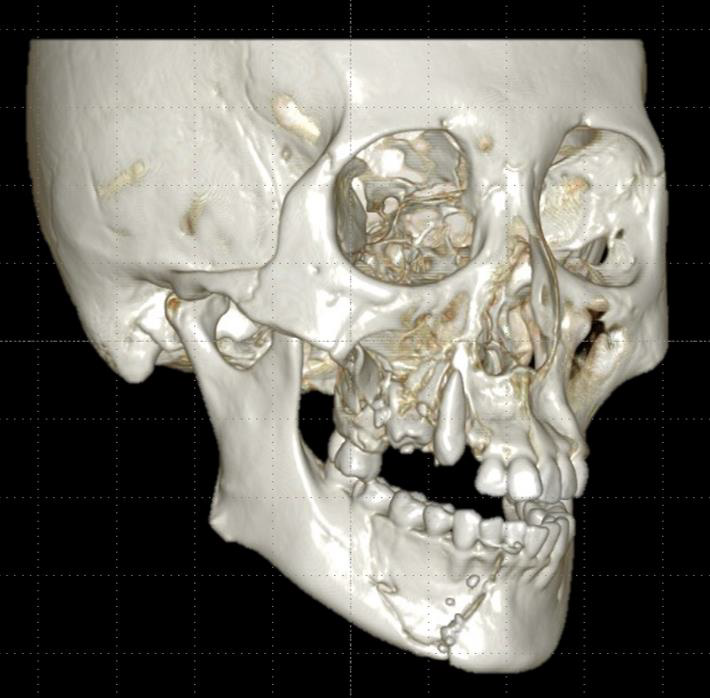
Nasal trauma is present in the history of many patients with exophytic papilloma[4]. In our case nasal trauma was also present. In childhood she suffered a nasal injury. Few years later nasal aesthetic surgery was done. The macroscopic appearance of exophytic papilloma is: grey–tan, exophytic, “mushroom-shaped” verrucous papillary proliferations (Figure 3). Until radiological signs of inverted papilloma was described, there had been no specific radiological sign for exophitic sinonasal papilloma [4,6]. On the preoperative CT slices bone destruction can be seen (Figure 1). Bone erosion can occur without malignant transformation, but is more likely when it is present.The pathogenesis of papillomas is not clarified. Some evidence suggested that SP may be associated with HPV infection, in particular HPV 6 and 11 and sometimes 16 and 18 [4,5,7]. It is not known how HPV is transmitted to the sinonasal tract, although sexual transmission is suspected. Exophytic papillomas frequently contain low risk HPV DNA, but rarely high-risk HPV [3,5]. In the literature HPV 6 and 11 are mostly associated with exophytic papillomas [3,5]. In one third of sinonasal papillomas HPV 6 and 11 are present [6]. In our case no HPV DNA was identified in the removed tissues. In the international literature we found 6 cases of exophytic SP with malignant transformation. These cases are presented in a table for better comparability Table 1 [7-10].

Vibrational Cooling Effect of Zeolite on Molecular Desorption Studied by Time-Resolved Laser Desorption Ionization Mass Spectrometry-https://biomedres01.blogspot.com/2020/09/vibrational-cooling-effect-of-zeolite.html

More BJSTR Articles : https://biomedres01.blogspot.com
Vibrational Cooling Effect of Zeolite on Molecular Desorption Studied by Time-Resolved Laser Desorption Ionization Mass Spectrometry

Vibrational Cooling Effect of Zeolite on Molecular Desorption Studied by Time-Resolved Laser Desorption Ionization Mass Spectrometry

Introduction

Laser desorption ionization (LDI) mass spectrometry is one of the most reliable techniques for analyte detection. By using matrix molecules that absorb laser photons, “soft ionization,” a process that does not decompose an analyte during ionization, becomes possible. This technique, called matrix-assisted laser desorption ionization mass spectrometry (MALDI MS), has been widely applied to the analysis of biopolymers, such as proteins or peptides, as its soft ionization potential enables observation of analyte-related ions with very few fragments [1-3]. However, MALDI MS has several drawbacks, including low ionization efficiency, protonated analyte peak suppression by alkali metal ion contaminants, and inapplicability to low molecular weight compounds due to the dissociation of matrix molecules. To overcome these drawbacks, many attempts have been made, including matrix-free techniques [4-7], application of nanometer-sized particles [8-10], and the use of a co-matrix with conventional organic matrix molecules [11-14]. Recently, we have revealed that zeolite is one of the most promising additives for MALDI MS. Zeolites are crystalline aluminosilicates with three-dimensional frameworks and nanometer-order cages. Zeolites have high catalytic activity due to the charge imbalance at the Si-O-Al bridging sites, and those sites are compensated typically by such cations as H+ and Na+. Hydroxyl (OH) groups having Brönsted acidity exist in H+-exchanged zeolite. It is known that the Brönsted acid site is responsible for the catalytic activity of zeolite. By using zeolites in MALDI MS, it was found that they prevented the dissociation of matrix molecules and enhanced the peak intensity of the protonated analyte [15].

The mechanism of the peak intensity enhancement could be understood as efficient proton supply from the Brönsted acid sites and stabilization of intra- and intermolecular proton transferred states by the strong polarity of zeolite [15]. However, the reason why zeolite prevents the dissociation of matrix molecules is still not clear, although it could be assumed that the excess vibrational energy of matrix molecules would be released to the lattice vibrations of zeolite. In this study, we examined how the three-dimensional framework as zeolites influenced molecular desorption by time-resolved mass spectrometry. Time-resolved mass spectrometry is a pump-probe type measurement [16-18]. By pumping, vibrational excitation of intermolecular dissociative modes takes place. Then, the molecule undergoes ionization including protonation and cation adduction by the probe, and the ion is released to the gas phase for detection. Therefore, the time required for molecular desorption can be detected by changing the delay time between pump and probe. It was understood that zeolite suppressed molecular desorption because it acted as a heat bath, diffusing excess vibrational energy efficiently into its lattice vibrational modes.

Experimental

2,4,6-Trihydroxyacetophenone (THAP) and 1’-hydroxy- 2’-acetonaphthone (HAN) were purchased from Sigma. NH4+- terminated zeolite (NH4M20) was supplied by the Catalysis Society of Japan. For time-resolved mass spectrometry, a Ti: sapphire laser with a regenerative amplifier (Spitfire, Spectra Physics) was used to provide femtosecond pulses (800nm, 0.8mJ, 110fs). The repetition rate was 10Hz. The output was frequency-doubled (400nm) by an LBO crystal and then divided into pump and probe beams. The time delay between pump and probe was changed by the optical delay equipped in probe line. Mass spectrometry was performed under the condition that the spectrum could not be observed by single-beam excitation (pump or probe pulse only); pulse energy was 8μJ for the pump and 10μJ for the probe. Both pulses (pump and probe) were focused by a quartz lens and introduced into a laboratory-built time-of-flight (TOF) mass spectrometer. Solutions of THAP and HAN (1.0 x 10⁻² mol dm-3) were prepared with a mixture of acetonitrile and water (v/v=7:3). Ten microliters of each solution were pipetted onto a stainless steel sample target and the solvent was allowed to evaporate. Thus-obtained crystals of THAP or HAN were subjected to time-resolved mass spectrometry. For the measurement of THAP on zeolite, THAP (16.8mg) was first mixed with NH4M20 (67.2mg) (weight ratio=1:4) in a mortar and pestle and the mixture was suspended in a mixture of acetonitrile and water (v/v=7:3, 10mL; 1.0 x 10⁻²mol dm-3 for THAP). Then, ten microliters of the suspension was pipetted onto a stainless steel sample target. The obtained crystals of THAP/NH4M20 were subjected to time-resolved mass spectrometry.

Results and Discussion

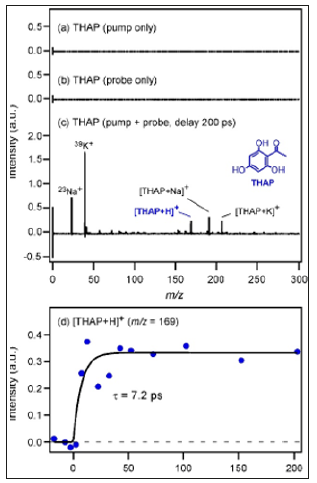
Figure 1a shows the mass spectrum of THAP crystals measured by pump pulse irradiation. No ions were detected by only the pump pulse irradiation. Figure 1b shows the mass spectrum of THAP crystals measured by probe pulses. No ions were detected also by only the probe irradiation. In contrast, when the sample was irradiated by the pump and probe pulses at the same spot at a certain delay time, the situation became different. Figure 1c shows the mass spectrum of THAP crystals obtained by the pump-probe measurement (probing at 200ps delay after pumping). The peak of protonated THAP, [THAP+H]+ , was observed at m/z=169, whereas the peaks of [THAP+Na]+ and [THAP+K]+ were observed at m/ z= 191 and 207, respectively. It is noted that this delay time was neither TOF nor the timing of ion extraction voltage (10ms) after a laser shot, but the timing between the pump and probe pulses. In addition, any time resolution of electronic devices was not used in this study. Figure 1d shows the time dependence of the peak intensity of [THAP+H]+. The peak intensity of [THAP+H+ gradually increased with increasing delay time.

Figure 1: Mass spectrum of THAP crystals measured with a) pump pulse b) probe pulse c) Time-resolved mass spectrum of THAP crystals measured with pump and probe irradiation (delay at 200ps) d) Time dependence of peak intensity of [THAP+H]+.

biomedres-openaccess-journal-bjstr

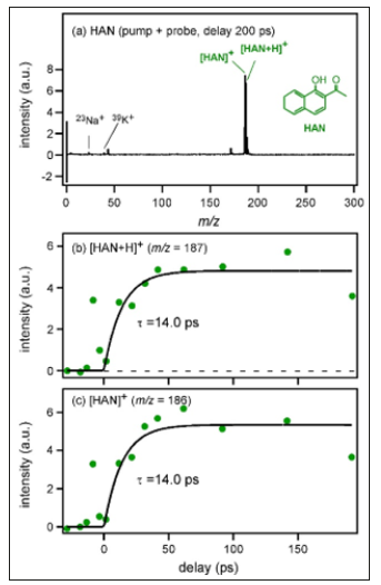
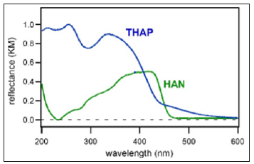
By fitting analysis using exponential functions, the time constant of the rise component was determined to be 7.2±2.3 ps. Figure 2a shows the mass spectrum of HAN crystals measured with pump and probe irradiation (probing at 200ps delay after pumping). Peaks of [HAN]+ and [HAN+H]+ were observed at m/ z= 186 and 187, respectively. The time dependence of the peak intensity of [HAN+H]+ is shown in Figure 2b. Although the S/N ratio was not good, the rise time was determined by fitting analysis as 14.0±9.4 ps. In addition, the time dependence of the peak intensity of [HAN]+ is shown in Figure 2c. Although [HAN]+ and [HAN+H]+ were different species, it was found that the intensity rise was also reproduced by the same exponential function having the lifetime of 14.0ps. Therefore, it became clear that the rise component represented not the ionization efficiency but the desorption time, which was determined by the environment where each molecule existed. HAN has a similar molecular structure to THAP but a higher molecular weight. In addition, it was found that the absorbance of THAP at the excitation wavelength for time-resolved mass spectrometry (400nm) was almost equal to that of HAN as shown in diffuse reflectance spectra of THAP and HAN crystals (Figure 3).

Figure 2:Time-resolved mass spectrum of HAN crystals measured with a) pump and probe irradiation (delay at 200ps). b) Time dependence of peak intensities of [HAN+H]+ c) [HAN]+

biomedres-openaccess-journal-bjstr

Figure 3:Diffuse reflectance spectra of THAP (blue) and HAN (green).

biomedres-openaccess-journal-bjstr

Therefore, the different appearance of the mass spectra is due to the difference in desorption and ionization properties of the two crystals. If we consider the 3N-6 rule for molecular vibrations (N is the total number of atoms in the structure), the number of intramolecular vibrational modes of HAN would be larger than that of THAP. By pumping, THAP or HAN was photoexcited to the S1 state. In the deactivation of the S1 state to the S0 state, photon energy was changed to intramolecular vibrational energy. After the intramolecular vibrational relaxation (IVR) process, those energies dissipated into the intermolecular vibrational modes. For the desorption of a molecule, multi-quantum excitation and breaking of every intermolecular vibrational mode are necessary. Therefore, it is possible that HAN requires much more time for IVR and vibrational energy dissipation into the intermolecular modes than THAP because the number of intramolecular vibrational modes of HAN is larger than that of THAP, leading to a longer desorption time. The desorption time difference between HAN and THAP could be explained also by taking those dimers into consideration. If a HAN dimer and the 3N-6 rule for the molecular vibrations are assumed, the calculated number of intermolecular vibrational modes would be 6, which is equal to that of a THAP dimer.

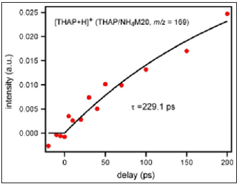
Figure 4:Time dependence of peak intensity of [THAP+H]+ from THAP/NH4M20 measured by time-resolved mass spectrometry.

biomedres-openaccess-journal-bjstr

However, it is generally known that intermolecular vibrational modes often couple with intramolecular ones. Therefore, the number of intermolecular vibrational modes of HAN is larger than that of THAP practically because HAN has more intramolecular vibrational modes than THAP. The desorption time was determined by how rapid desorption-related intermolecular vibrational modes were excited. As HAN crystals as well as the dimer had more intermolecular vibrational modes than THAP crystals, it could be understood that much more time was necessary to excite those modes in HAN than THAP as the pump input energy (400nm, 8μJ) was equal for both measurements. Then, how desorption proceeded when the molecules existed on a material with a high vibrational density of state was investigated. In this experiment, NH4+-terminated zeolite was used instead of H+-type zeolite in order to extract the influence of only the three-dimensional framework on the desorption; ammonium ions blocked the active sites of zeolite, which led to minimum electrostatic attraction between the molecule and zeolite. (Figure 4) shows the time dependence of the peak intensity of [THAP+H]+ obtained from THAP/NH4M20.

As the active sites of zeolite were blocked by ammonium ions and efficient proton supply to THAP was hindered, the peak intensity was almost one-tenth of that observed for THAP crystals (Figure 1c); only the effect of the three-dimensional framework on the desorption could be extracted. Although the S/N ratio was not good, the time dependence of the peak intensity was fitted by exponential functions and the rise component was determined as 229.1±133ps. For the desorption of THAP on zeolite surface, intermolecular vibrations between THAP and zeolite must be excited. However, as those vibrations were directly connected to zeolite lattice vibrations with high density, it became difficult to accumulate desorption energy in the intermolecular vibrational modes between THAP and zeolite. The rapid diffusion of vibrational energy by the zeolite lattice was responsible for the long desorption time (229.1ps) of THAP. Therefore, it was understood that zeolite offered such condition as diffusion of excess vibrational energy causing matrix as well as analyte fragmentation. Although molecular desorption became difficult by using zeolite, the peak intensity of analyte-related ions became strong when H+-type zeolite was used15 since ionization by efficient proton (cation) supply from Brönsted acid site was possible.

Conclusion

The desorption times of the protonated species of THAP ([THAP+H]+) and HAN ([HAN+H]+) from their crystals were determined to be 7.2±2.3 and 14.0±9.4ps, respectively, with a pumpprobe type time-resolved mass spectrometer. This desorption time difference could be explained by considering the number of intermolecular vibrational modes in THAP or HAN dimers. It was clarified that a long time was required for the desorption of HAN, which had many intermolecular vibrational modes. Then, the influence of the three-dimensional framework on the desorption process was investigated. The desorption time for THAP adsorbed on zeolite was 229.1±133ps. For the desorption of THAP on zeolite surface, the intermolecular vibrations between THAP and zeolite must be excited. As the intermolecular vibrations between THAP and zeolite were directly connected to zeolite lattice vibrations with high density, it became clear that desorption energy accumulation in those modes was difficult, leading to the long desorption time.

Toward an Exosome-Based Therapeutic Strategy in Regenerative Dentistry-https://biomedres01.blogspot.com/2020/09/toward-exosome-based-therapeutic.html

More BJSTR Articles : https://biomedres01.blogspot.com
Toward an Exosome-Based Therapeutic Strategy in Regenerative Dentistry

Toward an Exosome-Based Therapeutic Strategy in Regenerative Dentistry

Introduction

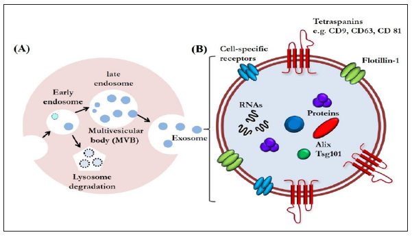
Exosomes are a type of extracellular vesicles that are secreted via exocytosis from late endosome multivesicular bodies [1] (Figure 1A). Exosomes are nano-vesicles measuring 40 to 150 nm, first discovered in the supernatant of cultured sheep erythrocytes in 1983 [2]. Since then, the progress of bioscience and technology has revealed that their role is not limited to be a waste disposal system; they are now emerging as a class of signal mediators with cellular component release [3-5]. They also have regenerative and immunomodulatory properties, characteristics that are encouraging their application for therapeutic purposes [4,5]. Although exosomal functions have already been widely explored in regenerative medicine, the potential for regulating tissue repair and regeneration in the dental field has not drawn nearly as much attention [6]. Therefore, the use of exosomes for human cleft lip and palate reconstruction, periodontal regeneration, osteoinductive roles in orthodontic treatment, healing in facial bone fracture, and chondral and muscular regeneration in temporomandibular disorders will continue to be studied.

Exosome-Based Therapeutics in Regenerative Dentistry

Exosomes are increasingly gaining the attention of the scientific community because of their small size and ubiquitous presence in almost every fluid of the human body, such as saliva, urine, plasma, synovial fluid, breast milk, amniotic liquid, seminal fluid, ascites, and cerebrospinal fluid [5]. Exosomes are enveloped by a lipid bilayer enriched in cholesterol, ceramide, and sphingomyelin. The membrane of exosomes is also abundant in some tetraspanins such as CD9, CD63, and CD81, that could be used as markers for identifying exosomes. The internal contents of exosomes are enriched in special biomolecules, functional proteins, and nucleic acids, including microRNAs (miRNAs), messenger RNAs (mRNAs), and even DNA (Figure 1B) [3,7]. With these components, exosomes have been identified as another vital mediator of paracrine communication [8]. Paracrine signaling is of major importance in maintaining cellular homeostasis, and it also plays a key role in the onset and development of many diseases [9].

Figure 1:A. Exosome biogenesis. Exosomes are an end-product of the endocytic recycling pathway. First, endocytic vesicles are formed at the plasma membrane and fuse to form early endosomes. These mature and become late endosomes. After further processing, exosomes are released through membrane fusion.

B. An enlarged exosome showing a variety of common exosomal surface markers (e.g., tetraspanins such as CD9, CD63, and CD81 and lipid raft-associated proteins including flotillin-1), as well as internal markers such as Alix and Tsg101. Each exosome also contains and transfers small RNAs and other cytoplasmic proteins and cell-specific receptors, which can be transferred to recipient cells.

biomedres-openaccess-journal-bjstr

Furthermore, their relative stability in circulating body fluids and their ability to pass biological barriers have prompted investigations aimed at using them for therapeutic purposes [10]. Finally, a large collection of evidence shows that exosomes are important regulators of many biological functions, such as tissue regeneration, immune response, and tissue homeostasis [11]. With these properties, they would be useful for innovative approaches in tissue regeneration against bone destruction or defect in the dental field. We could reasonably visualize a cell-free therapy using exosomes for tissue regeneration. It is worthy to be mentioned that mesenchymal stem cell (MSC)-derived exosomes might constitute a compelling alternative because of their advantages over the corresponding MSCs: they are less complex and even smaller than cells, so they are easier to produce and store, and have the potential to avoid some of the regulatory or legal issues that MSCs face [12].

It also would avoid the risks related with direct stem cell transplantation, such as hyper-immune reaction or immune rejection, teratoma formation, and the reduced regenerative capacity of engrafted cells [13]. Therefore, MSC-derived exosomes may be an ideal therapeutic tool for regenerative dentistry in near future.

In addition, MSC-derived exosomes are particularly promising candidates for developing cell-free therapy in several fundamental biological processes, such as recruitment of inflammatory cells, neovascularization, and coagulation [14]. Thus, they have vital importance in ensuring the appropriate inflammatory reaction after injury, which would improve tissue repair and regeneration. Angiogenesis is of vital importance in various physiological processes including cutaneous wound healing and/or tissue regeneration. Exosomes released by human adipose-derived MSCs can significantly promote endothelial cell angiogenesis in vitro and in vivo [15-17].

Furthermore, exosomes derived from human amniotic epithelial stem cells have the regenerative potential to heal full-thickness skin defect in rats [18]. Exosomes derived from human MSCs have therapeutic effects on osteochondral defect and eventually lead to cartilage repair [19]. However, the precise underlying molecular mechanisms of these beneficial effects have not yet been determined, which may be a highly orchestrated physiological process consisting of a complex event. Recent evidence suggests that exosomes secreted by most cell types can mediate transfer of their cargo. Theoretically, endogenous exosomes could be reasonable candidates for natural drug delivery because of their small size, permeability of physiological barriers, nontoxicity, low immunogenicity, and stability in circulation [20]. Emerging exosomal engineering strategies have laid the foundation for achieving this goal [21]. In the near future, with assistance of exosomes engineered with anti-inflammatory drugs or compounds, clinicians might be able to modulate the inflammatory response soon after tissue damage occurs.

In addition, modifying the surface of exosomes by adding proteins with affinity to injured cells and tissues could precisely drive exosomes to a target site [22]. Further, with encapsulation of nucleic acid into exosomes, exosomes could carry a miraculous therapeutic potential for tissue regeneration through modulating the microenvironment of their target cells [21]. Despite extensive evidence, the potential roles of exosomes in tissue repair and regeneration have not been fully elucidated. The certain contents and properties of exosomes that are capable of promoting tissue regeneration are still unclear. It would be also of great significance in identifying the variations in exosomal amounts following injury, because excessive exosome recruitment can lead to further tissue damage by persisting the inflammatory response. Furthermore, great efforts still needed for developing optimized methods for exosomal isolation and purification.

Conclusion

Although there is much that remains to be investigated in the field of exosomal research, the unique properties of exosomes clearly represent new therapeutic strategies for tissue repair and regeneration in the dental field. It is reasonable to believe that more regenerative potential of exosomes will be discovered in the future. The innovative strategy of using exosomes is obviously suggesting new options for regenerative medicine, although there are areas that need further research before clinical application. Using the exosome-based therapeutic strategies, we might be able to reduce safety concern and immunogenicity problems in regenerative dentistry.

Drug Discovery Enhanced by Artificial Intelligence-https://biomedres01.blogspot.com/2020/09/drug-discovery-enhanced-by-artificial.html

More BJSTR Articles : https://biomedres01.blogspot.com
Tuesday, September 29, 2020

Drug Discovery Enhanced by Artificial Intelligence

Drug Discovery Enhanced by Artificial Intelligence

Introduction

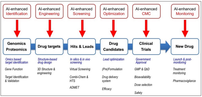
The Fourth Industrial Revolution, which has been widely publicized through the Davos Forum in 2016, has had a profound impact on the industry. Especially in the pharmaceutical industry, which requires a lot of resources and time, it is expected that a new breakthrough that maximizes efficiency will be achieved through the 4th Industry Revolution. Artificial intelligence (AI) and big data analysis technologies, which are leading technologies in the 4th Industrial Revolution era, are expected to have a great impact on the new drug discovery (Figure 1). Many pharmaceutical companies have performed the rational drug discovery through various omics and structure-based drug development. Now they are making great changes through the fusion of AI and the previously developed technologies for drug discovery [1-3].

Figure 1: Cycle of rational new drug discovery and application of artificial intelligence.

biomedres-openaccess-journal-bjstr

The traditional drug discovery is a high-risk, high-return industry that is costly and time-consuming, although it can bring enormous benefit if successful. As of 2016, an average of more than 10 years and $2.6 billion are required to develop one new drug [4]. For this reason, in the field of a new drug discovery, pharmaceutical companies are pursuing a strategy to reduce the risk of failure such as open innovation and increase the possibility of success. AI could be attracting attention as a key to dramatically reduce costs and time in the pharmaceutical industry. So far, the AI is mainly used in the early stage of drug discovery and search for candidate compounds in the pharmaceutical industry, but AI will be used for various purposes in order to drastically reduce cost and time for new drug discovery.

AI-Enhanced Identification

Rational drug discovery, which identify and validates the targets of a new drug discovery based on large amounts of genetic and protein information such as genomics and proteomics, has been the mainstream of pharmaceutical industry. Compared with the conventional screening method based on random screening, it was able to utilize time and resources relatively efficiently. The target selection method based on omics is a crucial process for selecting a valid target from numerous candidates. Therefore, selection and validation of new drug targets can be facilitated with the help of AI. In addition, AI can be actively used to improve the efficiency of drug repurposing, which uses existing drugs already marketed in other diseases, and Watson Drug Discovery developed by IBM have presented the increased efficiency of the drug repurposing using AI [5].

AI-Enhanced Engineering

Because the selected targets for a new drug discovery are proteins in most cases, engineering and obtaining threedimensional structures of proteins are important keys to increase efficiency of the entire steps of drug discovery including virtual screening step. Also, those are essential elements of structurebased drug discovery (SBDD) used nowadays in most big pharma, and the three-dimensional structures of proteins are mainly determined using X-ray crystallography and nuclear magnetic resonance spectroscopy (NMR) methods. To develop a new drug based on the protein target, it is necessary to search for active site and validate the target protein for efficacy confirmation. AI could be helpful for selecting mutation site for protein property modification during in vitro and in vivo experiments. In the process of designing or engineering bio drugs including biosimilar, which is growing rapidly in the new drug market, it is a crucial process to transform proteins, so it can be used like application of AI to protein engineering process. For example, Atom wise company actively use AI in drug design through target engineering as it has developed the first AI technology available for three-dimensional drug design [6].

AI-Enhanced Screening

Traditionally, searching active compounds is a painful process in drug discovery because it is necessary much time and resources in screening step. Modern rational drug discovery uses molecular docking as a core technique to perform virtual screening and computationally select compounds that are effective among large libraries of compounds. Many docking algorithms have been developed and used for this purpose, but the virtual screening through the in silico method does not yield perfectly hits or lead compounds. Therefore, if the AI method such as machine learning is applied to the existing molecular docking method focusing on the scoring function, the efficiency of screening the active compound can be maximized. Many pharmaceutical companies are focusing on utilizing AI now of virtual screening along with drug repurposing. In addition to structure-based virtual screening, the AI in the screening step can be used for predicting and verifying the ADMET parameters used in pharmacokinetics and can also affect the efficiency of the drug repurposing mentioned above

AI-Enhanced Optimization

AI can also be utilized in the process of optimizing the obtained lead compounds to be commercialized. It is important to develop an effective drug delivery system through a formulation process that optimizes the lead compounds to derive drug candidates. Recently, a variety of material engineering methods have been introduced for the development of a target-oriented drug delivery system. In a typical drug delivery system using an amphipathic substance such as micelle or liposome, a proper combination of drug and other substances is required. To make such a drug delivery system, there are many cases of material composition and environment selection, so that it is possible to shorten the time and resources for drug candidates by preferentially selecting composition ratios and environments that are likely to be successful with the help of AI [7]. In addition, AI can be used to increase the accuracy of toxicity prediction, preclinical experimental design and analysis of experimental results.

AI-Enhanced CMC

Drug candidates that are very close to the new drug will eventually reach clinical trials, the biggest obstacles to drug development, and will require approval of administrative procedures, such as FDA CMC (Chemistry, Manufacturing, and Control), to be licensed as commercial drugs. Along with successful clinical trial results, there is room for AI to be used in the manufacturing and licensing step to be approved as a new drug. Selection of subjects using AI can increase the efficiency and reliability of clinical trials as appropriate screening is the key to successful clinical trials. If AI is used to optimize clinical trial design, the accuracy of clinical outcome analysis can be improved. Although it is still in the early stage, efficiency-oriented production processes that utilize data such as smart factories during GMP (Good Manufacturing Practice)-based drug manufacturing are likely to be necessary for AI utilization.

AI-Enhanced Monitoring

In the monitoring process after the new drug is released as a commercial product, AI can be used to improve the efficiency of pharmacovigilance. Using AI in the pharmacovigilance step will improve compliance and accelerate customized drug guidance. For instance, GPVAI of Genpact company is currently providing solutions for OCR (Optical Character Recognition) and AI to analyze, monitor and control information related to worldwide drug side effects reports.

Conclusion

New drug discovery is a vast resource and time consuming field, and a wide variety of specialized skills must be integrated in each step. Therefore, in order to increase the efficiency of a new drug discovery, it is necessary to improve the efficiency of all steps during drug discovery, and AI can be very helpful for whole steps in a new drug discovery. So far, AI has been used mainly in the step of searching for candidate molecules in addition to drug repurposing, but it is certain that the necessity of AI will increase in the future. Also, if AI succeeds in improving the accuracy of go / no-go decision support during drug discovery, it is possible to make dramatically improvement of the efficiency of a new drug discovery

Of Parasites and Their Hosts-https://biomedres01.blogspot.com/2020/09/of-parasites-and-their-hosts.html

More BJSTR Articles : https://biomedres01.blogspot.com
Of Parasites and Their Hosts

Of Parasites and Their Hosts

Mini Review

Host Parasite Relationships

Parasitism is an association between two different species of organisms, where only one partner which is always the parasite derives benefits metabolically. The other partner is the host and gets nothing in return but harm, damage or death [1]. A parasite lives in a particular way called parasitism. Parasitism as a one-sided partnership in which the host receives no benefits but instead some degree of harm, or death is an ingratitude and is synonymous the parasite committing matricide! An ‘ideal’ parasite must not even dare kill its host because the death of the host means the death of the parasite. An “ideal” parasite does not even exist because the slightest effects of all parasites are detrimental to their hosts [2]. Caullery [3] considers that “parasitism is a case of balance of power; the parasites and the host form a functional balance system which is placed in opposition to the external environment. Both the parasite and host make the best of a bad job. The host reacts to keep the parasite away or to a minimum while the parasite attempts to live as un-obstructively as possible, despite all the host’s reactions which threatens the parasites’ existence.

Parasites Metabolic Dependency on the Hosts Could be in Several Ways:

a) Nutritional: As in Flukes, tapeworms such as Diphyllobothrium latum

b) Developmental stimuli: As in P. intergerrinum and Opalina ranarum , (parasites of frog),

c) Digestive Enzymes: As in tapeworms such as Taenia sp and Echinococus sp

d) Shelter: As in endoparasites such as Plasmodium sp , Entamoeba sp, Ascaris sp e.t.c

e) Movement: As in ectoparasites such as lice, fleas, ticks and mites

f) Control of maturation: As in P. intergerrinum and Opalina ranarum

Parasitism has prolonged effects on Both Hosts and Parasites Such as:

a) Parasites have mechanisms for locating their host like chemotaxis and active penetration such as Schistosome cercariae and hookworm larvae

b) Transmission of parasites to the new host may be associated with daily or circadian rhythms such as cell division and migrating patterns [4].

c) Parasites have to survive in their host using several mechanisms like encystations, transformation or tail loss as in schistosome cercariae.

d) Parasites have surfaces that play important roles in nutrition. There is usually a major nutritional interface with the host such as the digestive-absorptive epithelium in platyhelminthes which has enormous increase in surface area by development of folds and microvilli such as tubercules, spines and pores of flukes and tapeworms [5].

e) Damage to host can be as a result of immune response (immuno-pathology). This can be more damaging than the infection itself [5,6].

f) Parasites exhibit host specificity and are adapted to infect specific species but cannot invade certain species or strains of host, for example, the malaria parasite Plasmodium yoelli can alone affect mice while Plasmodium falciparium and P. malariae are human specific [7].

Effects of Parasites on their Hosts: Parasites May Injure their Host in a Number of Ways

Toxins: Apart from disease and death, parasites can produce poisonous substances in the form of secretions, excretions or other products such as proteolytic enzymes and pigments. These can harm or sensitize the host, for example, Schistosome cercariae, Entamoeba histolytica and Malaria parasites [5].

Mechanical Effects: Mechanical damage due to big size or number of parasites occurs such as the hydratid cysts of Echinoccocus species. Intestinal obstruction, blockage and entangling of worms also occur such as in Ascaris lumbricoides. Parasites can perforate vital organs when migrating as in hookworms and Ascaris. Adhesive structures of parasites also cause mechanical damage as in Taenids.

Absorption of Food: Parasites can deplete the host nutritional level to reach disease level for the host. For example, Diphyllobothrium latum, the broad fish tapeworm absorbs a great quantity of vitamin B12 reaching to megaloblastic anaemia. Hook worm absorbs iron daily leading to iron deficiency anaemia.

Destruction of Host Tissues: Skin penetrating parasites cause skin destruction such as the larvae of hookworm and Schistosomes which causes swimmer’s itch and the larvae of myasis producing flies. Microfilariae of Onchocerca volvulus causes skin onchodermatitis, nodules, leopard skin and lizard skin. The skin lesions become the site of secondary bacterial infection (6, 7).

Ingestion of Host’s Body Constituent: Some parasites such as hookworms and microfilariae of filarial worms feed on the body fluids (blood and lymph), and epithelia cells causing blood and fluid loss.

Gigantism: Some parasites such as larval stages of trematodes enhance growth of their snail intermediate host [1].

Parasitic Castration: Sex reversal and parasitic castration occurs when gonadal tissues of intermediate hosts such as in crabs and snails are destroyed [1, 3].

Effects of Hosts on the Parasites (Host reaction): These Effects are not as Apparent as Parasites Effects

Tissue Reaction: These are host defense mechanisms. The tissue reactions are localized in the vicinity of the parasite’s invasion. They usually disappear after the invading organism has been eliminated, for example, inflammatory reactions, nodules, induction of abnormal growth, hyperplasia, metaplasia and neoplasia (tumors) and hypertrophy such as in Trichinella spiralis and Onchocerca volvulus infections [5].

Immunity: This is a generalized effect on the body. It can originate in organs or systems remote from the vicinity of the infection. It persists for a long time even after elimination. This is a physiological response directed against the survival of the parasite. Anti bodies are produced to destroy parasites in some cases. Cell mediated immunity by complement activation occurs [6, 7] such as in helminthes infections.

Host Specification: Hosts have their own specific parasites. Some parasites are naturally adapted to certain species of hosts. For example, Trypanosoma brucei brucei infects only cattle but Trypanosoma brucei gambiense and Trypanosoma brucei rhodesiense infects humans.

Adaptations: Certain parasites develop certain morphological and drastic modifications to enable them fit to the host. For, example blood flagellates have torpedo shape to enhance swimming. Giardia lamblia has adhesive discs and Taenia solium have hooks to hold fast to the intestinal wall of host.

Host Resistance: Host insusceptibility is the unsuitability of a host as physiology, behavior, and structure Host parasite interactions may also influence host resistance. For example, Plasmodium vivax is not popular among West African because of the duffy factor in their red blood cells [8].

Conclusion

It is obvious that parasites have undermined the health status of individuals and jeopardized the economic development of nations in tropical Africa leading to poverty [8]. Parasitism is also the major cause of low productivity of livestock and poultry in the tropics. Parasites are ‘unwelcomed strangers’ because the body reacts and builds a resistance against them. The immune of the hosts fights back against parasites but unfortunately parasites fight back using several mechanisms of evading the host immune system. Only the host with the most vibrant immune systems can fight back successfully. However, the battle continues.

Multidisciplinary Approach in Breast Cancer-https://biomedres01.blogspot.com/2020/09/multidisciplinary-approach-in-breast.html

More BJSTR Articles : https://biomedres01.blogspot.com
Neurological Manifestations of COVID-19: A Brief Update on the Cuban Context

  Neurological Manifestations of COVID-19: A Brief Update on the Cuban Context Introduction Despite of severe acute respiratory syndrome cor...