Showing posts with label Journals on Medical Research Openaccess journals on surgery Journals on Biomedical Science Journals on Medical Drug and Therapeutics Journals on Biomedical Intervention. Show all posts

Wednesday, April 6, 2022

Pathogenic Microorganism Detection in Invasive Pulmonary Aspergillosis(IPA) Patients Using Bronchoalveolar Lavage Fluid


Pathogenic Microorganism Detection in Invasive Pulmonary Aspergillosis(IPA) Patients Using Bronchoalveolar Lavage Fluid

Introduction

The insufficiency of detective methods during early stage of Invasive Pulmonary Aspergillosis(IPA) has lead to severe misdiagnosis [1]. Surveys and researches had been conducted to proved that the uncertainty and improper treatment could result in relatively high mortality rate [2,3]. Bronchoalveolar lavage (BAL) is the method which can collect secretory fluid in the respiratory tract and alveoli. The fluid can be separated for further analysis so that the content of the components can be quantified. It is an common method in locating the lesion site in clinical practice. Compared with other methods, for example, BALF has fewer chance of contamination rather than collecting sputum specimen. Therefore, it can improve the specificity of etiological diagnosis. At present, this technology is mainly applied in the clinical diagnosis of pulmonary bacterial infection, but there have been few reports on the evaluation of its pathogenic detection for the diagnosis of fungal infection. In this study, BALF was collected from 100 suspected patients suffering from invasive pulmonary Aspergillus infection. Three pathogenic detection methods, smear microscopy, fungal culture and PCR were used to quantify the Aspergillus examples. Then the results of these three methods would be valued under the clinical criterion. To provide assistance for the clinical diagnosis of pulmonary aspergillosis.

Method

Collection and Storage of the BALF Specimens

The ethical approval for this study was obtained by First Affiliated Hospital of Sun Yat-sen University. Before bronchoalveolar lavage fluid were collected, the patients involved in this research received detailed explanation and understood the benefits and potential danger, and had signed the consent letters for bronchoscopy procedures. The subject inclusion and exclusion criteria used in this study refer to the consensus of Chinese experts on the detections of pathogens in the bronchoalveolar lavage of pulmonary infectious diseases, which was proposed by respiratory disease branch of the Chinese Medical Association in 2017 [4]. The procedure of collecting bronchoalveolar lavage was as follows: a) 1-2mL of 2% lidocaine was injected through the biopsy hole for local anesthesia, after the bronchoscope reached the lung segment of the lavage site.

b) The top of the bronchoscope was incarcerated in the opening of the target bronchial segment or sub segment. A dose of 20-50mL sterile normal saline at 37 ℃ or room temperature was injected through the biopsy hole of the bronchoscope. After each injection, the appropriate negative pressure (less than 100cmH2O) was used to recover the liquid, and the total recovery rate was secured to be more than 40%. The intensity of negative pressure during lavage was under severe control to avoid the collapse of distal airway caused by excessive negative pressure.

c) 8-10mL of BALF specimens were collected with a sterilized plastic collector. And the specimens were placed in an incubator with ice cubes and immediately sent to -80℃ refrigerator for preservation. The examples, one hundred hospitalized patients were collected from the different hospitals (47 patients from the Guangzhou Thoracic Hospital, 44 from the Guangdong Province People’s Hospital, and 9 patients from the First Affiliated Hospital of Sun Yat-Sen University, respectively) during the period ranging from June 2018 to October 2019. These patients had been clinically suspected of having pulmonary Aspergillus infections in accordance with the standards set in EORTC/MSG criteria [5].The IPA patients were diagnosed by pathology or detecting the Aspergillus hyphae or fungal culture by tissue biopsies or BALF. All patients were checked with bronchoscopy and BALF were collected before the antifungal therapy, then the BALF were immediately transferred to a -80℃ refrigerator in the laboratory facilities. The bronchoscopy biopsies had also been performed, and pathological diagnoses had been made by the pathology departments of each hospital.

Detection of the Specimens

After being collected, The BALF examples were thawed and divided into three parts for the application of smear microscopy, culture, and PCR product analyses. Smear microscopy and culture methods were conducted in the microbiology laboratory facilities of the First Affiliated Hospital of Sun Yat-Sen University. The smear microscopy was performed using fluorescent staining techniques. In the experimental processes, 5 mL of BALF were digested with digestive fluid (dithiothreitol). Then the examples were selected and centrifuged at 3,000 r for 15 to 20 minutes. The supernatant was discarded and the sediment remained. The sediment was placed on a glass slide and one drop of fluorescent staining solution would be added. The slide was observed under microscopeby technicians. After the residual sediment was treated again with digestive fluid, it was inoculated in Sabouraud culture medium and cultured at 30℃ for one week. After the culture was completed, the colony characteristics were observed and identified according to the characteristics of colony and conidia.

BALF PCR

Primers: After the BALF had been sent for microscopy and culturing, the remaining specimens were sent to Sagene Biotech Company Ltd for PCR detection. Then, the internal transcribed spacer (ITS) primers, which serve in Aspergillus identification, were redesigned based on the methods and sequences provided by the relevant literature [6,7] (ITS-F:GCCTGTCCGAGCGTCATTG;ITSR: TTAAGTTCAGCGGGTATCCCT; size of amplified fragments: 224 bp). The primers and the experimental reaction system were verified by the pre-experimental processes. The primers were found to have good specificity, and thus could be used for the identification of Aspergillus.

DNA Extraction: A DNA extraction kit (Sagene, China) was used to extract the DNA from each specimen. Following the extraction, gel electrophoresis was performed. 3 μL of DNA was taken and added to 1Μl of loading buffer. Electrophoresis was conducted at 120 V for 25 minutes for the purpose of analyzing the quality of the extracted DNA.

Establishment of the PCR Reaction System

a. Reagents: The reagent types and volumes are shown in the Table 1.

b. PCR Reaction Conditions: The following were the PCR reaction conditions in this study: 98℃ for 1 minute; 98℃ for 10 seconds; 60℃ for 5 seconds; 72℃ for 5 seconds; and 72℃ for 2 minutes, for a total of 40 cycles.

c. Preliminary Analysis: For the preliminary analysis, 3 μL PCR product was selected to perform the 1.2% agarose gel electrophoresis (Figure1) and preliminarily analyze the size of the amplified fragments, in order to determine whether or not the targeted fragments had been successfully amplified. d. Sequencing (Abi 3500) of the PCR products: Sequencing of the PCR products was conducted for the specimens which have been successfully amplified to the target fragments. Then, an NCBI BLAST comparison was performed on the sequencing results (Picture 1).

biomedres-openaccess-journal-bjstr

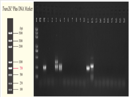
Figure 1:ITS amplification electrophoresis map of aspergillus.

biomedres-openaccess-journal-bjstr

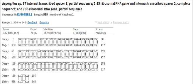
Picture 1:Example of NCBI BLAST comparison results of amplified fragment.

biomedres-openaccess-journal-bjstr

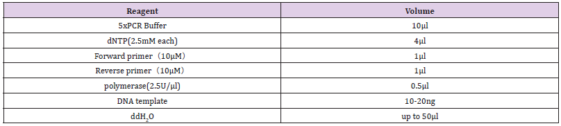
Table 1: The reagent types and volumes during the PCR reaction system.

Statistical Data Method

SPSS (IBM 22.0) software was used for the statistical data in this study.

a. A Chi Square Test Method was used to compare the distributions of the three detection methods among the proven and clinical diagnosis groups. The differences were considered to be statistically significant when p<0.05.

b. A Kappa Consistency Test Method was used for the pairwise comparisons of the results of the three detection methods as a whole and among the groups, respectively.

c. The sensitivity (Sn), specificity (Sp), and positive likelihood (PRL) ratios of the three detection methods for IPA diagnoses were calculated.

Results

biomedres-openaccess-journal-bjstr

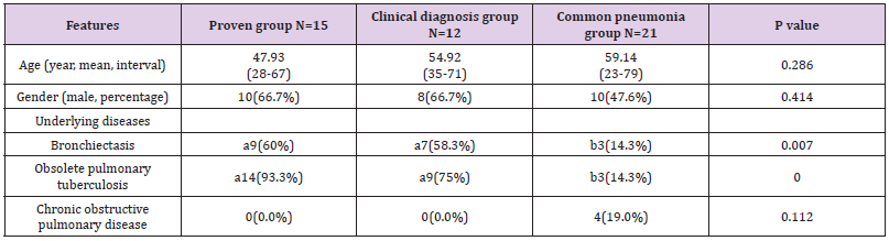
Table 2: Clinical baseline data of patients.

In the present study, 100 patients with suspected clinical pulmonary Aspergillus infections were registered. Through clinical examinations including pathology, radiology, etiology. Patients’ data were collected (Table 2). According to the diagnosis standards of IDSA(2016) [5], 15 patients were diagnosed by pathological data(proven group), and 12 cases were diagnosed by radiology, etiology, and other clinical examinations. The other 73 patients were diagnosed as having tuberculosis, common bacterial pneumonia, vasculitis etc. Among those patients, 21 patients with common bacterial pneumonia were used as the control group. The positive number with smear microscopy, culture, and PCR in BALF was 6 (40.0%), 7 (46.7%), and 6 (40.0%) in the proven group respectively; and 5 (41.7%), 3 (25.0%), and 9 (75.0%) in the clinical diagnosis group, respectively. In addition, the total number of patients with positive results as diagnosed using the abovementioned three methods was 0 in the common pneumonia group, as illustrated in Table 3.

biomedres-openaccess-journal-bjstr

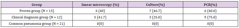
Table 3: Number of positive cases in the diagnostic tests.

χ2 = 2.187 and p = 0.335 could be obtained, there were no significant differences in the distributions of the three detection methods among the two groups (p>0.05). It was determined that the calculated χ2 and p values were 0.182 and 0.913 (proven group) and 6.241 and 0.044 (clinical diagnosis group). The statistical results showed that in the clinical diagnosis group, the BALF culture and PCR results were significantly different (p<0.05). Furthermore, in the proven group, there were no significant differences observed between the three detection methods (p>0.05).

 A Kappa Consistency Test Method was adopted in this study for the pairwise comparison of the results of the three detection methods as a whole, and also within the different groups. The results are shown in Table 4. As shown in the table, there was a weak consistency observed between the microscopy and culture methods. However, the results of both were found to be inconsistent with the PCR method.

 In accordance with the above-mentioned results of the Internal Kappa Consistency Test, the consistency between the smear microscopy and culture methods among the three test methods was weak. However, the results of the PCR were observed to be very inconsistent with those of the other two methods. However, it was speculated that if the microscopy, culture, and PCR methods were combined for diagnosis procedures, the diagnosis efficiency could be significantly improved. the calculation results are shown in the last row of Table 5.

biomedres-openaccess-journal-bjstr

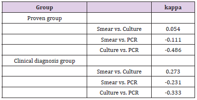
Table 4: The kappa consistency test for the three diagnostic tests.

biomedres-openaccess-journal-bjstr

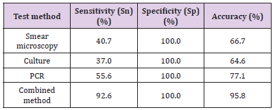
Table 5: Diagnostic value indicators of each test method.

Discussion

The molecular biological diagnosis method (represented by PCR) is found to be more sensitive and rapid than the traditional culture method. It could directly detect the nucleic acid molecules of the pathogens without the steps of isolation and culturing, which proved to be helpful for the early diagnoses of the disease [8]. It is known that PCR can be theoretically detected regardless of whether the pathogen are active or inactive. The positive results of the PCR were significantly greater in number than those of the culture method. However, there were no significant differences observed when compared with the smear microscopy (p> 0.05). The reason was that the IPA patients had received antifungal therapy, therefore, the positive rates of the culture method were lower than PCR. This showed fungal culture method is less the diagnosis efficiency than PCR, when antifungal therapy were conducted. However, we found that some patients were positive with BALF smear microscopy or fungal culture, but negative with PCR method. A possible cause of this is the fungal composition. The outer sides of Aspergillus (which belong to eukaryote) are wrapped by a layer of compact and hard cell wall components. The complete cell wall will inevitably reduce the extraction effects of nucleic acid substances [9].

Therefore, the interpretations of PCR results should be made in combination with other factors, such as clinical manifestations, other examination results. In our study, it was found that when smear microscopy techniques were performed for the lower sediment of BALF, the concentrations of pathogen components in those specimens were relatively high. The positive rates of smear microscopy were 40.0%, while that of common sputum was only 4.5% [10,11]. This showed the lower sediment of BALF was very high efficiency for microscopic examinations . In summary, the three examined diagnostic methods were found to have high specificity, but insufficient sensitivity. In the present study, considering the low degree of overlap in the results of the three detection methods, it was speculated that if a combination of microscopy, culture, and PCR methods was applied in diagnosis processes.

Conclusion

The pathogenic detections of bronchoalveolar lavage fluid (BALF) have been determined to be an ideal diagnostic method for Aspergillus infections. Because of the limitations of the different detection methods, The combination of BALF smear microscopy, culture, and PCR could significantly improve IPA diagnosis efficiency.


For more Articles on : https://biomedres01.blogspot.com/

Tuesday, April 5, 2022

Updates on Urgency of Treating Stroke

Updates on Urgency of Treating Stroke

Introduction

It is a proof of concept that time is muscle in treating myocardial infarction and even if for any reason you missed the boat in timely management of heart attack, we postulate that probably you might have a second chance which is heart transplant if patient meets the requirement, and this is not an excuse for not treating the heart attack very urgently [1]. The situation in stroke is more serious and more urgent [2] as you need to exclude stroke mimics in a very short time to make a therapeutic decision, in a large vessel stroke [middle cerebral artery stroke]. There is a loss of 2 million nerve cells per each minute delay [3] which is correlated with loss of 1.8 days of healthy life [3], After saucerful treatment of heart attack you can still run a marathon in contrast of stroke that you might need to lose few brain cells to be in a vegetative state. Most of strokes are ischemic, around 80%, less than 15% are hemorrhagic, and less than 2% are venous stroke (cortical or cerebral venous stroke) [4]. Treatment of each type of stroke is quite different, and all types of strokes is emergency and needs to confirm the diagnosis.

Mini Review

In this review, i am going to discuss the updates of investigation and specific physical signs and symptoms which help to reach a correct diagnosis in a very short time, and iniate urgent treatment. Firstly, diagnosis of stroke and ruling out stroke mimics is of para-amount importance, Seizure, manganous Aurea, cardiogenic and metabolic syncope, encephalitis, demyelination, infective endocarditis and embolic stroke, metabolic encephalopathy, need to be ruled out some tips to exclude mimics.

a) Seizure, loss of consciousness, patient can’t remember what happened, post ictal state, Shaking, bite on the lateral side of the tongue, if patient has history of seizure, usually most seizure is stereotyped, patient can have Todd’s paralysis which usually resolve in few hours [5].

b) Migranous Aurea, history of migraine, fortification spectra, Aurea could be in language and pronunciation, sensory, motor, usually it is slowly progressing, each one modality happened in one time then followed by another modality, patient could have nausea [6], patient with basilar migraine might have ptosis or Horner syndrome [7].

c) Syncope, quite sudden, patient lose his consciousness, medical history usually includes cardiac problems, familial history of cardiac syncope or sudden death (familial cardiomyopathy, Q-T syndrome).

d) Loss of consciousness usually last for seconds, if it is more than few minutes, other diagnosis should be considered [8], most of these patients have car accidents during syncope.

e) Transient Global Amnesia, it is sudden, temporary episode of memory loss, recall of recent events vanishes, sometimes patient can’t remember anything about the event and keeps asking same questions [8].

f) Encephalitis, usually patient will be confused and have diseases or sepsis underlying problems like chronic liver disease, respiratory failure, brain infection.

g) Demyelination, crescendo is slowly, commonly affecting females, usually resolve in few days, could be manifested as a flare of multiple sclerosis or could be precipitated by infection, if associated with infection, clinician should treat the infection and not to give methyl prednisolone [9].

Sometimes confirming the diagnosis of stroke will be difficult, here we are presenting other tools which help the diagnosis of stroke and the pathology of stroke to help starting an early treatment.

1. The definition of TIA as clinical symptoms which last less than 24 hours is outdated [10] because MRI can confirm a diagnosis of stroke even if the clinical symptoms last less than 60 minutes [11].

2. Restriction diffusion in DWI can diagnose stroke even after one minute of having the symptoms and cytotoxic oedema can be confirmed by ADC [12].

3. Biomarkers in Ischemic Stroke, S100B and specific enolase are proteins measured from serum and correlate very well with the volume of stroke 24-27 hours [13]. As we treat ischemic stroke with Alteplase [single chain recombinant tissue plasminogen activator] in therapeutic window which is four and half hours, CT perfusion and CTA are a standard of care in Australian hospitals which usually show the volume of infarct tissue (core) and the volume of hypo perfused region that can be salvageable with reperfusion(penumbra), even the severely hypo perfused region with good collateral below the occluded vessels, still have a high chance of reperfusion [13]. More than third of stroke are excluded from lysis because of the unknown time of symptoms. It became a best practice in Australia to consider the mismatch between DWI-Flair Mismatch.

Is a surrogate marker of the time of onset of stroke [14] specially in wake-up stroke?. Endovascular therapy is a standard of care for proximal large vessel occlusion, therapeutic window is up to 12 hours [15]. Venous stroke is an emergency, it affects mostly females, usually presented with seizure, decreased level of consciousness due diffuse encephalopathy, signs of increased intracranial pressure, headache, cranial nerve palsy, It is underdiagnosed because of the rarity of the stroke.

Predisposing Factors

Female gender, contraceptive pills, dehydration, inherited thrombophilia, myeloproliferative diseases, Rare diseases such as paroxysmal nocturnal hemoglobinuria. Helpful serum markers are positive D dimer, Uncontracted CT shows dense clot sign in cerebral vein or sinus, CT venogram is diagnostic. Due to increased intracranial tension, CT might show cerebral oedema, mass effect and intracranial hemorrhage which can progress to subarachnoid hemorrhage and massive hemorrhagic infarction [16]. Treatment is mainly anticoagulation, patients who do not respond to anticoagulation, treatment should be escalated to endovascular thrombolysis [17]. Hemorrhagic stroke is a life-threatening emergency, usually patient manifest with decline in consciousness and very mild neurological loss, seizure and signs of intracranial hypertension. Mortality is high in the first 90 days. Types of hemorrhagic stroke either lobar hemorrhage due to amyloid angiopathy, hypertensive bleed in Basel ganglia, hypothalamus and brain stem, subarachnoid hemorrhage due to ruptured Aneurysm, anticoagulation, AVM misinformation, cocaine abuse CTA is diagnostic as it shows Spot sign which is marked attenuation due to active bleed [18]. There is no definite cure of hemorrhagic stroke, patient should be managed in ICU to observe and treat seizures, intracranial hypertension, systolic hypertension with aim under 140. Treating fever, sepsis, hyperglycemia, endovascular treatment for ruptured aneurism, Reversing anticoagulation, prophylaxis for thrombosis with pneumatic compression in the first few days followed by low molecular weight heparin, craniectomy for evacuation of Hematoma, external ventricular drain for intraventricular hemorrhage causing hydrocephalus [19].


For more Articles on : https://biomedres01.blogspot.com/


Monday, April 4, 2022

The Role of Information Management in the Process of Digital Health Confrontation with Covid-19

The Role of Information Management in the Process of Digital Health Confrontation with Covid-19

Introduction

A successful decision, under normal circumstances where the capacity of human thinking is normal, depends on the ability to analyze, quality information, accuracy, and focus. However, in critical situations, the capacity of human thinking decreases, and lack of time aggravates the severity of the crisis. In such a situation, providing quality information is a hard task that would never accomplish undoubtedly without the help of information and communication technology. Information management, on the other hand, is the process of collecting and processing data in order to provide quality information. The role of this process becomes more prominent with the help of information technology and it operates successfully in critical situations [1,2]. The purpose of this study is to review how to prevent infodemic using information management and digital health.

Background

With the outbreak of the SARS viral pandemic in 2003, human society faced a phenomenon called Infodemic and has been living with this phenomenon along with the persistence of viral pandemic diseases such as MERS and COVID-19, COVID-19. The infodemic, introduced by the United Nations and the World Health Organization on March 31, 2020, following the SARS Cov-2 crisis, has cast a frightening shadow over human society [3,4]. Infodemic is literally a portmanteau. In fact, it consists of the words information and epidemic and means the universal spread of true and false information about any phenomenon, including disease. In this case, rumor and fear are intertwined so inextricably that complicate learning a subject. This concept itself includes two concepts, namely misinformation, and disinformation. Misinformation is false information about a subject, regardless of its being intentional or unintentional. Moreover, disinformation refers to misleading or false information that is deliberately provided to deceive the audience. Historically, the term has been coined and used by some countries’ security agencies to deliberately diffuse misleading information about the military, which is sometimes propounded to boost military power [5,6].

The bitter and costly experience of coexistence with viral diseases has clarified it for human society that in order to ensure its national security it needs to equip and rehabilitate health care professionals more than equip and reorganize its military [7]. In addition, the utilization of information and communication technology provides considerable preparation for the management of future pandemic-viral diseases due to the nature of pandemicviral diseases that can be managed by observing the principle of physical distance. On the other hand, the nature of information and communication technology is such that makes it possible to track patients and provide remote health care services to people at risk [8]. Noteworthy to mention is that digital health can not only facilitate the management strategy and response to pandemic diseases, but also can curb and manage the annoying phenomenon of infodemic, which severely increases the conditions caused by the outbreak of Covid-19 based on educational and informational application [9,10]. The application of digital health (which is actually e-health with a greater focus on health) can facilitate the management strategy and response to pandemic diseases. This perspective provides a framework for the use of digital technologies in the management and response to pandemics. It also presents the methods that successful countries in using digital technologies have adopted and implemented for the planning and management of pandemic diseases, including surveillance, screening, triage, diagnosis, monitoring, contact tracing, and health care [11].

Results

The Necessity for Information Management in a Pandemic Crisis

In October 2020, The British Academy and the Royal Society both made an announcement about the infodemic phenomenon that the spread of Covid 19 vaccine would confront a flood of misinformation that would fill the knowledge void. They believe that the existing infidelity has five features including distrust of science, distrust of pharmaceutical companies and the government, spreading outspoken words, using emotions, and expanding echo chambers. For this reason, the Singaporean government has praised the action of the Penal Code (POFMA) on natural or legal persons who spread lies [12]. It is noteworthy that some countries, such as China, India, Singapore, and South Africa, have enacted punitive laws against individuals, organizations, sites, and social media outlets that have intentionally or unintentionally published false information about the Covid pandemic. Other countries such as Taiwan, South Korea, and Switzerland, demonstrated their commitment to democracy and the free flow of information through interaction with and with the help of the people and managed the destructive phenomenon of infodemic. In the meantime, some countries, like the United Kingdom, acted in a double standard [13-19].

The various thematic areas plagued by the pandemic of false news include the cause of the disease, misleading statistics about its prevalence and casualties, the economic effects of the Corona crisis, journalism and their discrediting; medicine (symptoms, diagnosis, and treatment), society, and social beliefs (Panic), politicization (discrediting the politicians), Internet scams, and personal life of celebrities [20]. Since 2003, some countries have gained experience in controlling infestations, preventing the circulation of false information, and strictly monitoring all social media in the country, by undergoing the outbreak of SARS and the vigilance of the pandemic. They have managed the flow of data and information from both the people and the government (a health system), and in fact, by consolidating the digital health and information management, they have been able to prevent the problem of infodemic and the spread of fake news and rumors into the society. International organizations have taken effective actions to counter the destructive effects of spreading lies, public ignorance, and the lucrative activities of some social media. Among them, the World Health Organization has introduced a framework that includes a five-step process for managing infodemic, which includes: identifying evidence, translating knowledge and science, amplifying actions, quantifying impact, and coordination and governance [21].

UNESCO, in particular, has tackled the spread of false information through the provision of Open Education Resources, Networks of fact- checkers, and the provision of media and information literacy resources. It has also used digital technologies such as artificial intelligence to combat the existing pandemic in response to the crisis [22]. What follows is a more detailed description of the framework provided by the World Health Organization for the management of the infodemic phenomenon. At the stage of identifying evidence, all scientific findings that can have a positive effect on the health of individuals and the society should be collected, examined, and evaluated. False and misleading information also needs to be identified in collaboration with international organizations such as the World Health Organization. Governments and public health institutions are responsible for the circulation of accurate and quality information in society since they are trusted by the people. The government is obliged to check the contents of social media through the relevant and designated institutions and identify all fake news and false information.

In the translation phase of science and knowledge, health authorities should translate their scientific messages into simple and general language and into practical messages that change behavior in a way that can be easily used by everyone so that all classes of the people and even its politicians understand easily. This makes the community trust the health authorities. Cultural bias should be considered and applied to the content of the message. Local and ethnic translation is also required. The step of strengthening the action is done at the national level aiming at building trust and disseminating the right information to the right people at the right time. The government must optimize the use of social platforms through reputable and approved agencies and use all necessary media, including text, video, and infographics. Frequent and accurate messaging in culturally friendly formats is essential. Also, the timely correction of incorrect information and, if necessary, the use of the method of denial should be considered. Governments and other relevant actors need to reach out to key communities to understand their concerns and information needs, and it is best to provide advice and messages that can help these communities identify their audiences. Through this process, communities of any kind, whether religious, professional or etc., can reinforce appropriate public health messages in a way that is user-friendly leading to correct changes in behavior.

Active contacts and dialogues should be established with private sector employers, telecommunications companies, the food and agriculture sector, religious or humanitarian charities, medical and health professional associations, and the media. Community health workers, the first line of health care in many low-resource environments, must be equipped with the right information, graphics, and narrative to mobilize in communities. Strategic partnerships with the social media platforms of technology platforms and stakeholders, as well as universities and civil societies, all of which reinforce and monitor information, are also needed. Through strategic partnerships with health authorities, these platforms can prioritize and prioritize relevant information and advice, ensuring that it is seen by citizens. In the quantifying impact phase, it is necessary to collect, organize and analyze data with the participation of all key institutions to help measure and describe the information epidemic. In addition, the process of managing fake news and social media as well as the circulation of accurate and quality information and the impact of messages and interventions should be pursued.

The coordination and governance phase emphasizes the principle of public participation and the need to continue monitoring all activities. To achieve this important coordination between stakeholders including the World Health Organization and its member countries, scientific and public health institutions, telecommunications companies and private communication institutions, government communication institutions, search engines, civil society, universities, and academies, health care workers on the front lines, and all institutions to the lowest level of local mutual support groups are required [22].

Conclusion

Some countries, being Prudent and informed about global issues, understood the outbreak of newfound pandemic-viral diseases and the possibility of their continuation and have tried to improve their digital technologies and focused on digital health which is based on using smart phones since 2003. The use of digital technologies as well as digital health in critical outbreaks of pandemic diseases that require physical and social distance is a smart and successful plan to manage the disease. On the other hand, in addition to paying attention to the recommendations of international organizations such as the World Health Organization and UNESCO, these countries have opposed the publication of false news by creating a mechanism for the exchange of accurate data and information that is in the cycle of society and government, as well as implementing methods based on ICT.

Some countries have controlled the phenomenon of infodemic by enacting punitive laws on spreading lies about the pandemic disease, and others by interacting with the people and preventing the situation from escalating. Gathering reliable information and identifying fake news, monitoring the activity and content of social media, educating people on how to use social media and avoiding spreading rumors false information, and news, creating a system of collecting data from people, and distributing reliable information from the government (which is considered as a system of data exchange and reliable information) are among the important measures taken by the governments of successful countries in managing the crisis of Covid-19.


For more Articles on : https://biomedres01.blogspot.com/


The Resistance of the Cantilever Bridge by the Use of Dual Resinous Cement

  The Resistance of the Cantilever Bridge by the Use of Dual Resinous Cement Advantages for Dental Bridges with Resin-Fixed Dental Prosthese...