Wednesday, March 31, 2021

Airway Remodeling in the Asthma Model: Is there Standardization in this Evaluation?

Airway Remodeling in the Asthma Model: Is there Standardization in this Evaluation? 

Introduction

Asthma is a chronic inflammatory disease characterized by obstruction of airflow causing respiratory difficulties at various intensities [1]. It represents a major cause of physical disability, affecting 300 million people worldwide [2]. Airways inflammation has an important role in the pathophysiology of asthma, which is characterized mainly by the TH2 type immune response. The inflammatory process in the airways of individuals with asthma promotes significant changes in the respiratory structures, induced by repeated lesions and repairs [3]. This review aimed emphasize the importance of histological evaluation of airway remodeling in the asthma experimental model.

Airway Remodeling

Airway remodeling represents a late and permanent response to tissue injury, mainly caused by primary irritants. It is characterized by tissue and cellular changes in the bronchioles, such as increased collagen deposition, hyperplasia and smooth muscle hypertrophy, increased vascularization, basement membrane thickening and epithelial layer and epithelial desquamation, goblet cell metaplasia, mucus production and hypersecretion [4]. Inflammatory cells participate of this process in different intensities (Figure 1) [5]. In the normal airway, the distribution of collagen and other components of the extracellular matrix is in equilibrium due to regulation of synthesis and degradation. In asthma, interruption of homeostasis results in increased deposition of extracellular matrix, especially in the reticular lamina, lamina propria and submucosa [6]. Smooth muscle thickening is primarily responsible for bronchoconstriction of the airways and persistent airflow obstruction. Muscle layer thickening may be caused by hyperplasia, hypertrophy or migration and differentiation of progenitors such as myofibroblasts [7]. Further to the center of the bronchiole is located the epithelium, which is considered a physical barrier that protects the body against environmental influences. Its function is dependent of cell integrity and protein interaction in junctional intercellular complexes, especially the Tight Junctions. Failure in this complex makes the epithelium permeable to allergens and other agents to airway tissue [8]. In the case of asthma, due to the metaplasia and hyperplasia of the goblet cells, the mucus hypersecretion has contributed intensively to the morbidity and mortality due to this disease, because before an inflammatory process, the increased mucus production causes the narrowing of the airways and asphyxia, which may result in death [9]. The currently available treatments minimize the symptoms in the patients, reducing the inflammation. However, there is still no therapy that prevents or reverses the changes resulting from airway remodeling.

Figure 1: Lung histological sections of Balb/c mice. A. Airway showing aspect of normality. B. Airway with an intense lung remodeling. Magnification: 200x.

1) Presence of inflammatory infiltrate,

2) Muscle thickening,

3) Epithelial thickening,

4) Epithelial cells desquamation into the lumen of the bronchioles.

Asthma Experimental Model

Preclinical models have been widely used in the search for understanding the pathophysiological events responsible for the development of asthma. Several aspects of this complex disease have been investigated experimentally, and it is hoped that further progress in this area will facilitate the development of new effective therapeutic approaches [10]. However, most of the studies used the murine, in which the Balb/c [11,12] and C57BL/6 [13,14] lineage is highlighted. Balb/c and C57BL/6 mice are isogenic animals that develop a strong TH2 immune response with increased IgE levels after exposure to ovalbumin (OVA), a gold standard allergen for assessing the allergic response [15,16]. The experimental asthma models in mice do not accurately reproduce the human disease, since the clinical signs are difficult to investigate. However, animal models have aided in understanding the pathophysiological mechanisms inherent to the disease and in identifying new targets for therapeutic interventions and potential treatments [17].

Airway Remodeling Evaluation

Usually, at the end of the experimental asthma study, the mice are submitted to euthanasia mainly by anesthetic overdose [18] or cervical dislocation [11]. The lungs are then removed and fixed in formalin or paraformaldehyde. After this time, the lungs are transferred to cassettes and processed by histological techniques. Then, the remodeling could be evaluated in lung histological sections through the staining: Hematoxylin & Eosin (H&E), PeriodicSchiff Acid (PAS) and Masson’s Trichrome (MT) but, there are many variables in the procedures used (Table 1). The H&E staining allows the lung morphology evaluation, the presence and inflammatory infiltrate intensity in the peribronchiolar, perivascular and adjacent area, the smooth muscle layer thickening and changes in the epithelium, with emphasis on cellular desquamation for the lumen of the bronchioles. PAS can identify the mucus produced by goblet cells and the distribution of these cells. The MT identifies collagen deposition in the peribronchiolar and perivascular region in the histological sections [19]. Lung histological involvement is a relevant aspect of asthma, there is no established standard of assessment. We believe that all the criteria are important, but the standardization of tissue damage analysis could help researchers to reproduce protocols available in the literature and ensure the effectiveness of the experimental model developed. As each investigator uses a different method of airway remodeling evaluation, it is difficult to compare results involving new therapies for asthma, since the baseline condition of the disease may not be the same for the different conditions evaluated.

Table 1: Evaluation methods of airway remodeling in asthma induction model. Staining: Hematoxylin & Eosin (H&E), PeriodicSchiff Acid (PAS) and Masson’s Trichrome (MT).

Conclusion

Airway remodeling is a result of significant tissue changes induced by repeated injury and repair processes in individuals with asthma. There are several methods of histopathological analysis available today, but the implementation of a standardized methodology would help in the damages caused by the allergens and future assessments of possible therapies used to treat the disease.

More BJSTR Articles : https://biomedres01.blogspot.com/

Tuesday, March 30, 2021

Atrial Septal Aneurysm and Atrial Arrhythmia: A Review of The Literatures

Atrial Septal Aneurysm and Atrial Arrhythmia: A Review of The Literatures

Introduction

Atrial Septal Aneurysm (ASA). is a saccular deformity located in the atrial septum [1]. The first report on ASA was published by Lang and Posselt in 1934. It is a localized bulging of the interatrial septum and the diagnostic criteria is usually a protrusion>6 mm into the right atrium or left atrium or both atriums [2]. The pathogenesis of atrial septal aneurysm might be explained by an abnormal structure of the interatrial septum, by a change in the normal inter-atrial pressure gradient, or by both. Although ASA is a well-recognized cardiac anomaly, the clinical significance of ASA has not yet been fully elucidated. The relationship between ASAs and cardiac arrhythmias have been evaluated in many studies and case reports [3-7]. The prevalence of supraventricular tachycardia in a 50 cases study has been reported to be 40% by Schneider [5]. While the prevalence of supraventricular tachycardia has been reported to be 57% by Longhini [7] and 45% by Morelli [6] in the smaller series.

It is unknown whether the ASA itself or associated structural and functional abnormalities are related to the pathogenesis of atrial arrhythmias [4-6,8]. There are only few case reports or retrospective studies that analyse the association between ASA and atrial arrhythmias [9-16]. Some studies [6,15,16] showed a positive association between ASA and supraventricular arrythmias, whereas others [5,17,18] did not confirm the relationship. Deveci [15] reported that the prevalence of supraventricular arrhythmias was significantly higher in a group of 66 young patients with echocardiographic diagnosis of ASA without comorbidities in comparison to a control group of 62 healthy volunteers (43.9 vs. 8.1%, P<0001). In the study by Morelli [6], the group of 20 patients with ASA showed a higher prevalence of supraventricular arrhythmias with respect to the control group of 19 healthy individuals. On the contrary, the prospective study by Schneider [5], including 50 consecutive patients with ASA, did not show a clear association between supraventricular arrhythmias and the presence of ASA.

Similar results were also obtained in the study by Miga [17], in which the prevalence of ASA was not significantly different in a group of 30 infants with supraventricular arrhythmias compared with a control group of 30 infants without any arrhythmias (13% vs. 7%, difference not significant; n=30, respectively). Vincenti [19] have noted that nearly half of the paroxysmal atrial fibrillation attacks were induced following a supraventricular ectopic beat. The study of Deveci [15] showed that the most common supraventricular tachycardias in patients with ASA are frequent supraventricular extrasystoles, which enrolled greater than 2000 supraventricular extrasystoles were detected in 27.3% of ASA patients within 24 hours. It is confirmed that ASA can act as an arrhythmic focus, generating focal atrial tachycardias [20]. What is the proarrhythmia mechanism of ASA? Evidence given by two studies that sinus impulses are conducted in a heterogeneous and anisotropic manner within the atrial tissue due to an irregular physical structure and differentiated microstructure of the atrial myocardium [21,22]. In ASA patients right and left atrial appendage function are impaired, the biatrial dysfunction may cause arrhythmia [23].

Morelli [6] confirmed a clear etiopathogenetic relationship between a large ASA and re-entry related atrial tachycardia, they believed the re-entry mechanism could be dependent on an electro anatomical barrier and/or different electrophysiological properties between ASA and the remaining atrial septum. Deveci [15] believed that the heterogeneity of atrial macro- and microgeometry caused by ASA may lead to changes in electrophysiological dynamics of the atrial myocardium, which in turn leads to more frequent atrial extrasystoles and induces atrial fibrillation attacks. Deveci [15] also found that the P-wave dispersion was significantly increased in patients with ASA compared to the control group. Prolongation of conduction has been documented both by 12-lead surface electrocardiogram and by a signal-averaged electrocardiogram recording to have a longer P-wave duration [24]. In the opinion of Muser D and colleague, the presence of ASA is indeed a structural element favoring the occurrence of atrial tachycardia and this is probably due to the abnormalities of both functional and anatomical characteristics of the atrium, including dispersion of the action potential and appearance of re-entrant circuits [16]. In all, although the relationship between ASA and supraventricular tachyarrhythmias is also controversial, more and more evidences tend to support the proarrhythmia mechanism of ASA.

 More BJSTR Articles : https://biomedres01.blogspot.com/

Sport Injuries in Karate Kyokushin Athletes

Sport Injuries in Karate Kyokushin Athletes

Introduction

Background

Over the last several years the martial arts became an increasingly popular activity and benefited people of all ages. They can lower level of aggression, improve self-reliance, self-discipline and improve physical body [1], for example: cardio-respiratory and musculoskeletal systems, body composition [2,3]. Instead of positive profits, each of martial art style has many risks such as accidents and injuries. The risk of experience variable injuries is dependent on a style (technical aspects of each style), hours of training and a degree of competitiveness [1,4]. In karate style injuries are mostly associated with the karate striking surfaces for both sides- hands and feet and therefore they can be often damaged by attacking and other body parts can be damaged because of receiving a hit. In the past, karate competition was without any safety equipment. Doctrine of kyokushin style was to be hard and tough, so there was no protection equipment. In traditional form of kumite (karate competition), attacks to the head were prohibited and punishable even with disqualifications. Nowadays, to meet modern criteria of combat sports, protection gear is used.

It differs to certain tournaments and mastery levels which comes along with rank of a games. Allowance of protection also changes traditional rules, so attacking to a head is allowed now. This is crucial to quantitative analysis of the strikes, as head is mostly accessible by roundhouse kick and other type of high kicks. Common protection for such strikes is to protect head with forearms. But in some cases, striking feet may accidentally hit the elbow, when opponents try to protect himself, when kick trajectory is badly set. Foots are also vulnerable while performing low kicks, as opponents try to block with their shins and knees. Badly executed technique or bad aiming may also lead to injury of attacker instead of delivering damages to an opponent. In case of fists, hits also are blocked (still, hitting a face with hands are prohibited in this style). Protecting with forearms, especially elbows are may cause more damage to attacker than for blocker. Moreover, successful, powerful strikes may deliver severe damages to opponent when hits are constructive.

For that purpose, protection gear for hands, shin and foots, head and teeth and suspensor are allowed. But their use is not obligatory, and some of athletes tends to not use it as from subjective feeling they affect a movement or simply they believe that use of such equipment are not the way of this martial art. Usage of such equipment differs among athletes. Central organs such head or genitalia are more sensitive, and athletes are more prone to use protection for that areas, but choice of distal parts protection varies. Exact distribution of injuries with or without such equipment through an athlete carrier is not fully studied yet. Moreover, collision injury is one of possible type, but there are also injuries that comes with overuse of body part as accumulated damages to ligaments which may led to torsion or backpain. Strikes requires lot of full body rotation, so there is considerable torque applied to spine ligaments each times they strike. In bad circumstances, when overload accumulates and because of counterforce of opponent workload for back’s ligaments are to severe, it may lead to an injury or in light circumstanced to lower back pain for sometimes in case of only some torsion or microdamage. Finally, the aim of this study was to analyze the injuries occurring in top elite karate kyokushin athletes. From premises presented above, which describes circumstances of injuries and possible severity of damages, following hypothesis were formed:

a. Most of athletes are willing to use protection of central parts (breast, suspensors).

b. Women athletes are more prone to use protective equipment than male athletes.

c. Injuries were more frequent among distal part of a body than central ones.

d. Males suffers more injuries than a woman.

4. Material and Methods

Questionnaire survey designed by the authors was conducted amongst the group of 61 people participating in Kyokushin Karate European Cup in 2015 and XLIV Polish Karate Kyokushin Championships in 2017 selected randomly. The inclusion criteria for the study were: regular participation in karate training, at least three years of competition and consent to participate in the study. Exclusion criteria were: lack of consent to participate in the study, irregular participation in karate trainings and too short training experience (less than three years of competition). Then the 61 athletes were divided into 2 groups. The first group consisted of 40 males and the second consisted of 21 females. The largest group were athletes with black belts- 82% and brown belts 18%.

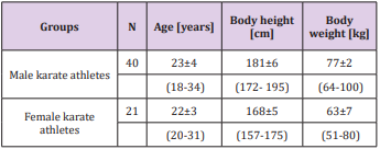
Among males, the mean age of was 23±4 years (18-34), mean weight of 77±2 kg (64-100), mean body height of 181±6 cm (172- 195). 36 individuals showed right laterality of the upper and lower limbs, ambidexterity was declared by 4 people. Among the male group the mean of training experience was 10,8±3 years.

These athletes practiced karate approximately for 5 times per week, additionally they practiced another kind of physical activity for more than 3 times per week. Among females in turn, the mean age of was 22±3 years (20-31), mean weight of was 63±7 kg (51- 80), mean body height of was 168±5 cm (157-175). 19 females showed right laterality of the upper limbs, left one - remaining 2 females. Prevailing right laterality of the lower limbs (18 females), while left and right laterality of the lower limbs declared 3 of them. The mean of training experience was 11,8±3 years. These athletes practiced karate approximately for 5 times per week. Also they practiced other physical activities for almost 4 times per week. Characteristics of the study groups are presented in Table 1.

Table 1: Characteristics of the study groups: number (N), age, body weight, body height (mean, standard deviation, minimummaximum range).

The method of diagnostic survey questionnaire technique was used. Karate athletes anonymously filled out the individual author’s questionnaire, consisting of questions about: gender, age, height, weight, laterality of the limbs, length of training experience, stage of karate training, severity of the trainee, frequency of other exercises, using protectors to avoid the injury, frequently recurring injuries, how many times during training or competition a particular part of the body was injured and about serious or permanent damage to the respondent’s health [Appendix]. Survey results were collected and subjected to statistical analysis using Microsoft Excel and Statistica v.10 (StatSoft). The mean, standard deviation, minimum and maximum values, Mann U Whitney test and severity rates of specific indications were calculated.

Results

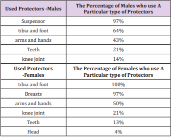
Karate athletes were asked to indicate what type of protectors they use to prevent or minimize the risk of injury. In the first group, the most common protector was suspensor (protector of the genitals), used by 97% of the karate practitioners. Other often used protectors were leg - the tibia and feet (64%), upper extremities and hands (43%). Several athletes have also used a teeth guard (21%) and knees protector (14%). All questioned female athletes to minimize the risk of injury applied at most tibia and foot protectors (100%), then -breasts protector (97%) , arms and hands (50%), knee (21%) and teeth (13%). One of the women used additionally head protector. There was no person who did not use any 
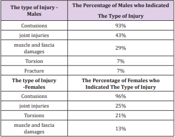
protection during exercises (Table 2). Karate athletes were asked to 
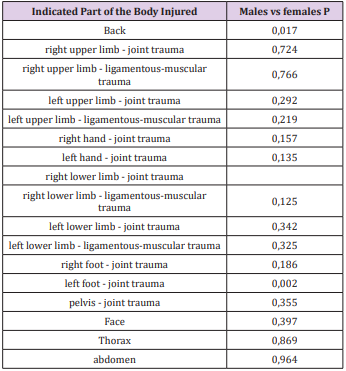
indicate the most common injuries. In the study group prevailed: contusions (91%), joint injuries (49%) with a total number of 548 different types of injuries, especially related to the left foot (23%) and right hand (19%).

Table 2: The most commonly used protectors by males, females: the percentage of athletes who indicated the type of protection.

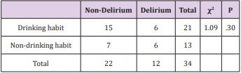
Athletes reported also selected cases of torsion (7%) and fracture (7%). There was not statistically significant difference in the types of injuries between study groups except of injuries of the left foot (p=0.002) and back (p=0.017). The results are shown in Tables 3 & 4. Subsequently, contestants were asked to indicate how many times during karate training particular part of the body was damaged and were asked about injuries that occurred in the body during the last year. The results are presented in Tables 5-7. 
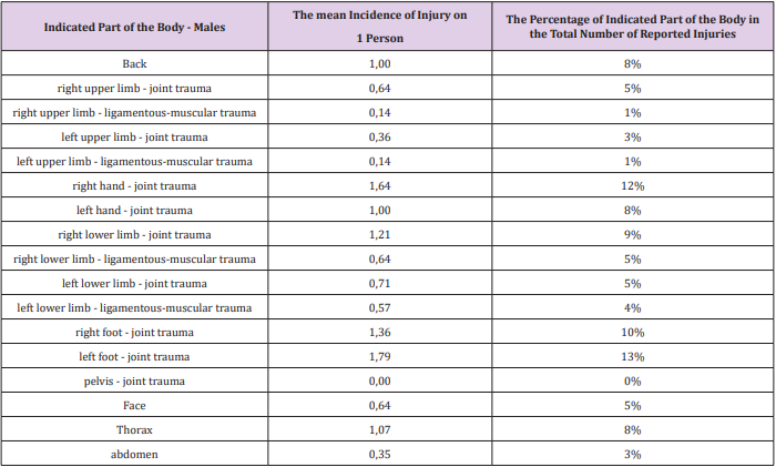
In the first group most injuries were related to the left foot (13%), right hand (12%) and right foot (10%). Reported cases of the right lower extremity injuries (hip injury, 9%), chest, left hand, back (all at 8%), right upper extremity (hip injury), face, right leg (muscleligamentous injury), left leg lower (joint sprain) (all at 5%), left lower limb (ligamentous injury-muscular, 4%), left upper limb (joint injury 3%) and abdominal (3%). Joint injuries were more frequent than muscle damages. There was no report of pelvis injury.

Table 3: The most common injuries in males and females the percentage of athletes who indicated the type of injury.

Note: From 2017-foot protectors and breasts protector are obligatory for women to participate in every one championships.

Table 4: The differences between most common injuries in males and females.

Table 5: The most damaged part of the body in males: number of males who have suffered trauma (N), the mean incidence of injury to one karate athlete, the percentage of males who identified the part of the body.

Table 6: The most damaged part of the body in females: number of females who have suffered trauma (N), the mean incidence of injury to one karate athlete, the percentage of females who identified the part of the body.

Table 7: The most damaged part of the body in the last year among males: number (N), the percentage of males who identified the part of the body.

There was no person who does not suffer any injury of the 
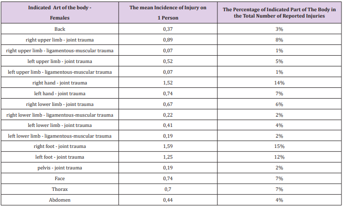
mentioned. The results are shown in Table 5. In the second group, among women while practicing karate the most commonly reported injuries were related to the: right foot (15%), right hand (14%) and left foot (12%). Reported cases of right upper extremity injury (injury to the joint 8%), left hand (7%), chest, face (7% each), right arm (joint sprain, 6%), left upper extremity (hip injury 5%), left lower limb (hip trauma - 4%), abdominal (4%), back (3%), right left leg (ligamentous-muscular injury 2%), and left and right upper limb (ligamentous-muscular injury 1%). Joint injuries were more frequent than muscle ones. Responses are shown in Table 6. Respondents suffered a total of 548 different types of injuries - 186 injuries among women and 362 injuries in men. Mean rate of injury for one person among men were: 0.78, in which the right hand was: 1.64, left hand was: 1.0, right foot was: 1.36, left foot was: 1.79; head was: 0.64. For women, the mean rate of injury for one person was: 0.62, including the right hand: 1.52, left hand: 0.74, right foot: 1.59, 
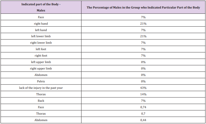
left foot: 1.25, head 0,74. In the past year in the group of males, 43% of them did not suffer injury. Most injuries underwent were right hand (21%), left lower limb (21%) and chest (14%). Reported isolate cases of injury to face, left hand, right leg, foot and back (all 
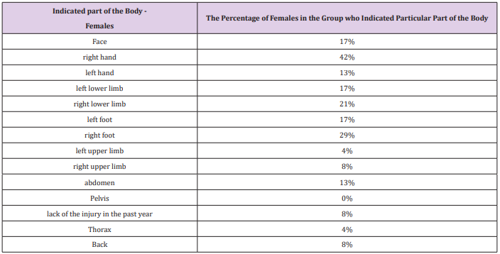
at 7%) (Table 7). In the second group, in the last year 8% of karate kyokushin female athletes did not suffer any injury. Similarly, as in male group, the most occurring was the right hand injury (42%). Subsequently - right foot (29%), right lower limb (21%), face (17%), left lower limb (17%), left foot (17%). There were several cases of abdominal trauma (13%), left hand (13%), right arm (4%), chest (4%) and back injury (8%). Responses are shown in Table 8.

Table 8: The most damaged part of the body in the last year among females: number (N), the percentage of females who identified the part of the body.

Lastly, athletes were questioned whether during training they encountered with serious or permanent damage to the health. 
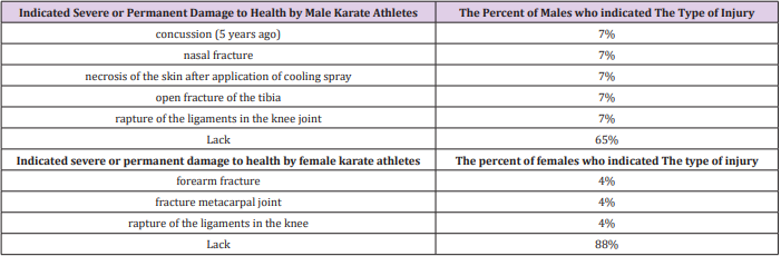
Serious and permanent injury occurred rarely. In the first group experienced them 35% of males and in the second group it was 12% of females. In the first group 65% of men during long-term training were not encounter with a severe or permanent damage to the health. The other karate athletes mentioned: bone fractures - tibia and nose, a concussion (5 years ago), damage to the ligaments of the knee joint and incompetent use of cooling spray resulting in necrosis of the skin. In the same group, 88% of females have no experienced serious or permanent injury. Other women from the first group mentioned: fractures-of the forearm, metacarpal bones and knee ligament damage. Respondents completing the survey were asked to answer questions about the frequently recurring injuries, how many times during training or competition a particular part of the body was injured without their mechanism of occurrence. The results are shown in Table 9.

Table 9: The most indicated severe or permanent damage to the health experienced during exercises, of the males and females: number (N), the percentage of athletes who indicated an injury.

Discussion

Arriaza and Leyes [3] analyzed the injuries sustained by athletes training karate: shotokan, shito-ryu, goju-ryu and wadoryu - taking part in three consecutive World Championships. The mean value of the injuries was at level 157.03/1000 competitors. Authors reported 891 injuries. The most common injuries were contusions of the large muscle groups (50.3%), facial traumas resulting in bleeding from nose (16.2%), cuts and abrasions (13.7%) and twisting of ligaments (3.5%). There were 796 (89.3%) minor injuries, 70 (7.9%) moderate and 25 (2.8%) severe injuries. Severe cases of injuries resulted in concussion, internal organs damages, third-degree sprains, eye injuries and various types of fractures. Most injuries were related to the face (72.5%), head (11.6%) and lower limb (6.4%). In our study in groups of karate kyokushin athletes strongly dominated minor injuries as contusions and joint injuries within hands and feet. The overall proportion of severe injuries was lower than in Arriaza and Leyers study.

These differences may be since athletes analyzed by the Arriaza and Leyers took part in the World Karate Championships (high level of competitors), while our competitors were taking part in the Polish and European Championships. Differences may be also related to the dissimilarities occurring between each style of karate. In the kyokushin style up to year 2017-foot protectors were not obligatory for women and only obligatory protectors were breasts protectors for women and suspensors for men. The authors emphasize that competitive karate is associated with a relatively high injury rate and note that cases of severe injuries were rare what is compatible with our study. Destombe et al. [5] analyzed the injuries sustained by the 186 French karate athletes for a period of 1 year. The study gropup consists of karatekas from three karate clubs in Brest, France but the author did not mention the style of karate. Total number of injuries was 83 (63 during trainings, 20 during competitions), average injuries stood at 44.6/100 players. The most common injuries were hematomas (53%), twisting (19%), muscle injuries (7%), fractures (7%), cuts and abrasions (7%). Predominated limb injuries were the upper (28.9%) and lower (35%), followed by head (26.5%) and trunk (9.6%) injuries. Authors stress that injury rate increases with time spent on trainings, rank of each competitor and years of practice what is similar to our study. Destombe et al. also emphasize that serious injuries in karate are rare. Minor injuries of the upper and lower limbs are dominating, what is compatible with our findings. Zazryn et al. [6] in turn, studied the incidence of injuries among professional athletes training kickboxing for a period of 16 years. Total number of injuries was 382, mean value of injuries stood at 109.7/1000 competitors. The most common injuries were superficial ones like bruises, lacerations and abrasions (over 64%). Predominated head, neck, face (51.6%) and lower limbs (39.8%) injuries, the lower leg (23.3%), face (19.4%) and intracranial injuries (17.2%).

The nature of kickboxing where kicking the opponent is the major movement and head is a prime object was associated with the distributions of body regions mostly injured by participants. The nature of kickboxing, whereby kicking the opponent is the prime movement and the head a prime target, is reflected in the distributions of body regions most commonly injured by participants. Therefore, the results obtained by authors differed from results received in karate kyokushin athletes (differences between karate kyokushin and kickboxing style). Gartland et al. [7] studied the incidence of injuries in muay thai kickboxing athletes. The survey was conducted among 152 people. Mean value of injuries stood at 13.5/1000 recreational competitors’ athletes (amateurs) and 2.79/1000 professional athletes. The most suffered injuries were bruises, lacerations and abrasions. Among the amateurs dominated injuries to the lower limbs (75%), trunk (15.9%) and upper extremities (6.8%). Head injuries accounted for 2.3% of all injuries.

Among professional athlete’s lower limb injuries (53%) also predominated. More often they experienced trauma to the head and trunk. The most frequently occurring injuries were: bruises, sprains, cuts and abrasions. Authors highlight that different martial arts are associated with particular injury pattern which may explain the differences in percentage distribution of injuries between muay thai kickboxers and karate kyokushin practitioners. However, worth noting is fact that, according to the authors, karate is linked to the traumas within the lower extremities, as it can be seen in our study. Kazemi et al. [8] analyzed the injuries suffered by athletes training taekwondo for a period of 9 years. Average injuries stood at 16.18/100 athletes. Predominated head injuries (19%), foot injuries (16%) and thigh (9%). Minor traumas as: contusions (36%), sprains (19%) and muscle strains (15%) were the most common. Differences between Kazemi et al and our study occurred in the distribution of rates and the leading locations of injuries.

Nevertheless, according to the authors, the most commonly reported injuries were bruises, injuries of joints and muscles, which is similar to our results. Kujala et al. [9] analyzed 54186 sports injuries sustained by judo, karate, football, ice hockey, basketball and volleyball players in the years 1987-1991. Average injuries stood at 142/1000 of karate athletes, 117/1000 of judo competitors, 94/1000 of hockey players, 89/1000 among football players, 88/1000 among basketball players and 60/1000 of the volleyball players. In team games 46-59% of injuries happen during competitions and tournaments, while in martial arts the figure was 70%. Injuries typical for each discipline predominated. Fractures and teeth injuries were the most common among players of hockey and karate, lower extremity injuries among football and volleyball players and upper extremity injuries in judo. Sprains, muscle injuries and contusions were the most common types of injuries. The most predominant areas of the injury among karate athletes, according to the authors’ injury were lower limbs (37.3%) - especially: knee (11%) and foot (10.7%), and upper limbs (26.3%) - fingers (9.3%). Other traumatic sites (36.3%) were head and neck (10.9%). Mostly injuries (sprains, strains, bruises, fractures) were related to the striking surfaces in karate-hand and feet joints, what is compatible with our study findings.

Zetaruk et al. [10] investigated the incidence of injuries among the 263 athletes trained martial arts. The survey was conducted among athletes training: karate shotokan (114 individuals), aikido (47 people), taekwon-do (49 athletes), kung fu (39 athletes) and tai chi (14 individuals). Most injuries were related to the athletes practicing taekwon-do (59%), aikido (51%) and kung fu (38%). The least frequent injuries applied to karate shotokan athletes (30%) and tai chi (14%). According to the authors karate shotokan athletes suffered 114 injuries, among which the lower extremity (22.8%), upper extremity (16.7%), trunk (14.9%) and head, neck injuries (9.6%) were dominating. The areas of suffered injuries in karate shotokan athletes are consistent with parts of the body that have been injured in karate kyokushin individuals even despite the differences in shotokan karate protector’s usage (obligatory to compete are tibia and foot, hands protectors for athletes and breast protectors for women and suspensor for men).

Also, Vitale et al. [13] investigated foot anatomy, anthropometric measures, and other background factors and information as possible risk factors of injury in barefoot athletes practicing judo, karate, kung fu, Thai boxing, or aikido. The results of 130 subjects showed that most of the athletes (53.8%) did not sustain lower lib injuries, 19.2% reported an overuse injury and 27.0% suffered an acute injury. No significant differences were observed in the injury rates in relation to style and kind of martial art. Interesting and worth mentioning was the fact that in this study older and heavier martial artist, performing more hours of barefoot training, were at higher risk of acute and overuse injury. This aspect has not been addressed in authors’ study and requires further research on a larger study group divided into weight categories in the future. Other study shows that even athletes who taking part in kata tournaments (without fighting) are suffering form and overuse injury with the occurrence of pain in the joints of the lower extremities and lumbar spine from performing the basic stances [17].

Over the years a reduction in average number of injuries in martial arts [11,12], including several acute injuries as concussions to muscle and tendon ones, has been observed. Perhaps, it is due to the widespread use of protectors, greater awareness in the treatment area, prevention of damage and stricter enforcement of the competitions. But it can also be caused by insufficient data. As Thomas et al. [18] indicate that studies need to adopt one injury definition, one data-collection form, and collect comprehensive data for each study for both training and competitions. More data are needed to measure the effect of weight, age and experience on injuries, rates and types of injury during training, and for competitors with high injury rates. RCTs are needed of interventions such as training and feedback of performance data to reduce injury rates. The reduction in average number of injuries can be also caused by the changes to the safety regulations (i.e. foot protectors in females) by members of the approvals board and medical ethics [14,15]. The Council for Medical Ethics discourages doctors from accepting to act as ringside doctor for combat sports that permit knockouts or participating in approvals or appeals boards for such events because these types of assignments in their opinion may violate the general duty of doctors to protect human health [15].

Moreover, finding of this study considering other research presented above are justifying a recommendation for coaches to differ a training or accept a physiotherapist in order to prevent injuries due to overload of ligaments and muscles as it is in other professional sports. Martial arts did not emphasize use of rejuvenation treatment for athletes as it is on other professional sports, maybe due to insufficient founding in comparison to for example football. Polish karate kyokushin athletes use all the rejuvenation treatment individually on their own [16]. Karate clubs do not provide such treatment. This study reveals some information about competitive athletes and those statistics should not be extrapolated toward all practitioners. As in many other martial arts, people are more concern about their health and tend to avoid injuries [19]. Such risk and special attention should be given to those, who prepare for competitions only.

Conclusion

a. Most of athletes are willing to use protection of central parts and striking surfaces.

b. Women athletes are slightly more prone to use protective equipment than male athletes.

c. Injuries were more frequent among distal part of the body (hands and feet) than the central ones.

d. Males suffers more back and foot injuries than women.

More BJSTR Articles : https://biomedres01.blogspot.com/

Establishment of Methylation Sensitive Suppression Subtraction Hybridization Analysis and Isolation Of 5’cpg Islands Fragments in Chinese Fir Genomic DNA

Establishment of Methylation Sensitive Suppression Subtraction Hybridization Analysis and Isolation Of 5’cpg Islands Fragments in Chinese Fir Genomic DNA 

Introduction

Chinese fir (Cunninghamia lanceolate (Lamb.) Hook) is the most important conifer species for commercial wood production in southern China Zheng et al. [1]. Chinese fir accounts for about 25 % of total plantation area in China and about 50 % of domestic plantation timber production in southern China. Now, the breeding cycle for Chinese fir get into third generation Pan [2]. However, most research about Chinese fir was forced on traditional breeding Zhang, et al. [3]. The study of molecular biology was still not completely clear. DNA methylation profiling had become an important technology in many areas of epigenetics research Zhang, Kimatu et al. [4]. Methylation profiling approaches were aimed at investigating DNA methylation patterns either across the entire genome and at different levels of resolution, or they were used to study methylation patterns at specific areas of interest, for example at promoters and other regulatory elements Fatemi et al. [5]. In genetics, CpG islands were genomic regions that contain a high frequency of CpG sites but to date objective definitions for CpG islands were limited. The usual formal definition of a CpG island was a region with at least 200 bp and with a GC percentage that was greater than 50% and with an observed/expected CpG ratio that was greater than 60% Gardiner Garden et al. [6].

At present, a variety of methods were used to evaluate the methylation status of genes: southern blot Bickle et al. [7], bisulfite genomic DNA sequencing Frommer et al. [8], restriction enzymePCR Kane et al. [9], methylation-specific PCR (MSP) Frommer et al. [8], methylation-sensitive single nucleotide primer extension (MSSnuPE) Herman et al. [10], electrochemistry Kuppuswamy et al. [11] and so on. However, each method was restricted to the evaluation of DNA methylation on gene-by-gene basis. Such an approach had given researchers a limited picture of complex epigenetic alterations in plants. Therefore, a compelling need existed to develop novel technologies for the next phase of epi-genome research. A powerful approach for studying the genetic nature of many biological processes was to characterize genes that vary in expression level during this process. Suppression Subtractive hybridization (SSH) Diatchenko et al. [12,13], was a technology that allowed for PCRbased Von Stein et al. [14-16] amplification of only cDNA fragments that differ between a control and experimental transcriptome. Differences in relative abundance of transcripts were highlighted, as were genetic differences between species. The technique relied on the removal of dsDNA formed by hybridization between a control and test sample, thus eliminating DNA or gDNA of similar abundance, and retaining differentially expressed, or variable in sequence, transcripts or genomic sequences. The purpose of this study was to explore the feasibility of enrichment methylation CpG islands of genome through combining the suppression subtraction hybridization with methylation in Chinese fir.

Materials and Methods

DNA Extraction and Enzyme Treatment

The materials were obtained from Yangkou Forestry Center (Fujian, China). Genomic DNA was isolated by standard methods using CTAB method Gu et al. [17]. The DNA derived from the buds of Chinese fir was divided into two parts. One aliquot was treated by methylase SssI (SM) as positive control with the conditions recommended by the supplier (New England Biolabs, Beverly, MA). The other was not treated by methylase SssI as negative control. The positive control generated in this way had 100% methylated cytosine in the test CpG sites, whereas the negative control had all unmethylated cytosine residues in the test CpG sites. Approximately 2–3 ug genomic DNA was restricted to completion with 50 U of MseI following the conditions recommended by the supplier (New England Biolabs). This enzyme restricted bulk DNA into small fragments. As its recognition site (TTAA) rarely occurred in GC-rich regions, most CpG islands remain intact after the restriction. The digests were purified with QIAquick column (QIAGEN).

Driver Preparing

The cleaved ends of DNA were ligated to unphosphorylated linkers. The linker sequences were H-24 (5’-AGG CAA CTG TGC TAT CCG AGG GAT-3’), and H-12 (5’-TAATCC CTC GGA -3’) Hou et al. [1]. The use of universal linkers allowed to subsequently amplifying all methylated fragments in ligated DNA samples. The ligation step began with the annealing of H-12 to H-24 (each 100uM) by mixing equal amounts of each oligonucleotides in a microcentrifuge tube and allowing the mixture to cool from 55 ℃ to room temperature over 1 hour. The annealed linker-primers (10ul/sample) were then added to the purified DNA, together with 2.0ul 10×T4 DNA Ligase Buffer, 2.0ul 10mM ATP, 1 ul T4 DNA Ligase (Takara), and water to 20ul. The ligation reaction was carried out at 16℃ overnight. The mixture was then held 65 ℃ for 10 min to stop the reaction. The ligated products were purified with QIAquick column (QIAGEN) and digested with the methylation-sensitive endonucleoase HapⅡ following the conditions recommended by the supplier (New England Biolabs).

The digest was used for PCR without further purification. PCR reactions were performed for only 20 cycles. At this level of amplification, especially if most unmethylated repetitive sequences were restricted away by the HapⅡ digestion, the proportions of PCR products for most templates were expected to be in the linear range. PCR reactions were performed in a 50ul volume, containing 0.4 uM H-24 primer; 4 U Taq DNA polymerase (Takara); 5% DMSO; 200uM dNTP; 2.5mM Mg2+ in a buffer provided by the supplier. The tubes were incubated for 3 min at 72 ℃ to fill in 5’-protruding ends of the ligated DNA and subjected to 20 cycles of amplification consisting of 1 min at 95℃ and 3 min at 70 ℃. The final extension was lengthened to 10 min

Tester Preparing

Adaptor1 (10uM) and 2 (Figure 1) were each separately ligated to 2ul of 1:6 dilution of SM-digested DNA (Tester) at 16℃ overnight. Samples were then heated at 72℃ for 5 minutes to inactivate. Then extracted with phenol chloroform and resuspended in 5.5μl H2O and digested with the HapⅡ.

Figure 1: Sequences of adaptors and adaptor primers used in MS-SSH. The illustration shows the detailed sequences of adaptors (i.e., adaptor 1 and 2) and adaptor primers (i.e., PCR primer 1 used in primary PCR, Nested PCR primer 1 and 2 used in secondary PCR) used in the MS-SSH system.

Two Subtractive Hybridization

In the first hybridization, 1.5μl HapⅡ-digested normal DNA (Driver) was mixed with 1.5μl of diluted adaptor1-ligated or adaptor 2-ligated SM -digested DNA (Tester) (about 15 copies of Driver DNA vs. 1 copy of Tester) in the presence of hybridization buffer. The samples were denatured at 98℃ for 1.5 min and immediately incubated in 68℃ for 6-8 hours. In the second hybridization, the two samples from the first hybridization were mixed in the presence of a freshly denatured (1μl of 1:2 dilution) Driver DNA. The sample was incubated at 68 ℃ for 12 hours. After adding 200μl of dilution buffer, the sample was incubated for an additional 7 minutes.

Two Suppression PCR (Nested PCR)

The primary polymerase chain reaction (PCR) was conducted in 20μl volume containing 1μl of the diluted subtraction mixture, 1× PCR reaction buffer, 10uM PCR primer1 (Figure 1), 0.2mM dNTPs and U Taq DNA polymerase (Takara). The reaction mixture was incubated at 75 ℃ for 5 minutes to extend the adaptors, followed by 94℃ for 2 minutes , then followed by 35 cycles at 94℃ for 30 seconds, 66℃ for 30 seconds, and 72℃ for 1.5 minutes. The primary PCR mixture was diluted 20 fold, and 1μl was used in secondary PCR with Nested primers for primers 1 and 2 (Figure 1). The conditions of the reaction were 94℃ for 30 seconds , 66℃ for 45 seconds , and 72℃ for 1.5 minutes for 30 cycles.

Construction of 5’CpG Islands Fragments Libraries

The products of the secondary PCR were verified by agarose gel electrophoresis (AxyPrep DNA Gel Extraction Kit), and then cloned into the pMD 19-T Vector (Takara, Dalian). The detection of recombinant clones was based on blue/white selection. All recombinant clones were picked and culture overnight in 3mL of LB medium containing 100mg/L ampicillin in 10mL centrifuge tube at 37 ℃. The inserts were amplified by PCR using Nested PCR primer 1 and 2. The PCR products were electrophoresed on a 1% agarose gel to confirm the amplification quality and quantity

Dot-Blot Hybridization

To further confirm positive clones, the inserts were amplified by PCR using the Nested primer 1 and 2; the PCR products were purified by Takara DNA Fragment Purification Kit Ver.2.0, and then dotted into Hybond-N+ nylon membranes (Amersham, Piscataway, New Jersy). The probes (MS-digested DNA and undigested DNA) were labeled by digoxigenin (DIG). Hybridization and detection were carried out following the DIG High Prime DNA Labeling Detection Kit protocol.

Sequencing of Bands

The positive clones were sequenced with vector primers. The Advanced BlastN and BlastX programs at the NCBI website (http:// www.ncbi.nlm.nih.gov/) were respectively used for homology analysis of the cloned DNA sequences that gave quality-reads Benson et al. [18,19].

Result

MS–SSH was an efficient way to isolate and enrich genomic CpG islands. The driver DNA was prepared by Linker–PCR combined with Mse I and methylation-sensitive restriction endonuclease (i.e., Hpa II) digestion of genomic DNA, and then the SSH procedure was performed to isolate and enrich methylated CpG islands contained in tester DNA. The amplicons were generated by the procedures outlined above (Figure 2).Firstly, The size distribution of MseIdigested genomic DNA ranged from 100 to 2000bp (Figure 3B). Secondary, the MseI-digested DNA was ligated to H-24/H-12 linkers combined with subsequent digestion of HpaII to prepare MseI–HpaII pretreated DNA by Linker–PCR amplifications (Figure 3D). Genomic fragments containing methylated sites were protected from the methylation-sensitive enzymes digestion and could be amplified by linker-PCR. Many of these linker-ligated MseI fragments were expected to be present in the sample due to aberrant methylation in the test sites, whereas the same unmethylated fragments were digested and not present in the normal application after PCR. Finally, the MseI–HpaII pretreated amplicons were prepared by MseI digestion of their corresponding products of Linker–PCR to remove the H24/H12 linkers at the ends of DNA molecules (Figure 3E).

Figure 2: Schematic flowchart of driver generation.

Figure 3: Amplicon generation of MS–SSH system. (A) Genomic DNA. (B) MseI digestion of genomic DNA. (C) MseI–HpaII pretreated DNA. (D) Linker–PCR products of MseI–HpaII pretreated DNA. (E) MseI digestion of the Linker–PCR products. White triangles indicate the H-24/H-12 linkers cleaved from both ends of the DNA molecules.

Isolation and Construction of CpG Islands

Firstly, the secondary PCR products of hybridization were evaluated by agarose gel electrophoresis (Figure 4A). Because there was no tester-specific DNA fragments contained in tester DNA, one visible smear mainly in the 200 to 1000bp range could be seen in the secondary PCR products of subtraction. After the tertiary PCR products of MS-SSH subtraction were purified, they were cloned into the pMD–19 T vector to prepare the CpG Islands library. To estimate the proportion of CpG Islands-like clones in the CpG Islands library, the size distribution of cloned inserts was firstly evaluated by PCR amplification of insert fragments using primers (i.e., Nested primer 1 and 2) that was flanking the cloned inserts. Altogether, 536 clones were randomly selected and analyzed, with 96.5% (517/536) of cloned inserts ranging from 200 to 1000bp and the remainder being longer than 1000bp (Figure 4B). The resulting insert fragments were ranged from 200 to 1000bp (Figure 4B). Finally, recombinant plasmids were digested by EcoR I and Hind III to verify the positive recombinants. For the above 536 clones, PCR fragments of 192 cloned inserts were further digested with EcoR I and Hind III, with 91.1% (175/192) containing one insert (Figure 4C). Thus, a putative subtracted MS-SSH library of Chinese fir was constructed.

Figure 4: Characteristics of the cloned inserts in the CGI library. (A) Secondary PCR products. (B) Representative amplified cloned inserts from the CGI library. (C) EcoR I/Hind III double digest of recombinant plasmids.

Differential Screening of MS-SSH Library

To characterize the differential gene expression of the identified 175 CpG Islands clones, a dot blot was performed, as both DNA subtraction and differential screening procedures utilized PCR amplified material. Several control DNAs were also utilized to validate this technique. Only 16 false-positive clones were detected and the rest were confirmed as ‘‘true’’ CpG Islands (Figure 5). As a more thorough evaluation of the CpG Islands library, the insert sequences of 159 randomly selected clones were determined by DNA sequencing. Subsequently, further sequence analyses were performed, including restriction site analysis, CpG Island Searcher (http://cpgislands.usc.edu/) to analyze the base composition (GC%) and frequency of observed CpG versus expected CpG (ObsCpG/ExpCpG) (Figure 6), and NCBI BLAST to search for homologous sequences in the GenBank database (Figure 7A). The results showed that 90.9% (145/159) containing inserts having one or more HpaII sites. In the 145 clones, bulk DNA (GC% < 40%, ObsCpG/ExpCpG < 0.6) was contained in 15 clones (9.4%, 15/159). The last 130 (81.7%, 130/159) were identified as CpG Islands clones because they satisfied the minimal criteria for CpG Islands length > 200 bp, GC% > 50%, ObsCpG/ExpCpG > 0.6), of which only 15 (10.3%, 15/145) were duplicated clones. All insert fragments in the 123 CpG Islands clones were single copy in genomic DNA based on the results of NCBI BLAST homology searching (Figure 7B).

Figure 5: Differential screening of clones from the identified CpG Islands clones. Quadruplicate colony PCR products were prepared and the membrane was hybridized with DIG-labeled DNA probers derived from (a) tester DNA pool and (b) driver DNA pool. T.C., tester control; D.C., driver control. Arrows indicate false-positive clones.

Figure 6: The result of CpG Detetion.

Note: CpGPLOT islands of unusual CG composion: EMBOSS-001 from 1 to 474 (Observed/Expected ratio>0.60; Percent C+percent G>50.00 and length>200.).

Figure 7: Characteristics of the cloned inserts in the CpG Islands library. (A) Size distribution of inserts of 159 randomly selected clones. (B) The results of NCBI BLAST homology searching.

Sequences Analysis of MS-SSH Library

Of the 69 aligned sequences, there are 19 genes with high similarity to known metabolism in GeneBank (Table 1), including oxidases, dehydrogenases, phosphatases, protein kinases, N-acetylglucosaminyltransferases, and proteins of unknown function. Among them, the MF1701 fragment has high homology with the pentatricopeptide repeat (PPR) protein, which is an RNA specific binding protein, and play important roles in plant growth and development, organelle formation, fertility restoration of cytoplasmic male sterility, RNA Editing and processing, signal transduction between nuclear and organelles, stress defense and so on. Table 1 Sequence of methylated fragments and database search.

Table 1: Sequence of methylated fragments and database search.

Discussion

DNA methylation was referred to as epigenetic phenomena that control various genomic functions without a change in nucleotide sequence Morris [20]. DNA methylation was associated with early embryo of plant organizations development Suzuki et al. [21], genetic imprinting Razin et al. [22], and chromosome inactivation Chan et al. [23]. Therefore, the detection method of DNA methylation was rapid development. Among various DNA methylation analysis methods, chip techniques have shown powerful abilities to isolate and identify the aberrant methylated CGIs. Such methods include MCA-RDA (Methylation in CpG island amplification-representational difference analysis) Toyota et al. [24], DMH (Differential methylation hybridization using CGI array) Yan et al. [25], MSO (Methylation specific oligonucleotide) Yu et al. [26], ChIP-on-Chip (Chromatin immunoprecipitation) Kuras [27]. Subtractive hybridization was a powerful technique that enables researchers to compare two populations of DNA and obtain clones of genes that were expressed in one population but not in the others Diatchenko et al. [13]. Although there were several different methods, the basic theory behind subtraction was simple.

First, we refer to the one DNA that contains specific CpG Islands as tester, and the reference DNA as driver. In this research, tester and driver DNA were hybridized, and the hybrid sequences were then removed. Consequently, the remaining unhybridized CpG Islands that were expressed in the tester yet absent from the driver (Figure 4). In some cases, the MS-SSH method greatly enriches for differential CpG islands; nevertheless, the subtracted sample would still contain some templates that correspond to CpG islands common to both the tester and driver samples Illingworth et al. [28]. Although this background may depend somewhat on the quality of DNA purification and performance of the particular subtraction, in chiefly arises when DNA species were differentially expressed in tester and driver. In general, a limited set of differentially expressed DNA and low quantitative difference in expression produces higher background, even if the sufficient enrichment of differentially expressed DNAs were obtained Ogino et al. [29]. With high background, picking random clones from the CpG Islands library for Southern blot analysis was extremely time-consuming and inefficient.

Therefore, differential screening should be performed before embarking on Southern blot analysis. In this study, it was appeared some un-CpG Islands, which may be related to the design of experiment (Figure 5). After M.Sss I enzyme treatment, all CpG sites of genomic DNA were methylated, and could not digested by HapⅡ in the tester. Nevertheless, the unmethylated CGIs in the driver were digested by HapⅡ. Therefore, the discrepancy fragments between tester and driver could amplify through the suppression PCR. Cytosine methylation influences gene expression, a hypothesis based on the frequency of methylcytosine and on the proposed transmission of methylation patterns through cycles of DNA replication Finnega et al. [30-32]. The quality of CpG islands library was close relationship with the following experiment. Therefore, it is very importance for testing the quality of CpG islands library through HTF-HapⅡ tiny fragment analysis of tools-Sequence Analysis-CpG plot software in European Bioinformatics Institute (EBI). The construction of library in the research is very high of CpG content, also can be used to detect new genes and following experiment (Figure 6).

Conclusion

Development of the MS-SSH technology not only significance for the CpG islands database construction, also advanced for high-throughput comparative analysis the difference between the organizations methylation.

More BJSTR Articles : https://biomedres01.blogspot.com/

Monday, March 29, 2021

Factors Associate with Delirium of Liver Surgery Patients in the Intensive Care Unit of a Medical Center of Southern Taiwan

Factors Associate with Delirium of Liver Surgery Patients in the Intensive Care Unit of a Medical Center of Southern Taiwan

Background

Intensive care units (ICUs) have a specific and closed environment, and they provide 24-hour care to patients. Delirium is a common problem in patients of ICUs and defined as the altered state of consciousness accompanied by a change in cognition that develops over a few hours or days and tends to have a fluctuating course Sendelbach [1]. The incidences of delirium in the different ICU patients were reported as following. In foreign countries, according to the findings of a systematic review of 18 studies, 50% of liver transplant Italy patients whose lengths of ICU stay were 0.5– 32 weeks developed postoperative cognitive dysfunction (POCD) Aceto et al. [2,3]. Examined 144 adult USA patients who received liver transplantation during a period of 6 years; they revealed that delirium occurred in 25% of the patients, with the corresponding average duration being 4.56 days. Another study showed that 50% of 90 USA patients undergoing hematopoietic stem-cell transplantation developed delirium Fann et al. [4]; additionally, a study demonstrated that 10.03% of 281 Canada patients who received orthotopic liver transplantation (OLT) developed delirium in the ICU during a stay of 2 days Lescot et al. [5].

Furthermore, Wassenaar et al. [6] reported that the incidence of delirium in 2914 German patients in ICUs was 23.6%. In Taiwan, Wang et al. [7,8] have reported that 58.3% and 31.5% of patients with and without drinking habits, respectively, experienced at least one episode of delirium at a hospital in central Taiwan after living donor liver transplantation (LDLT). Wang et al. [9] also reported that delirium was diagnosed in 47.4% of 37 patients after LDLT; specifically, delirium occurred approximately 7 days after surgery, and the mean duration was 5 days; the length of the ICU stay of patients with delirium was significantly longer than that of patients without delirium. Furthermore, Lin et al. [10] reported that 7.8% of 71 patients developed delirium following hepatectomy for the treatment of hepatocellular carcinoma (HCC) at a hospital in central Taiwan. Regarding the risk factors, in foreign countries, Bhattacharya et al. [3] reported that in 144 USA patients who underwent liver transplantation, the development of delirium was associated with age, long duration of hospitalization, high likelihood of alcohol assumption, and high frequency of acquired hospital infections. Additionally, opioid doses, current and prior pain, and BUN levels were revealed to be risk factors for severe delirium in USA patients after transplantation Fann et al. [4].

Furthermore, intraoperative transfusion of packed red blood cells is also a predictor of the development of delirium in Canada patients in ICUs after OLT, which is associated with an increased length of hospital stay and mortality Lescot et al. [5]. Finally, Wassenaar et al. [6] identified nine predictors of delirium in 2914 German patients in ICUs, namely age, history of cognitive impairment, history of alcohol abuse, blood urea nitrogen (BUN), admission category, urgent admission, mean arterial blood pressure, use of corticosteroids, and respiratory status. In addition to the above risk factors, among the liver transplant patients, the incidence of neurological complications was 20%, and these complications are crucial factors affecting morbidity; furthermore, hyponatremia was a risk factor for neurological complications following liver transplantation Lee et al. [11,12]. In addition to hyponatremia, alcohol-related Wernicke encephalopathy might be life-threatening for liver transplantat Australia patients Zuccoli et al. [13]. In Taiwan, Wang et al. [7] explored the predisposing factors for delirium in 54 ICU patients with alcoholic cirrhosis after LDLT; they reported that hepatic encephalopathy was a crucial risk factor for delirium.

On the basis of a study of 37 patients with HCC at a hospital in central Taiwan, Wang et al. [9] proposed that the risk factors associated with delirium included history of alcohol abuse and preoperative hepatic encephalopathy. Lin et al. [10] also identified that advanced age and low postoperative hemoglobin (Hb) levels were potential risk factors for postoperative delirium following hepatectomy in 71 patients with HCC at a hospital in central Taiwan. The consequences of delirium in ICU included the prolong the weaning time from ventilation and the tube insertion rate, occurrence of complications, the duration of hospitalization, care needs of the patients, medical cost, and an increase in the death Chen et al. [14-16]. Nevertheless, Olson [17] proposed that delirium is among the most common problems in ICU patients but is often overlooked or under-recognized for the critical care nurses to identify early. If the health care professionals could identify delirium in ICU patients early, they can prevent the subsequent confusion, disorientation, and unusual or irritable behaviors in the delirium patients. Consequently, the hospitalization days and death rate of ICU patients with delirium could be decreased, and the function of these patients might be improved.

Purpose of the Study

This study explored the factors associate with delirium in liver surgery patients in the ICU of a medical center in southern Taiwan.

Methods

This study used purposive samples of liver surgery patients admitted to the liver ICU of a medical center in southern Taiwan (IRB approval number 201601364B0) between November 25, 2016, and July 24, 2017. The inclusion criteria of liver surgery patients included GCS > 7, without vision and hearing impairment, no history of dementia, drug addition, psychiatric illness, CNS diseases, and agreement of participating into the studies by the patients or family members before the surgery. The data of samples were collected when they were transferred into liver ICU after 4 hours. Although CAM-ICU has identified as the validity assessment scale for ICU delirium patients Gusmao Flores, et al. [18], the Richmond Agitation and Sedation Scale (RASS) has been reported to effectively identify different levels of delirium and correctly assess fluctuations in delirium in patients over time as well as has been also tested in 38 ICU patients Ely et al. [19]; therefore, RASS was applied to assess delirium of the liver surgery patients in this study.

Before delirium-related data were collected, a delirium assessment scale with RASS was developed by the principal investigator and one nursing practitioner (NP) in the liver ICU, and the content validity of the delirium assessment scale, which was evaluated by three ICU medical professionals and one NP, approached 0.975. The assessment consistencies of the RASS, pain scores, and delirium assessment scale were evaluated by one head nurse and one NP through the assessment of the data of nine liver transplant patients in the ICU. The internal consistency coefficients of the RASS, pain scores, and the delirium assessment scale were .984, .997, and .992, respectively. The delirium assessment scale was used for assessing the samples by NP twice a day during the morning at 9 am and evening at 6 pm. The rest of data in the delirium assessment scale were recorded by following the chart of each sample retrospectively.

Results

Sample Characteristics

The sample characteristics of the 34 ICU liver surgery patients with and without delirium were age, marital status, education levels, occupation, delirium, drinking habit, liver surgery, and length of ICU stay. The patients’ ages were 21–75 years with average age as 51.2 years and 79.4% married. Their education ranged from the elementary level to college level, with more than 60% of the patients having graduated from high school, 32.5% ran businesses, and 38.2% had a drinking habit. Additionally, 35.3% of the patients exhibited delirium, and 38.3% was liver transplant patients and 61.7% were other different types of liver surgery. The length of ICU stays ranged from 1 to 27 days, with the average length being 12.4 days. Table 1 lists the demographics of the 34 liver surgery patients.

Table 1: Demographics of liver transplant patients (N=34).

Factors Associate with Delirium

Delirium and Age, Number of Hospitalizations, Length of ICU stay

Based on the correlation data from Table 2, delirium was not correlated with the age or number of hospitalizations of the liver surgery patients, but it was significantly correlated with the length of ICU stay. Hence, a longer length of ICU stays indicated a higher likelihood of delirium for the liver surgery patients (rbs= .39, P =.03).

Table 2: Correlation between delirium and other factors.

Note: P <.05

Delirium and Drinking Habit

Based on the data of delirium and drinking habit from (Table 3) and the chi-square test results, the 22 liver surgery patients without delirium (7 and 15 without and with drinking habits, respectively) and 12 patients with delirium (6 and 6 without and with drinking habits, respectively) did not show a significant relationship between delirium and drinking habit (χ2 = 1.09, P = .30).

Table 3: Relationship between drinking habit and delirium.

Note: P < .05

Comparison of Variables Between Delirium and NonDelirium Groups

When this study compared the delirium and non-delirium groups of the liver transplant recipient patients by using the independent t test (Table 4), only Hb levels (t = −2.71, P = .01) and BUN levels (t = −2.67, P = .01) were significantly different between the two groups. The delirium group exhibited lower Hb and higher BUN levels than the non-delirium group. However, the count of white blood cells and levels of glucose, Na, K, C-reactive protein (CRP), and creatine did not differ significantly between the groups. Nevertheless, the levels of glucose and CRP were higher than the standardized levels in both groups.

Table 4: Comparison of variables between delirium and nondelirium groups.

Note: P < .05

Discussion

The incidences of delirium for the liver surgery patients in the literature from 10.03% to 58.3% based on the data of different countries depending on various types of liver surgeries and chronic illness as well as drinking behaviors and other related factors. In this study 35.3% of 34 liver surgery patients had delirium which is in the middle range of incidence according to the literature information. Additionally, the average length of ICU stay was 12.4 days of 34 liver surgery patients in this study which is longer than the hospitalization days comparing with the literature data ranged from 2-7days. The reason for the liver surgery patients in this study stayed in the liver ICU longer than the rest of cases in the literature is because there are many liver transplant patients in this study comparing with the other studies in the literature. Liver transplant is a big and complex surgery process for patients to justify their bodies to fit into the new organs into their own systems that should take more time to adapt.

Because the patients in this study were aged <65 years, age was not significantly correlated with delirium. However, most studies have identified age as a risk factor for delirium. Half of the patients were businessmen with drinking habits due to their work environment; nevertheless, drinking habit was not significantly correlated with delirium in this study because of the small sample sizes and lower percentage of 34 liver patients who had drinking habits. In addition, 38.2% of liver surgery patients in this study with drinking habits were not necessary to be the alcoholism; even studies have indicated that alcohol consumption is a significant factor associate with delirium in liver surgery patients. Furthermore, the results regarding Na levels are also different from those in previous studies that have indicated that hyponatremia was a risk factor for neurological complications following liver transplantation Lee et al. [11,12]. In this study, only the levels of Hb and BUN were critical factors correlated with delirium; these findings are similar to those of previous studies Fann et al. [4-6,10].

The universal and consistent finding is that the longer lengths of ICU stay were significantly correlated with the development of delirium in liver surgery patients. This finding is similar to those of Wang et al. [9], who revealed that 37 HCC patients with delirium had a significantly longer length of ICU stay than patients without delirium. In addition, Bhattacharya et al. [3] reported that the development of delirium in 144 USA patients who received liver transplantation was associated with long lengths of hospitalization and a high frequency of acquired hospital infections. The liver transplant patients with longer lengths of ICU stay were considerably more likely to have had hospitalized infection, which also caused an increase in their blood sugar levels. In this study, the glucose and CRP levels were higher than the standardized levels for patients who stayed in the liver ICU. However, the higher glucose and CRP for the liver surgery patients could be the medication of steroid after surgery to maintain the functions of major organs in the whole system of the liver surgery patients.

Conclusion and Suggestions

In conclusion, according to the findings of this study, among 34 liver surgery patients, 35.3% had delirium, and the average length of ICU stay was 12.4 days. Only the length of ICU stay was significantly correlated with delirium in the 34 liver surgery patients, which indicates that liver surgery patients with long lengths of ICU stay might easily develop delirium. Additionally, the levels of Hb and BUN are critical factors associate with delirium for the liver surgery patients in ICUs. However, in this study, the glucose and CRP levels of the liver surgery patients were higher than the standardized levels in the delirium and non-delirium groups. Health care professionals could monitor the length of ICU stay and levels of Hb, BUN, glucose, and CRP for controlling delirium of liver surgery patients in ICUs. In addition to data monitoring, psychiatric and psychosocial assessments and interventions after liver surgery are crucial for the quality of care outcomes Grover et al. [20] because 50% of liver transplant patients developed POCD, according to the findings of a systematic review of 18 studies Aceto et al. [2]. The authors suggest that health care professionals should develop non-pharmacological nursing interventions for solving the psychiatric and psychosocial problems of liver surgery patients instead of using pharmacological procedures.

More BJSTR Articles : https://biomedres01.blogspot.com/

The Utility of a Digital Platform to Help Specialists Training in The Management of Headaches: An Italian Experience

The Utility of a Digital Platform to Help Specialists Training in The Management of Headaches: An Italian Experience 

Introduction

The primary headaches, especially migraine, are an increasing epidemiological disorder with an important impact on the public health. Migraine is at the seventh place among the more disabling disorders, more than demyelinating diseases or epilepsies. The prevalence is around 7.7%, involving a range of age between 2 and 70 years. However only in Italy less than 30% of patients have been visited by family physicians, 14% consulted a headache specialist and only 4% followed a prophylactic therapy [1]. Migraine is considered an “iceberg disease” where the unknown disease to doctors is greater than known disease [1,2]. These specific features are undervalued, likely due to an underestimation of the problem by health policies [3,4], with a deep negative impact on the practice of general doctors (unrecognition and/or undertreatment of primary headaches, over-reliance on emergency departments etc), pediatricians and headache specialists (superficial approach to a complex disease, reduced adherence to guidelines, poor communication among pediatric and adult headache specialist with general doctors or pediatricians or other specialists) [4-6].

For these reasons it is very important for specialists to have an adequate training for the headache management, however the reported epidemiological, clinical and treatment data suggest that it is far from being optimal [1]. The increasing development of digital instruments (digital platforms, web sites, social networks, and similar) seems to be a great help, but is it really true? Our recent studies [7-8] have pointed out that, after 2-4 years of experience, a digital platform dedicated to headaches, even though free and easy to use, stimulates the passive, but not active use of the platform. These studies have also shown that there is no difference in its use between headache specialists and other specialists. We observed a paradoxical contrast between the increasing interest on this instrument (exponential increase of subscription to platform for headaches) and its consequent poor use by the registered specialists [7-8]. Here we want to understand if this behavior depends on the topics offered in the platform and we want to compare this behavior to that of similar platforms dedicated to other health topics. We also did a synthetic analysis after more than 4 years from the functioning of the platform.

Methods

We have already previously described in a more extended way what this platform consists of (for more details refer to this reference [8]. In summary: with the technological support of an industrial sponsor (Janssen) the digital platform has been active online since 1 October 2014. The platform is easily accessible to doctors via free registration and approval of the group’s administrators. It is divided in two sections: a section for pediatric headaches and a section for adult headaches. The uploaded resources consist of different materials concerning headache: a folder including more than 100 articles dedicated to migraine with aura, a lot of books and over one hundred PowerPoint’s presentations dedicated to primary episodic headaches, chronic, secondary headaches and neuralgias, three atlas of Neuroradiology specific for headaches, collections of artistic images on migraine and neuroimaging pictures related to headaches, various clinical material for headache monitoring and treatment such as medical records, diary cards, models of diet, physical exercises etc. Furthermore, members can discuss clinical cases or submit new cases or topics [9,10].

Users can choose to receive notifications for each new contribution by email. We have also created a WhatsApp group to alert members about each new contribution and to encourage real-time communication. From 1 January 2018 an app to access the platform from smartphone is available. The system allows the administrators to monitor the platform members’ activity: number of access, number and types of uploaded and /or downloaded resources, submission of clinical cases and participation to discussions [11,12]. We took into account the evolution of the platform’s activities over time, both in upload and in download, we observed the more downloaded topics and measured the average download for user. Finally, we asked to the technological supporter the descriptive data on the use of other digital platforms, to compare our experience with other experiences.

Results

After two years, the platform members were 37 (14 in common for the two sections), while at the fourth year they were 67 (44 in both sections). 74 resources were uploaded during the first two years and other 64 during the following two (almost 90% only by one member). In the first two years the downloads were 486 (average: 13,1 downloads per member), active members were 22/37 (59.5%), 5 clinical cases were included in the platform; in the following two years the downloads were 940 (average: 14 downloads per member), active members were 49/67 (71%), no further clinical cases were submitted. WhatsApp group members were 61, however 33/61 have actively participated with more than two interventions. The smartphone app has been downloaded by 19 members (19/61), 12 members have not downloaded it for several reasons, other members have not answered to the administrator’s questions about the knowledge and use of the app. To this day the section of the platform dedicated to adult headaches is made up of 60 members, among them the active users (users who accessed the platform at least one time) are 50 (83%); the total number of the resources available is 123 which have been downloaded 700 times. On average, each user has done 14 downloads.

The section of the platform dedicated to pediatric headaches is, up to date, made up of 67 members, among them the active users are 52 (77%); the total number of the resources available is 148, which have been downloaded 1012 times. On average, each user has done 19 downloads. 53 users are enrolled in both the sections of the platform. As it is possible to observe from the activity performance charts, in the last 24 months there has been an increase in the number of downloads together with the upload of new resources or immediately after the new uploads, while summer months are characterized by a remarkable decrease in the number of downloads, uploads and new contributions.

Discussion and Conclusion

After 4 years the monitoring of the platform activity shows that social networks dedicated to headaches are appreciated by the scientific community because there has been an increase of about 80% in subscriptions to the platform. However, the activity does not appear to significantly increase (same average of downloads and uploads per member, a slight increase in the number of active members). Neither the possibility of a WhatsApp group nor of a smartphone app seems to modify this trend. These data confirm the limits in the efficacy of these digital instruments in the headache training for specialists. The other platforms show that discussions and downloads are much less than the number of subscribers, similarly to the activity of our platform. In Table 1 we summarized the activity of other digital platforms dedicated to different disorders (number of members, uploads and downloads). The analysis of the data implemented in this study allows us to evaluate with greater attention what are the strengths and weaknesses of using a digital platform in the scientific study of headaches and what is the development of interactions between registered physicians, also comparing it to data relating to digital platforms specific to other diseases.

Table 1: we summarized the activity of other digital platforms dedicated to different disorders (number of members, uploads and downloads).

We therefore have the opportunity to state that the interest aroused by our digital platform is greater than that of specific platforms for other diseases, based on objective parameters such as number of subscribers, interactions and downloads. In addition, the data show that the items that most capture the attention of registered colleagues are those of the fastest use and those closely related to real clinical cases. This data probably highlights the usefulness of the platform as a tool for sharing and comparing, representing a possible aid in complex clinical cases in terms of approach and differential diagnosis. Furthermore, we have highlighted that PowerPoint presentations are of particular interest, probably representing a quick and exhaustive tool for deepening knowledge. Less interest showed those contents that require more time reading and attention or those related to less common aspects of diseases. In Table 2 we reported the most downloaded items. In order to increase the use of these instruments and to improve the specialists training it would probably be necessary to make their use mandatory or to encourage it through institutional and economic credits.

Table 2: we reported the most downloaded items.

More BJSTR Articles : https://biomedres01.blogspot.com/

The Skin Aging Process and Anti-Aging Strategies

  The Skin Aging Process and Anti-Aging Strategies Introduction Appearances play a vital role in society as it is how one communicates to ot...