Thursday, January 30, 2020

Combination of Salinity and Sodicity Levels Facilitates Screening of Medicinal Crop Linseed (Linum Usitatissium)

Combination of Salinity and Sodicity Levels Facilitates Screening of Medicinal Crop Linseed (Linum Usitatissium)

Abstract

Salts lessen germination, delay emergence, and retard seedling growth of linseed (Linum Usitatissimum L.). In this research experiment, we designed to find out the effects of (4 dSm-1+ 13.5 (mmol L-1)1/2, 5 dSm-1 + 25 (mmol L-1)1/2 , 5 dSm-1 + 30 (mmol L-1)1/2, 10 dSm-1 + 25 (mmol L-1)1/2 and 10 dSm-1 + 30 (mmol L-1)1/2) on biomass yield of linseed to screen against salinity tolerance using biomass yield characteristic. Highest biomass yield (45.53 gpot-1) was attained by 4 dSm-1+ 13.5 (mmol L-1)1/2 treatment. Biomass yield was decreased as well as the toxicity of salts was increased. Lowest biomass yield (27.75 gpot-1) was produced at 10 dSm-1 + 30 (mmol L-1)1/2. 5 dSm-1 + 25 (mmol L-1)1/2 treatment performed better results i.e. the least reduction % over control (20.25). Salinity- sodicity showed serious effect on the growth reduction from 20.25% to39.05%. This reduction gap was affected by the negative effect of salinity and sodicity on Linseed growth. Salinity- sodicity showed severe impact on the growth reduction from 20.25 to39.05%. Based on the findings, linseed (Linum usitatissimum L.) was able to grow the highest at 4 dSm-1+ 13.5 (mmol L-1)1/2 treatment.

Introduction

Linseed (Linum usitatissimum L.) is a cool temperate annual herb with erect stems. Although there are several utilization purposes, it is cultivated commercially for its seed, which is processed into oil and a high protein stock feed after oil extraction Sankari [1-3] and for its fibers, which are made into linen and other cloths El-Nagdy et al. [4]. In addition, linseed varieties with oils suitable for culinary use are available Hosseinian et al. [5]. Seedling establishment is generally slow and seedlings have poor competitive ability. In arid and semi-arid regions where rainfall is insufficient to leach salts out of the root zone, the salinity is a major problem which limits plant growth Khajeh-Hosseini et al. [6], since evaporation tends to exceed rainfall Kaya et al. [7]. Out of 20.2 million hectares of cultivated land in Pakistan, 6.8 million hectares are affected with some degree of salinity Anon [8]. The main approach of testing the linseed growth for salinity tolerance is growing it on the salt affected soils. Several researches on the classification of crop plants for salinity have been performed using various criteria such as reduction in plant growth Bassil and Kaffka [9-11], water stress day index Katerji et al. [12], biochemical activities Johnson et al. [13], ion balance Alian et al. [4,15], and yield reduction Natarajan et al. [16].
Linseed (Linum usitatissimum L.) is an important crop produced for natural textile fibre (linen) or oil for industrial application as well as culinary purpose. Recently the market has evolved around linseed as a functional food laden with health promoting properties further highlighting its importance and increased demand. The total world production of linseed reached approximately 2.56 million tons in the year 2014, with Canada (34 %), the Russian Federation (15 %), and China (13 %) being the main producers (FAOSTAT, 2016). In world germplasm collections, there are 46,513 linseed/flax accessions reported (with perhaps 10,000- 15,000 unique accessions), of which L. bienne (the wild progenitor of cultivated flax) is rarely represented (279 accessions only) in gene banks Diederichsen [17]. Linseed germplasm is also represented by cultivars, landraces, wild relatives and other wild ancestral species which breeders can exploit to improve cultivars for future climatic adaptations Heslop-Harrison and Schwarzacher [18] Diederichsen and Fu.
Further, the use of landraces for fibre flax breeding was described by Zhuchenko and Rozhmina [19]. Such studies have proven to be useful tools for efficiently preserving and using flax germplasm collections Diederichsen [17,20,21]. These primary evaluations of flax germplasm collections were followed by numerous secondary evaluations for different characters related to tolerance to biotic and abiotic stress factors Brutch [19,22] with recent focus of germplasm screening on monogenic traits, such as disease resistances Rashid [23]. Some work on the effect of salinity on germination and growth of medicinal plants include Linum usitatissimum, Trigonella foenum-graecum Ashraf et al. [10, 24-30] Ricinus communis Raghavaiah [31]. It appears that little information is available regarding the effect of salinity on the growth and productivity of medicinal plants.
Lepidium sativum L., Linum usitatissimum L., Plantago ovata Forssk and Trigonella foenum-graecum L. have been evaluated and proved to be moderately salt tolerant at germination and seedling growth stage Muhammad & Hussain [32]. Supplies of good quality water are falling short of demand for intensive irrigated agriculture in many arid and semi-arid countries due to increased pressures to produce more for the growing population as well as competition from urban, industrial and environmental sectors. Therefore, available freshwater supplies need to be used more efficiently. In addition, reliance on saline waters generated by irrigated agriculture or pumped from aquifers seems inevitable for irrigation Bouwer [33] Qadir et al. The same applies to salt-affected soils, which occur on 831.106 ha Beltra'n and Manzur [34]. Sodicity causes structural problems in soils created by physical processes such as slaking, swelling and dispersion of clay; as well as conditions that may cause surface crusting and hard setting Quirk [35].
Several major irrigation schemes throughout the world have suffered from the problems of salinity Gupta and Abrol [3638]. Generally, the worst salinity impacts occur where farming communities are relatively poor and face economic difficulties. In severe cases, salinization causes occupational or geographic shifting of the affected communities, with the male population seeking alternate off-farm income opportunities Abdel- [1,40]. As the agricultural use of salt-affected land and saline water resources increases, their sustainable use for food and feed production will become a more serious issue Suarez [41], Wichelns and Oster, 2006. In the future, sustainable agricultural systems using these resources should have good crop production with minimized adverse environmental and ecological impacts Qadir and Oster [42]. Salt-affected soils are reported to comprise 42.3 per cent of the land area of Australia, 21.0 per cent of Asia, 7.6 per cent of South America, 4.6 per cent of Europe, 3.5 per cent of Africa, 0.9 per cent of North America and 0.7 per cent of Central America.
Australia has the world's largest area under salinity which is reported equivalent to about one third of the total area of the continent. Recent estimates indicate that 6.74 million ha (CSSRI, 2006; NBSSLUP, 2006; NRSA, 2006) in India are affected by soil salinity and alkalinity. In the present scenario human use of poor- quality irrigation systems is a major concern for scientists around the world. Therefore, apart from the need for proper irrigation practices a concerted effort to understand the effect of salinity on plants, development of genetically engineered crop varieties and superior tolerant cultivars are essential to combat the world's salinization problems Tester and Davenport [43].

Materials and Methods

A pot study was conducted to evaluate the salt tolerance of Linseed (Linum usitatissimum L.) as medicinal plant under different saline and sodic concentrations at green house of Land Resources Research Institute, National Agricultural Research Centre, Islamabad, Pakistan during, 2017. The soil used for the pot experiment was analysed and having 7. 0 pH, 1.8 ECe (dSm- 1), 4.9 SAR (mmol L-1)1/2, 22.5 Saturation Percentage (%),0. 33 O.M. (%), 7.0 Available P (mg Kg-1) and 95.9 Extractable K (mg Kg-1). Considering the pre- sowing soil analysis the ECe (Electrical conductivity) and SAR (Sodium Absorption Ratio) was artificially developed with salts of NaCl, Na2 SO4, CaCl2 and MgSO4 using Quadratic Equation.10 Kg soil was used to fill each pot. 10 seeds of Linseed (Linum usitatissimum L.) as medicinal plant were sown in each pot. Fertilizer was applied @60-50-40 NPK Kg ha- 1. Treatments were (4 dSm-1+ 13.5 (mmol L-1)1/2, 5 dSm-1 + 25 (mmol L-1)1/2, 5 dSm-1 + 30 (mmol L-1)1/2, 10dSm-1 + 25 (mmol L-1)1/2, 10dSm-1 + 25 (mmol L-1)1/2 and 10 dSm-1 + 30 (mmol L-1)1/2). Completely randomized deign was applied with three repeats. Data on biomass yield were collected. Collected data were statistically analysed and means were compared by LSD at 5 % Montgomery [44].

Results and Discussions

Salinity adversely reduces the overall productivity of plants including crops by inducing numerous abnormal morphological, physiological and biochemical changes that cause delayed germination, high seedling mortality, poor crop stand, stunted growth and lower yields. So Biosaline agriculture (utilization of these salt- affected lands without disturbing present condition) is an economical approach. Therefore, a pot study was designed to evaluate the salt tolerance of Linseed (Linum usitatissimum L.) at various salt concentrations. Significant difference was found among treatments on biomass yield (Table 1). Highest biomass yield (45.53 gpot-1) was attained by 4 dSm-1+ 13.5 (mmol L-1)1/2 treatment. Biomass yield was decreased as well as the toxicity of salts was increased. Lowest biomass yield (27.75 gpot-1) was produced at 10 dSm-1 + 30 (mmol L-1)1/2. Germination and seedling emergence may be influenced by temperature, sowing depth and seedbed conditions like available moisture and salinity Couture et al. [45]; Kurt and Bozkurt [2]. Salinity leads to delayed germination and emergence, low seedling survival, irregular crop stand and lower yield due to abnormal morphological, physiological and biochemical changes Munns [15]; Muhammad and Hussain [31].
Table 1 also explored the % decrease in biomass yield over control. 5 dSm-1 + 25 (mmol L-1)1/2 treatment performed better results i.e. the least reduction % over control (20.25). Salinity- sodicity showed serious effect on the growth reduction from 20.25 to39.05%. This huge fissure was impacted by the negative effect of salinity cum sodicity on Linseed (Linum usitatissimum L.) growth. Such problems affect water and air movement, plant-available water holding capacity, root penetration, runoff, erosion and tillage and sowing operations. In addition, imbalances in plant-available nutrients in both saline and sodic soils affect plant growth Qadir and Schubert [46-50].

Conclusion


Based on the findings, Linseed (Linum usitatissimum L.) was able to how more salt tolerance at 4 dSm-1+ 13.5 (mmol L-1)1/2 treatment [51-56]. Therefore, Linseed (Linum usitatissimum L.) is suggested to be cultivated in soil salinity farmlands.
 
Assessment and Alleviation of Lumbopelvic Pain and Pelvic Floor Dysfunction - https://biomedres01.blogspot.com/2020/01/assessment-and-alleviation-of.html

More BJSTR Articles : https://biomedres01.blogspot.com

Effect of First-Line Antiretroviral Treatment in HIV- Positive Patients on Cd4 Cell Count Response in Boru Meda Hospital, Amhara Regional State, Deisse, Ethiopia, 2013-2018

Effect of First-Line Antiretroviral Treatment in HIV- Positive Patients on Cd4 Cell Count Response in Boru Meda Hospital, Amhara Regional State, Deisse, Ethiopia, 2013-2018

Abstract

Background: In limited resource countries, the effectiveness of first-line antiretroviral treatment in HIV-positive patients could be measured by its strong predictor, the CD4 count for the initiation of antiretroviral therapy and proper management of disease progress. Even though, in addition to HIV, there are many factors which can influence the CD4 cell count considered to be constant. Methods: A retrospective cohort study was conducted to examine the response of first-line antiretroviral treatment on CD4 count in HIV-positive patients at 0, 6, 12 and 24 months, who enrolled in the first month of 2013 and followed up to begining-2018.The covariance components model was employed to determine the CD4 count changes over time. Results: A total of 320 ART attendants were used to analyze their data. The majority 294(92.4%) of the respondents were started AZT based ART regimens, but there was no a significance difference among ART regimens for CD4 cell count.
The mean baseline of CD4 cell count (166 cells/ mm3) was positive associated (p<0.001) with CD4 count increment at time of follow ups and was increased to 274 cells/mm3 significantly (p<0.001) at six months of initiation of ART. Working functional status and younger age, also contribute for CD4 count significant change. Conclusion: The change in CD4 count was high at the first 6 month than 12 and 24 month. All ART regimens without significance difference that increased mean CD4 count at the study period. Hence, it can be concluded that ART is effective enough in improving the immune system and slowing the progression of HIV infection to AIDS.

Introduction

Human Immunodeficiency Virus (HIV) destroys specific cells of the body that defend the body against diseases are called CD4 cells [1,2]. HIV-positive patients with optimal antiretroviral therapy in the first 6-12 months in ARV naive and adherent patient with drug susceptible virus, CD4 counts are increase by > 100 cells/mm3. Immunologic failure is indicated by a fall in CD4 counts higher than 50% from the peak value or a return to, or below, the pre-therapy baseline, or by persistent CD4 < 100 cells/mm3 [3]. The CD4+ cell count thresholds for ART initiation were recently raised 350 to500 cells/ml in the United States, while from 200 to350 cells/ml in mid- and low-income countries [4]. The introduction of ART as a modality of treatment in HIV positives has resulted in a dramatic decrease in AIDS related morbidity and mortality and a great improvement in CD4 count of patients [5]. The aims of this study were to examine the relationship between patterns in CD4 count after initiation of combination antiretroviral treatment in HIV-infected patients.

Methods

Study Design and Period

A retrospective cohort study was conducted to assess the effect of ART on CD4 count among adult ART users enrolled in the first month of 2013 and followed up to begining-2018.

Study Area and Population

The study was conducted at Bour meda Hospital (BMH) ART clinic, located in Dessie town, Amhara regional state, which is far 411 km, away from Addis Ababa the capital city of Ethiopia. The hospital gives ART initiation to the newly diagnosed HIV-infected patients after HIV testing with follow up treatment. The study population included HIV positive adults who initiated current first line ART treatment in the BMH.

Sample Size and Sampling Procedure

All HIV positive adults who initiated ART in the hospital at study period and who had a complete CD4 count measurements at 0, 6, 12 and 24 months all were included. Therefore, 320 patients were taken.

Data Collection Procedures

The study completely used secondary data. Therefore, a data extraction check-list was adopts and modified the routinely collected data. A baseline WHO stage, functional state, weight and CD4 count data were identified and collected from the registration documents of ART attendants which were collected from each patient at the initiation of ART. Subsequent CD4 count data were also taken from the same sources that were collected from each patient almost every 6 month. Similarly, other characteristics, like socio-demographic (sex and age) and initial regimen were also collected from the registration documents of patients. The data were entered, cleaned, and analyzed using SPSS version 20 statistical software. The CD4 count measurement just before the initiation of ART was considered as a covariate so that there should be at least one CD4 count reading that would be taken as a response variable after the initiation of ART.

Ethical Considerations

Prior to study initiation, ethical clearance was obtained from Amhara public health institute. In addition, an official letter was written to BMH from Amhara Regional Health bureau and also permission was sought from BMH medical director to conduct the study. The confidentiality of data collected was maintained by omitting the Name and address of patient and prescriber.

Result

Socio-demographic, Initial regimens, baseline clinical and immunological status of the respondents. A total of 320 HIV positive records were analyzed. The prevalence of age of study participants in the age group <35 years were 150(47.2%) and more than hafe184 (57.9%) of them were initiated ART at WHO clinical stage III. Regarding the baseline CD4 cell count 102(32.1%) of the respondents were <100 cells/mm3. The majority 294(92.4%) of the respondents were started AZT based ART regimens. More than half of the respondents 198(62.3%) were female and most 246(77.4%) of them were Workable (Tables 1-3) and (Figures 1-5).
Figure 1: Cd4 pattern on ART regimen.
Mean CD4 cell counts: The Mean CD4 cell counts of female at base line and time of follow up (173.6±99.2, 286.1±158.8, 319.2±174.0 and 375.6±201.0) were higher than male (136.0±85.8, 255.6±126.6, 314.3±172.0 and 353.0±184.0). According to the age group between 46-55 the Mean CD4 cell counts at 24 month was 391.6±278.4 and almost closely similar at WHO stage III (390.7±202.6)

Discussion

This study was aimed describing that the response of ART to CD4 cell counts for patients on ART at BMH. In the present study, more than half of the patients initiating ART 198(62.3%) were females. A similar finding was reported by Lemma Derseh; from Ethiopia, Felege Hiwot Referral Hospital who stated ART were more likely to be females [6]. Latin America, sub-Saharan Africa, Asia [7] and South Africa were similarly reported [8]. In the developing countries females have high prevalence to HIV infection, due to socially and biologically weaker to infect by HIV [9]. Other authors reported that females more likely to be diagnosed for infection earlier, could attend voluntary counseling and testing [10,11], had also better response to ART [12], repeated-testing, and acceptance of linkage to HIV-care after a positive result as compared to males [13-16]. The CD4 count has been commonly used as the main treatment outcome signs for continuation of ART
The goal of ART is to achieve sustained viral suppression and increased CD4 counts in individuals on treatment. The responses of ART to CD4 cell is determine by viral and host factors, in which individuals having suboptimal increase of CD4 cell count with approximately 15-30%, there are no compliance to ART [6]. In the present study, there was a good immune recovery at the six month of therapy. The mean baseline of CD4 cell count (166 cells/ mm3) was positive associated (p<0.001) with CD4 count at time of follow ups and was increased to 274 cells/mm3 significantly (p<0.001) at six months of imitation of ART. According to the study in Nepal Public Health Laboratory the baseline CD4 count was 155cells/ mm3, which increased to 297cells/mm3 significantly higher than the present study [17]. According to ART management guideline three or more ART drugs are recommended worldwide for the treatment of HIV+ [7].
In this study, showed that the majority 294(92.4%) of the respondents were started AZT based ART regimens. Most of ART regimens that increase mean CD4 cell count at the time of point of follow up, especially at the first 6 month (minimum 95 cells/mm3; AZT+3TC+EFV---Maximum 202.5 cells/mm3; AZT+3TC+ABC), but there was no a significance difference among ART regimens for sustain increasing the CD4 cell count. Similarly reported by Patel et al. study, both NVP and EFV at any given point of time there was no significance difference in the rate of increase of CD4 count [9]. In contrast after 6 months of therapy there is significant rise of CD4 count in Nevirapine (p < 0.001) regimens than in Efavirenz (p > 0.05) regimens [6]. According to Baseline WHO clinical stages the majority 184(57.9%) started treatment in late stages of the disease (stage III). On the other studies conducted in Addis Ababa, yekatit 12 hospital 465 (52.4%) and south Africa majority of HIV+ started ART treatment at the stage of III and IV [6,18].
Regarding baseline functional states were showed that a significance change on CD4 cell count at each time of follow ups, also this is supported by other studies done in north- west and eastern Ethiopia [7,19]. Patients who started ART at working status had a good immune response in the study period of time than ambulatory/bedridden, similar finding also reported by other study [7]. The CD4 count measurements could affect by opportunistic infections at the start of ART and treatment period over time [20]. There was a significant (p<0.05) association between Baseline age and Cd4 counts at six month. At older baseline age the Cd4 count was decreased by -28.269 coefficients as compared to younger age. Different authors stated that younger age more reasoned out on CD4 cell recovery [21]. ART treatment response for older persons was poor [22] and come to the clinic late [23,24]. In contrast, other authors reported there was no any significance association between age and CD4 count increment [7,20,21,25,26].

Limitations

In this paper some limitations that should be considered. The study was retrospective design, so it is difficult to get some relevant variables full information from record book and patients' card especially clinical characteristics, nutritional status and educational status.

Conclusion

This study had shown, higher baseline CD4 count, contributed to great significant (p<0.001) increment of CD4 count. The change in CD4 count was high at the first 6 month than 12 and 24 month.Working functional status and younger age, also contribute for CD4 count change. All ART regimens without significance difference that increased mean cd4 count at the study period. Hence, it can be concluded that ART is effective enough in improving the immune system and slowing the progression of HIV infection to AIDS.


Analysis on Synthesis of Silica Nanoparticles and its Effect on Growth of T. Harzianum & Rhizoctonia Species - https://biomedres01.blogspot.com/2020/01/analysis-on-synthesis-of-silica.html

More BJSTR Articles : https://biomedres01.blogspot.com

Wednesday, January 29, 2020

Analysis on Synthesis of Silica Nanoparticles and its Effect on Growth of T. Harzianum & Rhizoctonia Species

Analysis on Synthesis of Silica Nanoparticles and its Effect on Growth of T. Harzianum & Rhizoctonia Species

Abstract

Silica nanoparticles have been prepared by sol-gel method and peptization process. The silica nanoparticles were obtained by hydrolysis of tetraethyl-orthosilicate (TEOS) in ethanol but variation in parameters improved particles size from 290 nm to 85 nm in sol-gel process and 190 nm by peptization process using nitric acid as electrolyte. Detailed study on these nanoparticles was done by characterizing nanoparticles using dynamic light scattering. Scanning electron microscopy, X-ray diffraction, FTIR analysis and UV-visible spectrophotometer. Antifungal effect of prepared nano silica was carried out in Potato dextrose agar (PDA) media using concentration of nanoparticle at 1wt% against trichoderma harzianum and rhizoctonia solani.
Keywords: Silica nanoparticles; Temperature; TEOS; DLS; Trichoderma harzianum; Rhizoctonia solani

Introduction

Nanoparticles with size ranging between objects and microparticles have attracted much attention. These particles with various specialized functions not only deepen our understanding of nature but also serve the basic for development of new advance technology. Nanoparticles are characterized by size-dependent properties both size and surface effects are important. By controlling these, it is possible in principle to design materials of required optical, magnetic, elastic, chemical etc. properties. There is increasing interest in the design and synthesis of topological structures composed of monocrystals of various size and shapes [1]. The sol-gel methods are the most general method of synthesis of silica nanoparticles. Appetence in the sol-gel processing of ceramic and glass materials started in the half of 1800s by Ebelman and Graham's researches on silica gels [2]. The sol-gel technique is in expensive and the silica gels manufactured are non-poisonous matters [3-8]. Stober supplied monodisperse and nonporous silica spheres with the hydrolysis of tetraethyl orthosilicate (TEOS) in strongly basic medium. Stober and Fink promoted chemical reactions which checked up the growth of spherical silica particles [9]. Bogush and Zukoski procure monodispersed silica particles with controlled hydrolysis of TEOS in ethanol [10]. Sung Kyoo Park provided silica nanoparticles from TEOS in ethanol in order that controlled particle properties using a semi-batch process [11]. Ryu had prepared amorphous silica by oxidation of silicon [12-13].
In the present work, we suggested a novel method for preparing amorphous silica nanoparticles through the sol-gel techniques at various temperature range from 780C to 650°C to obtain best particle size even we prepared silica nanoparticles through peptization process also, using electrolyte nitric acid / hydrochloric acid / sulfuric acid for comparative study on particle size analysis to obtain best particle size below 100 nm. Another study on performed for fungal effect on human body. Fungi are heterotrophic organisms which are able to reproduce sexually as well as asexually. About 100 infectious fungal agents have been detected in man. The mycoses or fungal infection can be of various forms as:
    a) Superficial-seen on the skin, the hair, and nails.
    b) Subcutaneous-infection reaching dermis or subcutaneous tissue
    c) Systemic-internal organs infected deeply
    d) Opportunistic-infection in immunocompromised patients
Several works have been highlighted very high bactericidal efficiency on different microorganisms but there is no detail explanation for antifungal testing with silica nanoparticles against fungus that causing human disease. In this study, the effect of silica nanoparticle was observed against Trichoderma Harzianum and Rhizoctonia Solani.

Trichoderma Harzianum

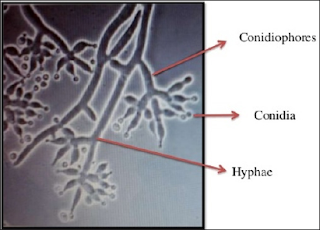
Morphology: Colonies were originally hyaline darkening to white with green tufts in most species. The form was irregular which rapidly grew and merged forming green carpet like appearance. The conidiophores were branched and hyaline. Phialides were divergent and flask-shaped. Conidia were generally green, smooth or roughened, ranged in shape from globose to ellipsoidal, and were produced in slimy heads Conidiophores were highly branched and demonstrated a pyramidal arrangement.
Pathogenecity: Trichoderma harzianum infections are opportunistic and develop in immunocompromised patients, such as neutropenic cases and transplant recipients, as well as patients with chronic renal failure, chronic lung disease, or amyloidosis. Peritonitis, pulmonary, perihepatic, and disseminated infections have so far been reported. A disseminated fungal infection was detected in the postmortem examination of a renal transplant recipient and confirmed in culture. The only other reported infection by this fungus caused peritonitis in a diabetic patient. Trichoderma harzianum were identified as causative agents of opportunistic fungal infections with increasing frequency. Trichoderma harzianum isolates are reported predominantly to cause health problems in humans ranging from localized infections to fatal disseminated diseases. Thus, it is very necessary to delete the negative pathogenic aspects of fungi by using proper treatment and care [14].

Rhizoctonia Solani

Human infections caused by Rhizoctonia are very rare, and first case report of keratitis by Rhizactonia was reported way back in 1977 [15]. Rhizoctonia solani is a most widely recognized strong saprophyte with a great diversity of host plants. It is a first ever case of extensive human mycosis caused by Rhizoctonia solani in a 65-year-old diabetic and hypertensive farmer, with a history of head injury caused by fall of mud wall. Necrotic material collected revealed septate fungal hyphae with bacterial co-infection and after mycological study & diagnosis it was confirmed the presence of rhizoctonia solani [16]. So, in this paper we tested anti-fungal effect of silica nanoparticles against these two fungi i.e. trichoderma harzianum and rhizoctonia solani in PDA media.

Experimental

Materials

Tetraethyl orthosilicate (TEOS) were purchased from Sigma Aldrich Chemicals Pvt Limited (India). Ethanol from Fischer Scientific (India), and Ammonium hydroxide from Qualikems Fine Chemical Pvt. Ltd. (India) was purchased. Potato dextrose agar (PDA) was purchased from HiMedia Laboratories Pvt Limited (India). Instruments-UV 1800 Shimadzu UV spectrophotometer, Malvern instruments-Zetasizer Nano S-90, Shimadzu 8400 spectrophotometer and Bruker D8 Focus X-Ray Diffractometer, Bruker Universal materials tester (CETR UMT-3) SEM-Zeiss microscopy.

Methods

Synthesis of Silica Nanoparticles through Sol-Gel Method

Silica nanoparticles were prepared according to the well- known Stober method by hydrolysis and condensation of tetraethyl-orthosilicate. 8 ml of TEOS was added in a mixture of 100 ml ethanol with 35 ml of DI water. This solution was stirred for 40 minutes and pH was maintained at 10 by adding ammonium hydroxide drop-wise. The synthesis starts with mixing and stirring of the components, requires a reaction time of about 2hr and is finished by centrifugation at 8000 rpm for 5-10 minutes and overnight of drying at temperatures at 100°C and calcinated at 650oC for 2 hours. Same process was used to obtain best particle size value of silica nanoparticle by changing in different parameters like temperature (78°C, 100°C, 650°C) and TEOS (4 ml,8ml,16ml) concentration.

Synthesis of Silica Nanoparticles through Peptization Process

In a typical peptization process, a specific amount of TEOS (8 mL) was added to 100 ml of ethanol and 35 ml of DI water under continuous stirring. After 20 minutes electrolyte i.e., nitric acid or hydrochloric acid or sulfuric acid was added to this solution and obtained silica gel containing salt. This silica gel was washed with DI water to remove salt and obtain a silica wet gel. After that DI water and NaOH was added to maintain pH at 10. Silica gel was formed, then heated for overnight at 150°C.

Antifungal Testing

Dissolved 39 gm of PDA in 1000 ml distilled water. Under stirring this solution was sterilized by autoclaving at 15 psi at 121°C for 15 minutes. Nanoparticles concentration was maintained in the media at 1%. Cooled it to room temperature prior to dispense. The fungi were inoculated on separate petriplates containing sterilized media with 1wt% of nanoparticles. After this placed these petriplates in BOD (Biochemical oxygen demand) chamber. Growth of fungus was observed for 7-10 days.

Characterization

Prepared nanoparticles were suspended in water and particle size was measured by Dynamic Light Scattering using Malvern instruments (Zetasizer Nano S-90). Fourier transform infrared spectroscopy of all prepared nanoparticles was performed with Shimadzu 8400 Spectro-photometer in powder form. X-Ray Diffraction was carried out ofall nanoparticles using Bruker D8 Focus in powder form. UV visible spectrum of prepared nanoparticles have been reported by UV 1800 Shimadzu UV spectrophotometer suspensions form prepared in water and SEM was performed using Zeiss scanning electron microscope in powder form. Antifungal effect was observed in PDA (potato dextrose agar) media.

Results and Discussion

Particle size measurement was carried out through dynamic light scattering. Particle size of prepared silica nanoparticles through sol-gel method was observed as mentioned in Table 1 and particle size of prepared silica nanoparticles through peptization process by using different electrolyte like nitric acid or HCl or H2 SO4 is mentioned in Table 2. Particle size of nano silica at different concentration of precursor and at different temperature up to 650°C was measured. To obtain particle size below 100 nm silica nanoparticle was prepared through two different process- sol gel and peptization process. Through sol-gel process particle size of nano silica was observed 154 nm at 650°C by 8 ml of TEOS that is minimum particle size as compared to 78°C and 100°C because at higher temperature gel structure breaks. After that we changed the concentration of TEOS, firstly reduced by % (4 ml of TEOS),obtained particle size was 260 nm at 650°C.After increasing the concentration of TEOS twice means 16 ml observed the particle size was 85 nm at 650°C.
Figure 2: Synthesis procedure of silica nanoparticle through sol-gel process.
Even silica nanoparticle was prepared through peptization process but obtained particle size was 190 nm, 310 nm and 277 nm using electrolyte nitric acid, HCl and H2SO4. (Figures 1-7) So further for all characterization were proceed with silica nanoparticle prepared through 16 ml of TEOS due to smaller particle size and this particle size shows better anti-fungal effect as compared to others. XRD analysis was performed for silica nanoparticle prepared through 16 ml of TEOS at temperature 78°C, 100°C and 650°C. The X-ray powder diffraction pattern of the nanoparticles were recorded using CuKa (1.5406 Ï) radiation at room temperature in the range 20 to 80° in 2θ scale. Amorphous nature of silica nanoparticle was confirmed by broad peak in XRD analysis at 20=15° (hkl=100) but XRD peak at 650°C was best as compared to 78°C and 100°C as shown in Figures 8 & 9.
Figure 3: Synthesis procedure of silica nanoparticle through peptization process.
In SEM image of silica nanoparticle have shown prepared by 16 ml of TEOS at 650°C. Scanning electron microscopy shows the spherical shape of silica nanoparticles. UV visible analysis was carried out in the region of 200-700 nm prepared by 16 ml of TEOS at 650°C and obtained absorption peak λmaxat 270 nm as shown in Figures 10 & 11. In FTIR analysis, shows the FTIR spectra of silica nanoparticles prepared through sol-gel process using 16 ml of precursor at 650°C. In the spectra of silica, the band around 1070 cm-1corresponds to assymetric stretching vibration of Si-O-Si bond whereas 3300 cm-1 and 1640 cm-1 bands have appeared for H-O-H stretching and bending of absorbed water. Another peak at around 910 cm-1 corresponds to Si-OH bond. Antifungal testing of silica nanoparticle was carried out in potato dextrose media against fungus- trichoderma harzianum and rhizoctonia solani as shown in the Figures 12 & 13. The presence of silica nanoparticle (1wt%) reduced the growth of T harzianum up to 80% as shown in Figure 13(ii). And growth reduction was observed up-to 70% in Rhizoctonia solani as shown in Figure 14. 

Conclusion

We successfully prepared silica nanoparticles using sol-gel synthesis and peptization process. Characterization like DLS, UV spectroscopy, X-ray Diffraction, SEM was successfully performed. Presence of silica nanoparticle shows significant growth reduction of both type of fungus- trichoderma harzianum and rhizoctonia solani. This nanoparticle can be incorporated in various nanocoating for fungal growth reduction. These coatings can be used at various places like hospitals, toilets etc.

Acknowledgment

We thank Prof. D.K. Avasthi, Director of Amity Institute of Nanotechnology, Amity University, Noida, Uttar Pradesh, for his continuous guidance, motivation and providing all facilities. Our sincere thanks to Dr.B.K.Goswami, Dr.Neetu Singh and Archana Singh from Amity Centre for Biomedical & Plant Disease Management for providing all kind of support for this project.

Dual-Resonance Long Period Grating in Fiber Loop Mirror Based Platform for Cheap Biomedical Sample Detection with High Resolution - https://biomedres01.blogspot.com/2020/01/dual-resonance-long-period-grating-in.html

More BJSTR Articles : https://biomedres01.blogspot.com
Assessment and Alleviation of Lumbopelvic Pain and Pelvic Floor Dysfunction

Assessment and Alleviation of Lumbopelvic Pain and Pelvic Floor Dysfunction

Abstract

Because of the time- and labor-intensive nature of the proposed study, a 5-week pilot test consisting of 10 subjects was implemented to determine if protocol modifications were necessary and if the full 60-subject study was warranted.
Abbreviations: Abbreviations: LBP: Low Back Pain; PFD: Pelvic Floor Disorders; PFM: Pelvic Floor Muscles; IRB: Institutional Review Board

Introduction

Low back pain (LBP) is a condition of localized pain to the lumbar spine whose etiology is commonly unknown [1]. Pelvic floor disorders (PFD) occur when the muscles that comprise the pelvic floor fail to properly contract. This can cause urinary incontinence, pelvic organ prolapse, fecal incontinence, or other sensory and emptying abnormalities of the lower urinary and GI tracts [2]. Current evidence shows that individuals with low back pain have a significant decrease in pelvic floor function compared to individuals without LBP [3]. Over 25% of all women and more than a third over the age of 65 experience PFD. Even though it is a physiological problem, the psychosocial impact of PFD can be much more detrimental to the patient's quality of life. Over the next 30 years chronic health problems associated with PFD are projected to increase by 50% due to the increasing numbers of women reaching age 65 [4]. PFD does not typically have one specific cause. Pregnancy/childbirth, age, hormonal changes, obesity, lower UTI, and pelvic surgery are major risk factors. Additionally, anatomical, physiological, genetic, reproductive and lifestyle components are probably PFD developmental influences [1,4].
The pelvic floor forms the inferior border of the abdomino- pelvic cavity [4].
It supports the abdomino-pelvic organs. The pelvic floor muscles (PFM) function as a unit instead of individually contracting. They play an important role in maintaining and increasing intra-abdominal pressure during functional tasks such as lifting, sneezing, coughing, and laughing to prevent urinary and fecal incontinence [3,5]. Men can also have disorders of the pelvic floor, however due the anatomy of the male pelvis, it is less common [6]. Current evidence supports exercise protocol with the common goal of regaining neuromuscular control of the pelvic floor and deep abdominal muscles in a functional matter [7]. There is also strong evidence for PFM training as conservative treatment for stress urinary incontinence [5,8]. Treatment should also include education on healthy lifestyle habits to promote optimal functioning of the lumbopelvic stability system. Examples of these habits include good posture, maintenance of a healthy body weight, proper diet, routine exercise, and refraining from smoking [6]. This purpose of this study was to evaluate whether the implementation of lifestyle modifications as well as a specialized exercise program would improve the symptoms of pelvic floor dysfunction and mild pelvic organ prolapse in women. These symptoms include low back pain, hip pain, pelvic pressure, pelvic pain with intimacy and/or the use of tampons, bladder and/or bowel leakage with laughing, coughing, sneezing, jumping, bladder and/or bowel urgency and frequency. The research addressed whether the interventions proposed lead to improvements in pain levels and quality of life. The study evaluated the severity of the participants' symptoms pre and post intervention (Table 1).

Design

Initially, a five-week pilot study that included 10 female subjects aged 44.1 + 8.4 years (mean + s.d.) was implemented to determine if a study of 60 female participants aged 35-65 years old was warranted and if protocol modifications were necessary The project was approved by the Institutional Review Board (IRB) from the University of New Orleans. The pilot test sample was a convenience sample. The subjects were selected because they exhibited symptoms of pelvic floor dysfunction as defined by the following 3 assessment questionnaires, as follows:
    a) Pelvic Floor Distress Inventory Questionnaire - Short Form 30 (PFDI-SF20)
    b) (assesses if the participant has certain bowel, bladder, or pelvic symptoms and how much they bother the participant.) [9]
    i. Exclusion from study if answer 'yes' on question 3, 4, 6, or 14 (indicating symptoms are too advanced for safe participation)
    ii. Exclusion from study if total score is greater than 200, out of possible score range of 0-300 (indicating symptoms are too advanced for safe participation)
    c) Oswestry Low Back Pain Disability Questionnaire
    d) (measures how participants' back or leg pain is affecting their ability to manage in everyday life.)[10]
    i. Exclusion from study if total score is equal to or greater than 41%, indicating 'severe disability' to 'crippled'
    e) Pelvic Floor Impact Questionnaire - short form 7 (PFIQ-7)
    f) (measures how much bladder, bowel, or vaginal symptoms affect the participant s activities, relationships, and feelings.) [9]
    g) Exclusion from study if 'quite a bit’ is answered in all 3 columns for questions 1 or 2 (indicating symptoms are too advanced for safe participation.
Subjects were excluded from the study if they are assessed to have greater than a 3 finger-width diastasis recti abdominal separation as measured by one of the co-investigators. The subjects were recruited from the greater New Orleans metropolitan area using informational fliers, Facebook video ads, email messages and community postings of the same flier. All subjects signed a letter of informed consent stating that the study was voluntary and confidential and that all results would be kept in a locked environment. Participation was voluntary and low-risk. Exclusion criteria ensured that those with advanced symptoms were referred for medical consultation. All participants had ample opportunity for questions to be answered as needed by the co-investigator clinicians during the live sessions. The subjects were not remunerated.
During the course of the 5-week pilot study one of the participants was dropped from the study due to protocol noncompliance, so only 9 subjects completed the pilot study. The pilot study design included pre- and post- research-validated quality of life assessments [11], In Body 570 body composition measurements [12], pelvic alignment assessments [13], diastasis recti assessments, and manual external pelvic floor muscle activation assessments [14]. Participants were instructed in specific lifestyle modifications and were taught an exercise program over the course of seven live group sessions, 45 minutes each, to be led by one or both of the coinvestigator clinicians. Participants were also instructed to perform a home exercise program at least 5 days per week and complete compliance forms to be turned in at each live group session. The two research clinicians were a licensed physical therapist and a clinical exercise physiologist.

Analysis

IBM SPSS Version 24 statistical programming was utilized and non-parametric, Wilcoxon "related-items" analyses were employed. A 0.05 level of significance was used.

Results

A comparison of the subjects’ results of the pre- and post- values from the 5-week pilot test indicated significant improvements of lower back pain (Oswestry, z(9) = -2.556, p < .05), significant improvements of the quality of life (subjective emotional gauge, PFDI -20, z(9) = -2.666, p < .05), and significant improvements of bladder symptoms (PFIQ - 7, z(9) = -2.536, p < .05).

Discussion

Although the sample of the pilot test was not large enough to eliminate statistical bias, the results warranted a continuation of the study. The full study of 60 female volunteers proceeded as planned with no modifications to the proposed study’s protocol. Researchers are currently recruiting volunteers for the full study.

In the Guidelines for Physicians and Urologists "Watchful Waiting" for Benign Prostatic Hyperplasia (BPH) should be Replaced with Thermobalancing Therapy that Treats Prostate Effectively, Cost-Effectively and Safely - https://biomedres01.blogspot.com/2020/01/in-guidelines-for-physicians-and.html


Tuesday, January 28, 2020

In the Guidelines for Physicians and Urologists "Watchful Waiting" for Benign Prostatic Hyperplasia (BPH) should be Replaced with Thermobalancing Therapy that Treats Prostate Effectively, Cost-Effectively and Safely

In the Guidelines for Physicians and Urologists "Watchful Waiting" for Benign Prostatic Hyperplasia (BPH) should be Replaced with Thermobalancing Therapy that Treats Prostate Effectively, Cost-Effectively and Safely

Abstract

Primary care physician is the first contact for screening for benign prostatic hyperplasia (BPH), conducting timely diagnostic work, and initiating relevant therapy. To protect men with lower urinary tract symptoms (LUTS) due to BPH from severe side effects and complications after standard BPH drugs and surgeries "watchful waiting” was introduced. Innovative Thermobalancing therapy with Dr Allen's therapeutic device, patented in the USA, has demonstrated the effectiveness of treatment of LUTS due to BPH by reducing the size of the enlarged prostate, urination symptoms and improving the quality of life. The results of 10-year follow-up and clinical trials confirm that there are no side effects after using Dr. Allen's device. Therefore, the guidelines for BPH treatment and management should be amended, and "watchful waiting” must be changed to Thermobalancing therapy.
Abbreviations: BPH: Benign Prostatic Hyperplasia; LUTS: Lower Urinary Tract Symptoms; TT: Thermobalancing Therapy; DATD: Dr Allen's Therapeutic Device; AUA: American Urological Association; QoA: Quality of Life; CP: Chronic Prostatitis; CPPS: Chronic Pelvic Pain Syndrome; EAU: European Association of Urology;

Introduction

Figure 1: Prostate volume measured by ultrasound decreased from 45.1 mL to 31.8 mL and the level of urinary symptoms by IPSS decreased from 14.3 to 4.95 in 124 men with BPH after Thermobalancing therapy. In the control group prostate volume increased and urinary symptoms scores worsened. Hence DATD reduces PV and decreases urinary symptoms significantly.
Benign prostatic hyperplasia (BPH) is a common cause of lower urinary tract symptoms (LUTS) in aging men, worsening their quality of life, and physicians are positioned well to screen for BPH and to manage it [1]. Thus, primary care physician has an important role with the identification and early treatment of bothersome urinary symptoms caused by BPH [2]. It is important as the prevalence of LUTS/BPH increases with age, and the burden on the healthcare system and society may increase due to the ageing population [3]. BPH is a nonmalignant enlargement of the prostate, therefore it requires safe treatment to alleviate symptoms, delay disease progression, and lessen the chance of needing surgery for BPH. The results of 10-year follow-up and a clinical trial on Thermobalancing therapy (TT) with Dr Allen's therapeutic device (DATD) in 124 men with BPH have shown its effectiveness to treat urinary symptoms due to prostate enlargement. According to this clinical study, after use of DATD as mono-therapy the decrease in urine symptoms, measured by International Prostate Symptom Score, was accompanied by a significant decrease in the volume of the prostate gland, see Figure 1 [4]. It should be noted that the use of DATD improves of the quality of life (QoL) in observed men. In addition, DATD provides a new safe natural treatment method, patented is the USA [5].

Watchful Waiting

Guidelines for the management of benign prostatic hyperplasia (BPH), according to the European Association of Urology (EAU), American Urological Association (AUA) and urological associations in other countries, includes pharmacotherapy, "watchful waiting”, surgical options and minimally invasive procedures [6]. The main reason for having "watchful waiting” for BPH is to protect men from severe side effects and complications after standard BPH drugs: alpha-blockers and inhibitors of the enzyme 5 alpha-reductase [7]; and surgeries [8]. For many years in "watchful waiting" are commonly used herbal medicines, such as Serenoa repens, Pygeum africanum, Urtica dioica, and dietary supplements and nutraceuticals, such as vitamin D, antioxidants, etc.
Despite widespread consumption, there is limited evidence of health benefits related to nutraceutical or supplement use and some of these products have the potential to produce significant toxicity [9]. Diet and physical activity are now considered important factors affecting prostate health in the aging male, However, whether physical exercise and modifications of dietary habit can really alter the natural history of BPH/LUTS remains to be determined [10]. The aim of this review is to summarize the management of BPH and is designed to aid the generalist with the pertinent information needed to provide excellent care for LUTS due BPH - the most common disorder for aging men.

New Understanding of Etiology and Pathophysiology of BPH

In the past decade, significant progress has been made in understanding the etiology and pathophysiology of BPH. It is becoming increasingly clear that treating the cause of BPH will lead to targeted therapy and will be useful for men who want to take a prophylactic approach. Thermobalancing therapy is based on a new understanding of the Origin of Diseases, which states: all chronic internal diseases have the same root, namely, a pathological activity of capillaries [11]. The development of prostate non-malignant chronic conditions, such as chronic prostatitis/chronic pelvic pain syndrome (CP/CPPS) and prostate enlargement or BPH, are also linked to the changes at the micro-vascular level [12]. The role of vascular dysfunction as possible mechanism of development of BPH leading to prostatic hypoxia, pelvic ischemia and increased pressure in the prostate gland were discussed by different researches [13-15].
According to the new understanding of the cause of BPH, the pathological activity of capillaries is in the core of the development of prostate enlargement, which is based on two functions of small blood vessels, namely constriction and spontaneous expansion of capillaries. Constriction of capillaries in response to different triggers, such as cold, infection and others, creates the micro-focus of hypothermia in the prostate tissue. In order to eliminate this focus of micro-hypothermia, blood flow increases through spontaneous expansion of the capillary network locally. The nervous system does not involve in it, so the expansion can continue and continue. Formation of new capillaries is responsible for the growth of the prostate tissue [16].

DATD, as Safe and Effective Treatment for BPH

DATD facilitates the treatment of the affected prostate by topical application of a special blend of waxes, which can collect the natural radiation of body heat, thus becoming a source of energy. This special wax mixture is called a thermoelement. DATD also consist of an elastic belt that holds the thermoelement in projection of the prostate gland for a long time. Thus, the thermoelement acts as a natural source of heat for the prostate. The neoprene belt keeps the thermoelement tight to the tailbone area and prevents heat disappearance (Figure 2). DATD by spreading energy towards local micro-hypothermia in the prostate tissue terminates it, stopping enlargement gradually. This leads to the elimination of pain and urine symptoms. [17,18].
Figure 2: DATD tightly attaches thermoelement to the coccyx area for a prolonged period of time.

Cost-Effectiveness of DATD

In the past decade, significant progress has been also made in understanding the demographics of BPH. The prevalence of this condition is increasing with the population aging and so does the economic burden, which is comprised of 3 components: direct costs for its treatment, indirect - lost earning and intangible costs - pain and suffering. In a rough estimate, the cost of treating BPH in the United States is $4 billion per year [19]. The investigation in Europe of annual cost of BPH medical treatment was lowest in the UK about $1000 and highest in Poland approximately $1500 [20]. DATD is one-time purchase for $200 that makes TT the most cost-effective therapy and in addition TT significantly improves QoL of men with BPH without side effects.

Summary and Conclusion

"Watchful waiting”, which was introduced about 20 years ago in the guidelines for doctors to protect men with BPH from side events, which follow up the use of conventional BPH drugs and operations. DATD has shown to be effective, safe and cost-effective for men with BPH [21]. TT with DATD is a completely new treatment in the world, therefore, the use of TT with DATD should rely on a strong collaboration between a patient, primary care physician and urologist [22]. Thus, TT and DATD should be recommended as monotherapy for LUTS / BPH, affecting millions of men around the world. In the guidelines for doctors, "watchful waiting” for men with BPH should be replaced with Thermobalancing therapy.

Acknowledgment

I am grateful to Prof Aghajanyan IG, the founder of Armenian Association of Urologists, for conducting the clinical studies on Thermobalancing therapy.


Effect of Histone Deacetylase (HDAC) Inhibitor on Gene Expression in LNCaP-MST and MCF-7 Cells - https://biomedres01.blogspot.com/2020/01/effect-of-histone-deacetylase-hdac.html

More BJSTR Articles : https://biomedres01.blogspot.com

Dual-Resonance Long Period Grating in Fiber Loop Mirror Based Platform for Cheap Biomedical Sample Detection with High Resolution

Dual-Resonance Long Period Grating in Fiber Loop Mirror Based Platform for Cheap Biomedical Sample Detection with High Resolution

Abstract

A dual-resonance long period grating (DR-LPGJ embedded in fiber loop mirror (FLM) was employed as a sample for the development of the biosensor application. The DR-LPGJ acts as a sensing part for biological molecules detection, whereas FLM enhances the rapid signal monitoring, which oversees the wavelength filtering properties. Herein, the DR-LPG surface was functionalized with biotin in terms of the avidin binding reaction detection. The detection mechanism is based on the alteration the refractive index of the DR-LPG surrounding monitored by the tracking interference dips changing. Moreover, by applying a broadband diode and photodetector we ensured the low-cost of the system.
Abbreviations: DR-LPG: Dual- Resonance Long Period Grating; FLM: Fiber Loop Mirror; LPG: Long Period Grating; RI: Refractive Index; APTES: Amino Propyl Tri Ethoxy Silane; EDC: 1-Ethyl-3-(3-DimethylaminopropylJCarbodiimide; PBS: Phosphate Buffer Saline; LOF: Lab-On-a-Fiber

Introduction

An optical fiber based sensor are very useful in many applications because of the compact size, electrical interference independence and suitability for remote sensing [1]. Recently, a significant effort has been devoted for the long period grating (LPGJ - based sensors for medical application [2,3]. The real-time detection, high sensitivity and accuracy are desire properties for the medical sensors in terms of the facilitate prompt disease diagnosis [3]. In spite of the high sensitivity of the LPG, the main asset is possibilities of modifying the optical structure with thin functional coatings in terms of facilitating highly selective and fast measurement in real time with the reported resolution of refractive index (RIJ change up to 10-6 [4]. To detect any molecular elements, the LPG surface needs to be functionalized by e. g. enzymes [5], gold nanoparticles [6], antibodies [7] or cross- linking bioreceptors [8]. The LPG is an optical periodic structure, where transmission spectrum consists of the resonance dips at discrete wavelength [9]. When it comes to achieve very sensitive structure, the LPG should work close to the turning point, where the dual-resonance appears.
Such dual- resonance long period grating (DR-LPGJ has been shown to be ultra-sensitive [10] regarding the micro-scale label- free detection [11]. The detailed theory of the DR-LPG structure can be found in [12,13]. To satisfy the need for very accuracy molecules detection and to maintain a stable temperature of the measurement, the DR-LPG can be combining with other optical elements [14]. Among various solution, the fiber loop mirror (FLMJ provides several advantages, including high extinction ratio, and low cost of final arrangement. The part of high birefringence fiber (Hi-Bi fiber] from FLM structure ensures ambient temperature changes controlling by the interference dips shifting/amplitude intensity changing observation. Moreover due to filtering properties of the FLM, it provides possibilities of control of the wavelength spacing between interference dips, limitation of the temperature and strain cross- sensitivities and consequently enhancement of the sensitivity of the specific sample detection [15].In this paper, for the first time of our knowledge, we present the combined structure of the DR-LPG in the FLM for biotin-avidin reaction as a potential platform for medical application.
In particular, the described optical platform can be applied as a sensor for limitless biomarkers or molecules, similarly to LPG which was already used to detect, e.g. bacteria [16]. It is possible due to the ultra-high sensitivity towards a refractive index of surrounding environment. Moreover, the introduction of a reception layer to the sensing system ensures its selectivity exclusively towards the desired analyte.
Here, we present a proof of concept of the application of our sensing platform by applying the well-known biological model, biotin-avidin complex. Esmond Snell discovered the biotin- avidin interaction during the investigation on "egg-white injury" disorder in 1941 [17]. Biotin calls also vitamin H, it is an crucial compound taking part in the metabolism of fatty acids and amino acids. The protein, avidin from raw egg white has capability to bind biotin causing its deficiency in mammalian bodies. The avidin- biotin binding belongs to the most robust noncovalent biological interaction with a dissociation constant 10-15 [18]. In the presented system, biotin is applied as a receptor, immobilized covalently to the surface, able to selectively capture of avidin dissolved in the solution.

Methods

The Experimental Set-Up

The DR-LPG was produced using amplitude mask technique, where the part of bare fiber was exposed on UV-radiation using KrF Excimer laser (Lumonics™ Lasers: Pulse Master ®-840) emitting at 248nm. The period of the used DR-LPG was 217 μm. The experimental platform used for monitoring biotin-avidin reaction is shown in Figure 1a and has been in details described in [19]. The transmission spectra of proposed platform are shown in Figure 1b, where the DR-LPG in FLM spectrum was adjust by a polarization controller in terms of maximum set-up response.
Figure 1: The scheme of the experimental platform for biotin-avidin interaction (a) and transmission spectra of DR-LPG (blue line) and DR-LPG in FLM (grey line) (b).

Receptor Immobilization Procedure

The DR-LPG surface was cleaned from organic impurities by soaking in hydrochloric acid (aq.)/methanol (1:1, v/v) mixture and next in concentrated sulfuric acid for 30 min, followed by plentiful rinsing with water. After that, the platform was dried in vacuum for 15 min and transferred for the aminization process. The DR-LPG was locked into the desiccator chamber together with trays of 30 μl of 3-aminopropyltriethoxysilane (APTES) and 10 μl of triethylamine for 30 min and/or 2h in the argon atmosphere [20]. In the next step, the biotins carboxylic group was activated by EDC/NHS to form an amide bond with amine groups of the APTES covered surface. For this, the biotin powder was dissolved in the mixture of water and dimethylformamide (1:1, v/v) containing 0.8 M 1-ethyl-3- (3-dimethylaminopropyl)carbodiimide hydrochloride (EDC) and 60 mM N-hydroxysuccinimide (NHS). The final concentration of biotin was equal 1 mg ml-1. Next, the APTES covered platform was immersed in the prepared solution for 1 hour in a sealed chamber. After this time, DR-LPG was rinsed with water and exposed to avidin for 30 min. Avidin solution was prepared by it dissolving in phosphate buffer saline (PBS) pH = 7.4 to obtain concentration equal 1 mg ml-1. Figure 2 shows the scheme of functionalised DR- LPG surface with a biotin-avidin interaction.
Figure 2: The scheme of the DR-LPG functionalised with a biotin (orange square) interacting with avidin (blue cross) from the solution: a) the whole platform, b) the cross-section of the platform.

Results

All presented measurement data was performed in a fresh PBS imitating human blood serum, and every step of functionalization process was followed by the DR-LPG excessive wash to remove weakly bonded biomolecules. This procedure allowed to obtain a reliable response. Moreover, to maintain stable physical conditions the temperature was monitored, and the DR-LPG was kept in a constant volume of PBS. The introduction of the sensor into a narrow tube allowed to control and suppressed unwanted solution evaporation. The spectrum was recorded three times in PBS. Firstly, after the surface sensor silanization process (Figure 3J, light blue lineJ. Secondly, after the biotin covalently attachment to the sensor (dark blue lineJ. And thirdly, afterword the sensor interaction with avidin (grey lineJ. The red dashed lines in Figure 3 mark the notches of the DR-LPG which corresponds with those from Figure 1b and only these minima are considered in regards of sensing properties of the device. When the ambient refractive index varying, the DR- LPG notches (around 1530 nm and 1615 nmJ shifting toward each other, and hence, the amplitude of the interference dips of the DR- LPG in FLM is changed.

Conclusion


We presented a potential application of the DR-LPG in FLM platform for label-free biochemical detection. The proposed solution fits squarely into technology named Lab-On-a-Fiber (LOFJ (more specifically Lab-around-FiberJ, which is dedicated to devices focused on the design and development of advanced fiber optic nanoprobes for biological applications. Hence, the LOF enables continuous monitor of detected samples. Adapting FLM in the sensor probe (DR-LPGJ influences on avoiding thermal and strain cross- sensitives, and this limitation dealing with pure unconventional light matter interaction in confined small-scale volumes. Despite of many asset of the FLM configuration, it ensures relatively high sensitivity due to the phase shift keying. Finally, throughout adoption of the laser diode, (broadband light source) photodetector and the commercially available optical passive elements with the telecommunication standards, the DR-LPG in the FLM configuration provides the low- cost usable sensor implementation. Therefore, we can conclude that proposed platform can monitor the biotin- avidin interaction and is good candidate to some other biological sample detection as a medical application.

Construct of Streptomyces Scabies Knock-Out Mutant of TXT Biosynthetic Gene txtB Via Intergeneric Conjugation - https://biomedres01.blogspot.com/2020/01/construct-of-streptomyces-scabies-knock.html

More BJSTR Articles : https://biomedres01.blogspot.com


Monday, January 27, 2020

Origin of Asexual Reproduction in Hydra

Origin of Asexual Reproduction in Hydra

 Introduction

Hydra's buds develop through the integrated activity of two different types of cells:
    a) Epithelial (aka epithelial-muscular) cells forming the didermic body wall (with extension into tentacles, hypostome, and foot) and
    b) Interstitial cells (aka amoeboid or basal cells) differentiating as cnidocytes (aka nematocytes, the cells that make cnidocysts, aka nematocysts), nerve, gland, and sex cells [1-3].
Over the years, I consolidated some ideas and data into a theory about how these different kinds of cells came to cooperate in budding [4-8]. I proposed that early in the Neoproterozoic Era a primitive two layered epithelial mat (resembling contemporary Placozoa [9-10]), was infected by amoeboid cells already equipped with an extrusion apparatus. The epithelia's attempt to reject the foreign cells failed but a symbiogenic relationship evolved and the duel system found a selective advantage in modified ejection. Ultimately, a mechanism for removing excess amoeboid cells was adapted for the production of buds. The adaptation hinged on three conditions:
    i. Amoeboid cells equipped with an extrusion apparatus were the ancestors of hydra's interstitial cells.
    ii. Hydra routinely produced excess cells that moved toward and accumulate in the budding region.
    iii. Excess cells form discrete modules that erupt as buds and are then "ejected."
a) Amoeboid cells equipped with an extrusion apparatus were the ancestors of hydra’s interstitial cells. I am hardly the first to point to the presence of stinging apparatuses in protozoans and to similarities between protozoan "cnidocysts" and cnidarian cnidocysts: the "peduncle," "rhizoid," and "perforator" cnidocysts of dinoflagellates [11-14], the trichocysts of trypanosomes [15], zooflagellates [16] and mastigophorans [17], the "apicoplasts" (apical complexes) of "Sporozoa," the "polaroplast," of microsporidians [18], and the "polar capsules" of myxosporidians [19-25]. Jiri Lom, the distinguished Czech protozoologist and parasitologist, suggested that these "homologies [were] perhaps too close to be considered only a convergency phenomenon" [26], and Pierre Tardent, the renowned Swiss coelenterologist commented "The wheel didn't have to reinvent itself" [27], i.e., cnidrians didn’t have to invent cnidocysts. It is a small step to extend Lynn Margulis' hypothesis of endosymbiosis [28] from mitochondria and chloroplasts to cnidocysts. Ancient eukaryotic amoeba could have been infected by monerans (presumably bacteria) already equipped with an eversion apparatus. The "guest” would then introduce genes to the "host" through unilateral horizontal gene transfer, and a permanent extrusion apparatus (a cnidocyst) would have evolved in the amoeba. What followed was a different sort of symbiosis -symbiogeny - the merging and mutual evolution of eukaryotes [5-6]. In the case of cnidarians, at some point in the evolution of multicellular eukaryotic life, probably prior to the Vendian Period 700 million years ago, an amoeboid protoctistan (to use Margulis' term [29], aka protozoan) already equipped with an extrusion apparatus gained access to a primitive didermic metazoan mat either by invading or being ingested. I suggest that, at the time, mechanisms for isolating different forms of metazoan life were not as restrictive as they became since and the efforts of the epithelia to reject the amoeba were feeble.
Thus, the protoctistan wound up sequestered in the primitive epithelium, and the two entered a symbiogenic relationship in which each symbiont evolved to mutual advantage. Inevitably, cooperation gave rise to the rudiments of the phylum Cnidaria. "Are cnidarians composite metazoans... metazoan chimeras" [30]? Did what begin as a protoctistan equipped with a cnidocyst - whether trespasser, guest, foreigner, invader, colonizer, or ingested prey - become Cnidaria’s interstitial cells, while the epithelial mat -host, victim, or predator -evolved into the cnidarian epithelia Had that happened, interactions between the two could have led to the further evolution of nerve and gland cells from amoeboid descendants, while the cellular mat could have evolved into the cnidarian body wall's epidermis and gastrodermis, tentacles with epithelial battery cells, hypostome, peduncle, and adhesive foot.
b) Hydra routinely produces excess cells that move toward and accumulate in the budding region. One of Hydra's attraction to biologists is that under optimal laboratory conditions, hydra cultures expand exponentially. The cell populations also expand exponentially [31-34]. Since the density of hydra’s cell populations is constant, except for transient increases in the budding region, hydras would seem to produce and get rid of excess cells. Rather than treating these cells as waste, however, symbiogeny capitalized on them by adapting them for asexual reproduction through budding. The movement of cells toward the budding region is well documented. Richard Campbell [31-35] calculated, that in H. littoralis, under optimal laboratory conditions, 85-86% of hydra's structural cells produced throughout the body cylinder (gastric, budding, and upper peduncle regions) migrate to the budding region. Similarly, in H. viridis, about 800 of a thousand parental gastrodermal [digestive] cells move to the budding region per day [34].
Likewise, interstitial cells and their differentiation intermediates make up more than half of all the cells in the adult animal moving to the budding region [36]. The remainder of a hydra's daily cellular production is lost at the animal's extremities, tentacles and foot. All the excess cells dedicated to budding are produced along the length of hydra’s body wall [31-37] in species- specific patterns of cell division. Paul Brien discovered "La zone de croissance sous hypostomiale" (Brien’s sub-hypostomal growth zone) in Hydra fusca [38], and Allison Burnett extended Brien's growth zone in H. viridis and H. pseudoligactis (H. canadensis) to the gastric and budding regions [39] where a high frequency of mitotic figures is also found. Campbell showed that in H. littoralis, a distal zone of elevated mitotic activity appears among epidermal epitheliomuscular cells (a.k.a. "Ectodermal epithelial cells") and gastrodermal gland cells (a.k.a. "Endodermal gland cells"), but cell proliferation peaks in the budding region for interstitial cells ("Ectodermal interstitial cells," a.k.a. basal cells, amoeboid cells) and gastrodermal epitheliomuscular cells (a.k.a. "Endodermal epithelial cells") [33, 35, 40-42].
Measured in mitotic figures and in the incorporation of tritiated thymidine, the epidermis supports a higher rate of cell division than the gastrodermis [33]and labeled epidermal epitheliomuscular cells move toward the budding region faster than gastrodermal digestive cells [32-34]. Whatever the cell type, and wherever along the body column cells are produced (i.e., both above and below the budding region) they converge on the budding region [33-35,40-43]. The mesoglea situated between the epidermis and gastrodermis is a substratum for cell movement rather than a glue holding the two epithelial layers together. Epithelial-muscle cells, with longitudinal muscle extensions seem to actively crawl on the mesoglea with the help of their muscle processes [44] and are seen to migrate over experimentally denuded mesoglea [45]. In contrast, the gastrodermal epithelial-muscle (digestive) cells with circular muscle extensions seem to become compressed and crowded into the budding region [46].
c) Excess cells form discrete modules that erupt as buds and are then "ejected." Neither the budding region nor buds are distinctive fountains of proliferating cells, meristems, or blastemata. Likewise, in H. viridis, the frequencies of mitotic figures in early buds lacking tentacles (stage I) and buds with tentacle rudiments (stage II) "could not be distinguished." Given the absence of mitotic figures in the hypostome, the "number of mitotic figures on the bud proper at the later stage" (stage III) is below that on the parent [32]. Moreover, cell divisions proceed at the same rate in freshly detached buds, during the initial growth period, and in budding animals [32,34,47-51]. The distinguishing characteristic of the budding region is the local production of new mesogleal components [52-54]. Indeed, "[a]t sites of tissue evagination... the mesoglea was dramatically remodeled and epithelial cells moved relative to the mesoglea" [43].
Thus, "no loss of ECM [i.e., extracellular material of the mesoglea] occurs before the time of bud emergence. Rather, the ECM is continuous at the sites of bud formation and what occurs is simply an increase in the expression of. [mesogleal components] as evagination of the bud progresses.. Before evagination of the bud occurs. upregulation of at least. [one mesogleal components] has already occurred. High expression of both basement membrane and interstitial matrix components occurs throughout all stages of bud formation" [54].
Hydra's excess cells funneled into the budding region form discrete bud modules that break with parental symmetry, jut outward, form a hypostome, tentacles, body cylinder, and feet, and ultimately detach as buds [8, 30-35]. In transgenic H. vulgaris [43] and grafted H. viridis [55], cells are literally seen moving out onto buds. "[C]ells located near the evaginating centre will end up in the oral/distal part of the bud; those located more distantly will move to a more aboral/proximal part of the bud" [46]. The further "[e] longation of the early bud is driven by recruitment of epithelial tissue from the mother polyp into the newly forming protrusion" [46]. Interstitial cells may play a special role in bud modules, since hydras partially or fully (?) deprived of interstitial cells, so-called "epithelial animals," have difficulty budding. Hydras’ interstitial cell population is reduced or eliminated in a variety of ways: treatment with colchicine, nitrogen mustard (NM), hydroxyurea, urethane, and lowered temperature [56-62]. Treated hydras suffer addition losses beyond interstitial cells: Specialized nerve and gland cells disappear, and the hydras neither move, capture prey, or ingest them. They do not restore the missing cells either. If they survive the initial treatment, "epithelial animals" frequently die from bacterial infections of slowly healing wounds inadvertently inflicted during forced feeding and evacuation. Surviving hydras (one in twenty) may enlarge, especially in their peduncle, and add thin supernumerary tentacles [56].
Photographs of "epithelial animals" show bloated hydras with stubby tentacles [56,58]. Like starved animals [63-66], "epithelial animals" bud initially, and they are capable of regenerating thin tentacles lacking cnidocysts, but without feeding the animals shrink in size [58]. Interstitial cell populations can be restored, however, in "epithelial animals" [67] and in clones of reaggregated cells from NM-treated hydras [68-70] through grafting with normal tissue. Along with the missing interstitial cells and cell linages [70], including eggs [71] and sperm [72], the hydras re-acquire normal morphology and behavior.
Under optimal laboratory conditions, hydras reach an equilibrium (a steady state) at which body size is constant and the rate of cell production is balanced by the rate of cell loss through budding (everything else being equal such as cell loss on tentacles and foot). At equilibrium, excess cells move to the budding region, join bud modules, and move off the parental body column into developing buds [30-41]. In contrast, starved hydras and "epithelial animals," deprived of interstitial cells and their products only produce buds initially while shrinking [64-66] and then cease budding. A residue of bud modules would seem to be fully determined at the initiation of starvation and interstitial cell destruction (albeit foot cells involved in detachment may be defective in "epithelial animals"). But starved animals resume budding when feeding is resumed, and "epithelial animals" resume budding when interstitial cells are reintroduced [67-72].
The failure of starved animals to continue budding is easily explained by the failure of these animals to fill bud modules, but the absence of budding in "epithelial animals" (deprived of interstitial cells and their products) suggests that epithelia alone are incapable of rejecting cells in buds. Thus, hydra's epithelia need a dose of interstitial cells to reject cells in buds. Interstitial cells would seem to provide an essential component of bud modules required for cell rejection or a trigger for the eruption of a bud from its module. The premise that budding evolved from a primitive epithelia's attempt to reject foreign amoeboid cells is consistent with these observations. Symbiogeny's constructive and creative roles in evolution might have modified cellular rejection into budding given budding's selective advantage [3-8]. "Natural selection... is not [after all] the only force governing evolution, nor had Darwin ever suggested that it was" [75]. The evolution of eukaryotes following the capture of mitochondria and chloroplasts certainly justifies Lynn Margulis’ claims for the creative consequences of endosymbiosis [28-29]. Likewise, the creative power of symbiogeny, is implicit in the comparison between pond amoeba and blood-borne magakaryocytes. The evolution of budding from the rejection of amoeboid cells by a primordial epithelial mat at the beginning of hydra's evolutionary history would seem another example of how creativity is captured by natural selection [5,73-74].

Liquid- Liquid Extraction of Zinc(II) From Acid Media with N-N-Heptylaniline as an Extractant: Analysis of Pharmaceutical and Commercial Sample - https://biomedres01.blogspot.com/2020/01/liquid-liquid-extraction-of-zincii-from.html

More BJSTR Articles : https://biomedres01.blogspot.com

Discrepancies of Pediatric Data of BNT162b2 mRNA Covid-19 Vaccine in the Assessment Report of the European Medicines Agency

  Discrepancies of Pediatric Data of BNT162b2 mRNA Covid-19 Vaccine in the Assessment Report of the European Medicines Agency Clinical Effic...