Tuesday, November 30, 2021

Ameliorative Role of Composite Flour Against Human Maladies

Ameliorative Role of Composite Flour Against Human Maladies

Introduction

Cereals as crops are grown throughout the temperate and tropical regions of the world and full fil the approximately fifty percent food energy requirements of population. Cereals in form of wheat, rice, maize, barley are significant source of minerals and bioactive compounds to dire human requirements. Cereals are mostly used as staple food in different regions of the world. Their grains mainly consist of three cereal grain consists of three main parts such as the endosperm (germinating seed), the germ (production of new plants), and the bran (provide protection to the grain). While, their concentrations are varied in different cereals grains with same general pattern. Wheat is the major crops after barley and oat among the cereals cultivated in the European countries and mostly consumed as staple foods [1]. Among cereals, wheat (Triticum aestivum) belongs to family gramineae and is considered second only to rice as the main human food crop. Commercially, Triticum aestivum ulgarev and Triticum turgidum durum are of most importance. It is one of substantial crop and cultivated approximately on 17% of the total land worldwide. Likewise, wheat grain is comprised of different types of tissues such as embryo portion (germ), endosperm which is fully covered by starch grains, aleurone layer (packed endosperm), and the pericarp [2].

Due to the presence of nutrients and phytochemicals, wheat become more important for the nutraceutical and functional properties. After milling, wheat has been used to prepare diverse food products such as bread for the supply of nutrients to human after milling. Wheat straw is also used as animal feed. Functional characteristics of wheat are influenced by composition, grain hardness and protein contents. Additionally, wheat also exhibits some anti-nutrional effects owing to the existence of antinutrients i.e. lectin and gluten that decrease the dysfunction and diseases in humans [3,4]. Environmental and climatic factors including temperature, humidity, topography, soil etc. affect the quality and yield of wheat productions. In industrial milling, bran is the by-product of wheat and significant nutritional profiling, health endorsing perspectives and further is also used to prepare multifarious food items [5]. Wheat is also prominent source of carbohydrates, protein, and fibers. The pericarp contains protein content (10%) whereas germ and aleurone layer comprises of 30% of the total protein. Likewise, endosperm contributes approximately 74% protein contents. In conclusion, aleurone and germ layers, endosperm relatively high protein contents [6].

Barley (Hordeum vulgare L.) is one of the most ancient cereal crops grown in the world today ranking fourth among cereal grains after wheat, rice, and maize. Barley is used as animal feed (65%), malting (33%), and human consumption (2%). It also prevents from cardiovascular disorder via decreasing the cholesterol concentrations and improving the glucose tolerance [7,8]. Mechanistically, barley flour markedly lowers the bile acids absorption, eliminates steroids, increases catabolism of cholesterol, reduces lipoprotein cholesterol secretion, enhances secretion of bile acids, and reduces the total body pool of cholesterol [9].

Chemical Composition

Considering wheat composition, wheat caryopsis is consisted of (14-16%) outer branny husk of the grain, (starch: 81-84%) central endosperm, and embryo portion (2-3%), respectively. Separation of endosperm from the embryo and bran layers are mainly done by conventional milling of wheat grains [2]. Barley flour contains higher amount of soluble dietary fibers especially ß-glucans, arabinoxylans and pectin. Barley grains is composed of higher quantity of ß-glucans as compared to other cereals. The ß-glucan contents vary from 10.89-19.7 g/100g on dry wt basis whereas this increased level is significantly used in food, feed and industrial applications [10].

The chickpea (Cicer arietinum L.) is cultivated in different regions of the world as important grain legume crop. It is also promising source of starch as major carbohydrates which is approximately 83.9% of the total carbohydrate. Chickpea seeds also consist of (21.70-23.40%) protein, and (41.10-47.42 %) carbohydrates. It also has high protein digestibility, consists of complex carbohydrates (low glycaemic index), enriches with minerals and vitamins. Chickpea flour is also free from anti-nutritional factors [11,12]. Additionally, it is also promising source of carbohydrates, minerals, proteins and vitamins and has significant role in daily nutrients intake of human. The carbohydrates and protein approximately constitute the 80% of the total dry seed mass. Moreover, dietary fiber in chickpea significantly lower the cholesterol level and also contain a variety of Anti-Nutritional Factors (ANF) including amylase and protease inhibitors [13].

Maize also known as Zea mays L. is an important Pakistani crop and it is the 3rd most cultivated crop pf Pakistan after wheat and rice. It is cultivated over a huge area in Pakistan. Mostly, maize is cultivated and produced in Punjab and Khaiber Pakhtunkhwa. Maize is consumed in different forms like in food grain form and used in different food products. It is also significantly use to form forage for utilization in livestock and poultry. It is very important as a commercial crop and contribute raw material to various industries in order to produce maize starch, maize syrup, maize oil, dextrose and maize flakes. The grain of maize constitute starch in the amount of 72%, protein in the amount of 10%, oil in the amount of 4.8%, fibre in the amount of 5.8 %, sugar in the amount of 3.0% and ash in the amount of 1.7%. In Pakistan, its area under cultivation is 992 thousand hectares and annual production of grain is grain 751 thousand tons [14].

Oat (Avena sativa) has multifunctional features and nutritional profile due to presence of components. It is a rich source of dietary fiber particularly minerals, b-glucan, and nutrients. Oats are very beneficial in the cure of diabetes mellitus and various cardiovascular diseases. The bran of oat is a rich source of vitamin B family, fat, minerals and protein, except b-glucan which is supposed to be beneficial fiber for the health of heart. This b-glucan has exceptional therapeutic properties and it is significantly important nutrient in human diet and nutrition. Diverse effects of b-glucan in terms of physiology are connected to viscosity, insulin responses, lowering the plasma glucose and enabling high movement of bile acids to lower intestine and cause high excretion of bile acid thus lowering the cholesterol level. Furthermore, it is beneficial in the treatment of celiac disease. The consumption of oat grains and oat brans in daily diet beneficial both as nutritionally and also used as therapy against many diseases [15].

Nutritional Aspects

Cereals belong to the family Gramineae and grown for their highly nutritious edible part or grain and frequently referred as grains [16]. Cereals have been consumed directly as staple foods and indirectly as feed for livestock long ago. Cereals are considered as important food sources, and foods made of cereals are supposed to be a primary energy source, vitamin B and protein, minerals for the population globally. In general, cereals are basically economical to harvest, can store and transport easily, if preserved dry it does not deteriorate readily. Cereals are used by humans since long from prehistoric times. Cereals are rich sources of macro and micronutrients and consuming high amount of cereals has significant impact by decreasing the risk of many diseases [17].

Cereals are rich source of carbohydrates and often known with the same name, because cereals consist of almost 75% carbohydrates. Starches being the chief part of cereals present in endosperm in starch granules. Starch granules contain different ratio of amylose to amylopectin which depends on the type of cereal and its variety. In general cereal varieties, amylose starch is present in the range of 25-27% although in many varieties of waxy (corn and rice) amylopectin is the chief starch present. Cereals composed of proteins in the range of 6-15% [18]. Wheat contain main storage proteins in the form of glutenins gliadins whereas rice contain glutelin (oryzenin) and maize contain prolamin (zein) while barley contain glutelins and hordeins while oats contain albumin and globulins [19]. Cereals also contribute significant quantity of good quality amino acids that are building blocks of proteins, while some amino acids are limited. Diet must provide essential amino acids and from these amino acids human body can make other nonessential amino acids. Limiting amino acids are those amino acids that are present in amount less than that is required. In case of cereals, lysine is the limiting amino acid while in rye the first limiting amino acid is the tryptophan. In rice, rye, barley and other high lysine cultivars like maize, sorghum contains favourable composition of essential amino acid. Mixture of cereals and plantbased foods like beans and rice can make up for such amino acids (limiting). Cereals contain minute quantity of lipids in the range of 1- 3% in barley, rye, wheat and rice, while oats contain 5-10% and corn contain 5-9%. This lipid is richest source of linoleic acid [20].

Cereals also contain vitamins and minerals. Cereals can also contribute to the intake of vitamin and mineral; though the content of micronutrient depends on the presence of amount of endosperm, bran and germ. Cereals do not contain vitamin B12, vitamin A & C, and no beta carotene despite of yellow corn. Though, cereals are source of significant amount of most B vitamin, particularly thiamine, niacin and riboflavin and contain appreciable amount of vitamin E. Cereals also contain minerals and are a rich source of potassium but are low in sodium like many plant foods. Whole grain cereals also comprise of iron, zinc and magnesium and minute quantity of numerous trace elements like selenium. Amongst the cereal grains, rice contributes highest level of selenium and provides about 10-13mg per 100g (Table 1) [21].

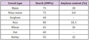
Table 1: Starch composition of cereals.

Milling of Cereals

Milling process is the process of grinding in which grind the grain into flour and significant step in post-production of grain. This technique is used to eliminate the husk, bran layers, and then produce an edible portion in form of powder with appropriate grain size. During milling, essential bioactive compounds are affected with minor alteration [22]. Categorically, there are two types of milling one is dry milling (separation of bran from the endosperm) and other is roller milling (separation of bran from wheat grain), respectively. the grains are subjected to tempering process by spraying of water to retain moisture and then shifted to tempering bin for conditioning before milling. Pericarp and germ portion of grains absorb water to soft the endosperm for extraction purposes. In addition, it also stops the breaking of bran during separation. The conditioned grains are shifted via metal rolls rotating in opposite direction during roller milling. Afterwards, the grains are transferred into three major portions such as bran, germ, and endosperm [23,24].

Regarding polyphenols, they are present approximately 20% of total phenols in bran portion of grains. The higher antioxidant activity of bran portion is due to the presence of phenolic compounds. The antioxidant compounds are significantly present in different concentrations in different parts of the wheat grains. Temperature considerably affected the rate of particle size reduction, granulometric particle distribution, and dissociation of diverse bran layers during grinding [25]. Bran layers exhibited the thermomechanical properties due to the presence of lipid compounds in the cuticles. On other side, bran was brittle, fragmented and then rapidly lowered the particle size whereas at low temperature, disruption of bran layers is low. The conversion of faster fragmentation of bran into fine particles and composite particles were done after cryogenic milling. Bran of cereals also work as an elastoplastic due to their higher high plasticity and extensibility of inner bran layers. Different bran layers also show different extensibility at ambient temperature and generated the wide range of particle sizes. The increased antioxidant potential is linked with presence of hydroxyl group of phenolic acids [26].

The replacement of wheat flour with defatted cashew kernel flour significantly enhance the fiber, fat, ash and protein contents, and energy value in the composite flour whilst lowered the carbohydrates and water contents, respectively [27]. In another study done by Igbabul et al. [28], they evaluated that product was prepared by wheat, plantain and soy in which higher protein contents significantly affect the rheological properties of bread. A peer group of researchers Méité et al. [29], they found that preparation of composite flour by mixing of wheat grains with soya bean in resulting enhanced the lipid content.

Water is an important criterion to evaluate the water quality, texture, and taste in various food products; they substituted the wheat flour with yam and vanilla flour. The low moisture contents in the flour main prevented from the growth of mico-organims and protected from the deteriorative quality of the product. In addition, low water contents enhanced the shelf life of the product [30]. Likewise, supplementation of cashed flour in wheat flour significantly increased the ash and fiber contents. The enhanced contents exert health endorsing functions such as facilitation in digestion process and prevention from the constipation. Composite flour also play role in preventing of cancer, diabetes, and cardiovascular diseases [31]. A study conducted by Brou et al. [32], they explored that substitution of cereal and legumes flours momentously enhanced the oil and water absorption potential. The enhanced water holding capacity were occurred due to higher amino acids, protein contents, conformational characteristics, and degree of interaction with water, respectively [32]. Therefore, higher water holding capacity leads to better quality of bread or chapattis by enhancing or improving the freshness of the product. In food industry, oil is absorbed in the flour through capillary process and thus acts as mouth feel and flavor enhancer [33]. Moreover, Diallo et al. [34] reported the oil absorption capacity of formulations in bread. The density of composite flour is decreased with enhanced substitution rate. Likewise, Giami [35] found that decreased density of flours linked with good formulation in infant foods. Moreover, group of researchers [36] investigated that supplementation of Treculia africana flour into wheat flour lowered the density whilst composite flours exhibited the higher mineral contents as compared to wheat flour.

Composite Flour

Composite flour is different from the ready-made mixed flour which is used in bakery products and by millers contain nonperishable constituents whereas the composite flour is the mixture of different flours of vegetables, wheat or non-wheat flour, rich in protein and starch which can be used for the production of leavened products breads, pastas, porridges, unleavened baked products, snack foods. Sometimes, only flour is used as replacement-for example, tortillas and wheat-less bread from sorghum, pastas from sorghum or maize [37]. It can be defined as combination of flours such as tubers enriched with starch (sweet potato, yam, cassava), cereals (barley, rice, buckwheat, millet and maize), protein rich flours (peanut and soy), and non-wheat flour, respectively [38]. Composite flour is promising and significant source of different acidic phenolic compounds such as ferulic acid, benzoic acid, sinapic acid, diferulic acids, and p- coumaric acid subsidiaries. Ferulic acid is the significant compound in tested grains with free, bound and dissolvable conjugated form. These compounds are present as bound phenolics and endure stomach and intestinal absorption to achieve the colon. It is also comprising of fibers, starches, proteins, B-complex nutrients, minerals i.e. Se, Zn, Mn, Fe, Mg, K, and P, respectively. Moreover, β-glucan and arabinoxylan fibers are linked with reduction in blood glucose level, coronary heart maladies, blood cholesterol, dyslipidemia, cardiovascular sicknesses, glycemic control, obstruction weight control, and ceaseless diseases [39].

Role of Composite Flours in Food Industry

Composite flour is the combination of different protein enriched foods and starches. The mostly used starches and protein to prepare the composite flour are jam, sweet potatoes as well as also peanut and soy, respectively. Different cereal and pulses are used to make composite flour including rice, millet, barley, maize, wheat, chickpea, and corn [37]. In recent years, consumers are diverted their attention towards ready-to-eat snacks due to lack of time and changes in lifestyle and eating habits. The production of composite flours to give a chance to the producers to attain and support their crops. Changes in pattern of life styles and shifting of large population to urban areas lead to enhance the consumption of flours to prepare the bread and other bakery commodities [40]. In developing economies, Oyeku et al. [41] documented that blended flours are good in nutrition and make them more economic due to presence of cheap ingredients. This phenomenon caused reduction in prices and are readily available. The supplementation of 30% legumes in composite flour significantly improve the quantity and quality of nutritious proteins that potential impact on nutritional status and economy of the country [42].

Composite flours play significant role by replacing wheat flour and are more economical by decreasing the wheat imports. This flour has role in confectionery products whereas deficient of essential amino acid in wheat and enrichment of threonine and lysine in pulse flours. Mixing of these flours make flour more nutritionally and economically. The composite foods functional characters are markedly enhanced with the increment of flours through addition of emulsion stability, swelling capacity and bulk density, accordingly. Composite flour is mainly used to enhance the nutritional values, and quality of the product in bakery products, in addition, it also prevents from the suffering from degenerative diseases associated with modern lifestyle [43]. They are also using to prepare the wheat flour by replacing the locally grown crops with higher protein contents. This substitution is also used to prepare the bread. The administration of corn (5%) in wholegrain bread to enhance the hypoglycemic role due to presence of starch, dietary fiber, and high amylase content. It also maintains the quality of bread through their organoleptic and physical properties [44].

Chickpea and wheat flour have been used to enhance the utilization of indigenous food crops whereas high protein contents with wheat flour to prepare bread. The 5% chick pea addition in whole wheat bread increases the glycemic responses owing to starch, high contents of amylase, and dietary fibers, respectively. Chickpeas bioactive compounds increase the satiation, lower the appetite and improve the glycemic index in diabetic people [45].

Health Perspectives

Hypoglycemic and Hypolipidemic

Diabetes is a chronic global disease burden affecting a large segment of population, worldwide. Various mechanisms have been involved in the progression of this human syndrome, such as pancreatic β-cell dysfunction, higher concentrations of free fatty acids, insulin resistance, leading to overproduction of reactive oxygen species, as well as pancreatic β-cell deficiency and apoptosis. The diabetes is promoted the cardiovascular disease which is linked with hyperglycemia, obesity, dyslipidemia, glucose intolerance, and hypertension [46].

In human, consumption of arabinoxylan fibers due to their soluble nature caused momentous impact on cholesterol and the glycemic index. The high viscous substances slowed down the stomach emptying and lower the motility of the small intestine that further delayed the glucose uptake. These compounds are proven effective to prevent and control the diabetes through decreasing the blood sugar level, enhancing the insulin sensitivity, decreasing the insulin resistance, and preventing from the damage of β-cells. There are linear relationships between consumption of β-glucan and reduction in blood sugar levels and glycemic index, and enhancement in insulin levels. Being potent anti- diabetic agent, arabinogalactans reduce the diabetes complications via decreasing the glucose and insulin resistance. Cereal β-glucans have physiological properties attributed to dietary fiber, such as swelling, water retention and junction, and diffusion suppression [47]. Ahmed et al. [48] recorded significant reductions in glucose levels by psyllium fibers in diabetic rats. They also found that isoflavones prevented from heart disease via maintaining physical property of arterial walls, suppressing the beta-lipoprotein -C oxidation, and proliferation of aortic smooth muscle cellular phone. Furthermore, p- coumaric acids lowered the blood lipoid levels and there was inverse relation between consumption of antioxidants and incidence of CVD [48]. The administration of wheat bran in diabetic volunteers caused momentous reduction in serum glycosylated protein levels, lipoprotein cholesterol, glycosylated albumin levels, and serum lipids levels as well as also decreased the concentrations of blood glucose. Likewise, utilization of arabinoxylan fiber markedly lowered the blood glucose level, insulin resistance, enhanced the insulin sensitivity, insulin efficiency, and provide protection from the damage of beta cells. Moreover, they also lowered the postprandial blood glucose, HbA1, low density lipoprotein and enhanced the high-density lipoprotein [49].

Composite flour has been associated with a reduction in the risk of obesity, type II diabetes, insulin resistance, hypertension, heart disease, and cancer [50]. It is enriched with higher fiber contents and recognized as to improve the glucose and insulin responses, and lower serum low density lipoprotein cholesterol through enhancing digest viscosity and embedding to bile acids in the small intestine. These bindings are linked with reduction in absorption of sugar and lipids. The insoluble fibers significantly lower the blood pressure and glucose levels in human volunteers [51]. In gastrointestinal tract, bacterial fermentation of nondigestible cereals components is directly responsible for the antiinflammatory role, anti-diabetic role and cardio-protective [52]. A study explored by [53], they investigated that whole-grain diet enhanced the diversity and number of intestinal microflora as well as also showed metabolic and immunological improvements in healthy subjects [53]. Likewise, composite flours of different cereals also have been found effective against multiple human syndromes such as obesity, diabetes, cardiovascular disease, diabetes and obesity in human [54].

Effect of Intake of Composite Flour on Renal Functions

From muscle activity, creatinine is a waste product in the blood. Creatinine level becomes higher in the blood after slowing down functions of kidney. In kidney patients, whole grains-based diet should be reconsidered. Low bioavailability of whole grain phosphate is linked with high phosphate content and phytase enzyme. This enzyme is responsible to release the phosphorus from the phytate. In kidney patients, the diet should be required to give fiber contents along with health benefits and provide sufficient protein without raising the phosphorus level. Prolamines are main wheat storage protein due to higher proline and glutamine amino acids. The prolamins categorized into three groups: (i) high molecular weight glutenins, (ii) low molecular weight glutenins, and (iii) sulfur-rich gluten subunits [55,56]. Wheat polyphenols provide protection against oxidative damage induced by carbon tetrachloride. These bioactive compounds in cereals mitigate the lipid peroxidation and the restore the healthy kidney tissue through inhibiting the free radical production and enhancing the antioxidant enzymes activities. They have potential to initiation of radical chain reaction by deactivating the reactive free radicals [57]. Cereals compounds are promising source of saturated fatty acids and unsaturated fatty acids which exhibited anti-inflammatory role by lowering free radicals’ development and Nicotinamide Adenine Dinucleotide Phosphate (NADPH) oxidase activity [58]. In addition, they also protect the body organs by reducing the pro- oxidation. In conclusion, therapeutic potential of these compounds has been found on acute renal damage [59].

Foods originating from animal sources meat, poultry, fish, eggs and dairy products are all high biological value protein foods; soya is the only high biological value food originating from a plant. Soya beans can be used as whole beans or soya sprouts, or processed as soya milk, tofu, tempeh, soya sauce or miso. The administration of wheat- soy breads (100%) and hard wheat bread (100%) have been found effectual to decrease the serum level of urea, creatinine and total bilirubin in experimental subjects after 4 weeks [60].

Oxidative Stress

Oxidative stress is imbalance between oxidative (production of free radicals in human body) and anti-oxidative systems of the cells and tissues. Thee free radicals embedded with lipids, proteins and nucleic acid which further linked with changed cell signaling, loss of energy metabolism, altered cellular transport mechanisms, cell cycle control, and genetic mutations as well as also lowered the biological activity, immune inflammation and activation [61]. Diabetic retinopathy remains the main source of visual deficiency in working-aged adults the world over. The diabetic macular edema and diabetic retinopathy are the serious vision compromising phases of the confusion. Hyperglycemia, oxidative pressure and provocative pathways are the vital segments that are ensnared in the prevalence of diabetic retinopathy [62].

Whole grain oats secure the body against age-related ailments, for example, and diabetes, cardiovascular maladies. The presence of micronutrients and fiber in different parts of wheat grain (germ and external layer) provide protection against oxidative stress. They also curtail the diabetes, obesity, and inflammations disorders. Whole grains are good and enrich source of bioactive compounds such as phenolic acids, zinc, iron, copper, selenium, manganese, phytic acid, lignins, lignans, and alkylresorcinols. All these compounds have effectual role against different types of human cancers and various syndromes. In vitro and in vivo studies, they also neutralizing or scavenging the effects of free radical production. In addition, it also enhances the cell reinforcement status due to the presence of choline, betaine, and sulfur amino acids [63-65].

Conclusion

Cereals are promising and prominent source of bioactive compounds and have been found effective to curtail various human disorders such as hyperlipidemia, obesity, diabetes complications, cancer insurgence, and cardiovascular disorder. They are good source of ferulic, p-coumaric acids polyphenols, vitamins, minerals, and arabinoxylans. The composite flour preparing by lentils, chickpea and guargum exhibited significant hypocholesterolemic effect via lowering cholesterol levels, low density lipoprotein and triglycerides concentrations, suppressing LDL-C oxidation, proliferation of aortic smooth muscle cells, and maintaining of the physical properties of arterial walls, respectively.

For more Articles on : https://biomedres01.blogspot.com/

The Solid Core (Trephine) Bone Marrow Biopsy Needle; Advantages of Employing A Biopsy Needle with Core Retention Design

The Solid Core (Trephine) Bone Marrow Biopsy Needle; Advantages of Employing A Biopsy Needle with Core Retention Design

Introduction

The technique of bone marrow trephine (solid core) biopsy was first introduced by Ghedini of Genoa in 1908 when he performed a surgical bone marrow biopsy on a patient to aid in the diagnosis of a hematologic disease. He obtained a sample of bone and marrow from the upper end of the tibia of an adult using a manual trephine [1]. The value of bone marrow trephine biopsy in the investigation and diagnosis of bone and bone marrow disorders has long been recognized [2-4] and has gained acceptance among hematologists and oncologists. This is specifically due to two reasons; (a) the development of new needles [5,6] ensuring that the biopsies can be carried out with minimal discomfort for the patient while imposing little or no damage to the biopsied tissue, and (B) improvements in the technique of processing bone marrow biopsies in plastic (methyl and glycol-methacrylate) which require no decalcification [7]. This approach provides thin (1-2 μm) sections of high histological quality that are stainable with Romanowsky type stains (Giemsa or May-Grunwald & Giemsa) providing excellent cytomorphologic detail of the haemopoietic tissue (Figures 1 & 2). It has also become apparent that in some cases additional diagnostic and prognostic information can be obtained from an adequately processed and properly stained bone marrow biopsy sections than from a dry film smears alone [8,9]. In general, both procedures are considered essential and complimentary [8]. An optimal clinical evaluation of the bone marrow requires an examination of air dried and well stained smears of the aspirated tissue along with a histopathological evaluation of adequately processed and properly stained core biopsy specimens. Currently several bone marrow biopsy needles are available. Unfortunately, not all of them provide good quality biopsy specimens for histological evaluation or are user friendly.

Figure 1: Plastic embedded bone marrow biopsy section from a normal adult showing elements of granulopoiesis. Note a myeloblast, two eosinophilic granulocytes, a basophilic granulocyte, a few neutrophilic myelocytes and their segmented forms. (Methyl methacrylate, May Grunwald & Giemsa stain).

Figure 2: Plastic embedded bone marrow biopsy section from a normal adult showing elements of erythropoiesis in different stages of maturation. Two megakaryocytes and a mast cell can also be seen. (Methyl methacrylate, May Grunwald & Giemsa stain).

The earlier trephine instruments such as Bordier’s [10], Nottar Labhard’s [11], Burkhardt’s [7] and Gidlund’s instruments [12] were large and bulky, contained multiple components, were cumbersome to use, and required an open surgical procedure. Above all they could not be repeated at the same site for at least 4-6 months. In the 1970s the Jamshidi bone marrow biopsy needle [13,14] presented a significant improvement in the technique of bone marrow biopsy procedure. This needle appeared to offer the best compromise between the larger Bordier’s, Notter-Labhardt’s, Burkhardt’s and Gidlund’s instruments and the other less satisfactory Sacker Nordin bone marrow biopsy needle [15]. However, a major drawback of the Jamshidi needle was that the biopsy specimens frequently slipped out of the needle during its withdrawal due to lack of a core retention device (Figure 3) and remained within the patient. This required a second or third attempt and further discomfort for the patient. Furthermore, the Jamshidi needle technique also required rocking or sculling movement or a change in the direction of the tip of the needle to secure the biopsy specimen. As a result, the needles were often bent and damaged.

Figure 3: Schematic representation of a bone marrow biopsy procedure using a Jamshidi needle. It demonstrates that as the needle is withdrawn, the biopsy specimen can slip out of the needle and remain inside the patient.

One of the most important drawbacks of Jamshidi trephine biopsy needles without any core retention device is the fact that as the biopsy needle is being withdrawn from the patient‘s body, the biopsy specimen contained within the lumen of the needle sometimes start to slip out of the needle (as illustrated in Figures 4-6). As a result, a good portion of the biopsy specimen is caught and remains trapped in the soft tissue and skin. In such circumnstaces it is impossible to extract all of the buried tissue. Furthermore, if and when this happens (see Figure 7) it can bode danger to the patient as there may be a chance of disseminating the disease outside the bone marow cavity. The problem of core loss was soon recognized and was overcome by a newly developed bone marrow biopsy needle with a core retention device [16, 17]. Following the introduction of this latter bone marrow biopsy retentive needle other manufacturers of bone marrow biopsy needles, including the Jamshidi needle recognized that this problem existed and soon developed their own version of needles to capture the biopsy specimen. Each of these needles features different methods of securing a core sample during withdrawal of the needle from the patient. Unfortunately, in the process of doing this, all of these needles significantly complicate the procedure by introducing multiple steps and an assortment of extra parts and components. Indeed, in some cases, these needles contain up to six or more additional accessories (Figure 8).

Figure 4: An illustration of a biopsy procedure using a Jamshidi needle. It does not have a core retention device. The needle has been introduced through the skin, soft tissue and cortical bone and into the marrow cavity. It shows the biopsy specimen (A) sitting within the lumen of the biopsy needle.

Figure 5: An illustration showing the biopsy needle [containing the biopsy specimen (A)] as it is being withdrawn from the patient’s body. It demonstrates that due to the lack of a core retention capacity the biopsy specimen has started to slip out of the needle (short thick arrow).

Figure 6: An illustration showing a portion of the biopsy specimen that is trapped in the skin and subcutaneous tissue (short arrow) and a portion of the biopsy that remains on the surface of the skin once the needle has been completely withdrawn (long Arrow).

Figure 7: A photograph of a portion of the biopsy specimen that is hanging on the surface of the skin (arrow) once the needle has been completely withdrawn.

Figure 8: A photograph of a newer Jamshidi needle. Note the number of components that are required to complete the biopsy procedure.

Furthermore, these core capturing devices such as sleeves (Figure 9a & 9b), coils (Figure 10) or marrow acquisition cradles (Figure 11) can induce considerable crush artifact, particularly at the edges of the biopsy specimen, when they are inserted into the needle and pushed forward over the biopsy specimen in order to grasp and retain it (Figures 12). This technique of capturing the biopsy specimen also reduces the amount of tissue (total width) that is obtained for histological evaluation. A newly developed needle by Islam and Moeller Medical has been designed specifically to protect the sample from loss as well as to deliver histologically intact, undistorted biopsy specimens (Figure 13). One of its advantages is that the intraluminal design offers a free space between the internal wall of the needle and the marrow core and this avoids crushing and compression of the tissue as well as plugging the lumen of the needle. It also helps easy deliver of the core through the proximal end of the needle. Another desired feature is the needle’s simplicity and the reduced number of parts and steps required to retrieve the tissue from the needle. In a recent comparative study [18] this needle was found to superior both in respect of the length of the biopsy specimens obtained and its retentive capacity. In addition, the artefactual change and tissue damage was minimal.

Figure 9:

a) Photograph of the sleeve/spoon that is used to capture the biopsy specimen for the J needle (Cardinal Health).

b) Photograph of the double sleeve that is used to capture the biopsy specimen for the Trap Lok bone marrow biopsy needle (MD Tech). The inset shows the distal end of the sleeves in greater detail.

Figure 10: Photograph of the coil that is used to capture the biopsy specimen for the Goldenberg needle (Ranfac).

Figure 11: Photograph of the sleeve that is used to capture the biopsy specimen for the Jamshidi (J) needle with marrow acquisition cradle (Care fusion).

Figure 12: A schematic representation of the capture of the biopsy specimen by the sleeve/marrow acquisition cradle (Cardinal, MD Teck, Care fusion). When the sleeve is rotated and pushed forward over the biopsy specimen it produces considerable crush artifact at the edges of the biopsy specimen thus reducing the total width of the biopsy specimen available for histological evaluation.

Figure 13: A schematic representation of the biopsy specimen as obtained with the Islam-Moeller biopsy needle. Note the internal step (A) that acts as a shoulder which prevents the biopsy specimen from slipping out of the needle as the needle is being withdrawn from the patient. Note the free space between the internal wall of the needle (B) and the marrow core (C). This free space allows expansion of the tissue, prevents crushing and avoids plugging of the lumen of the needle. It also allows easy delivery of the core through the proximal end of the needle.

For more Articles on : https://biomedres01.blogspot.com/

Expressions of Growth Factors in Autologous Derived Platelet-Rich Plasma and Platelet-Poor Plasma; Implication for Tissue Reparation and Wound Healing

Expressions of Growth Factors in Autologous Derived Platelet-Rich Plasma and Platelet-Poor Plasma; Implication for Tissue Reparation and Wound Healing

Introduction

Growth factors (GFs) released from activated platelets initiate and modulate healing in both soft and hard tissues [1-5]. A recent therapeutic strategy to promote wound healing is through preparing autologous platelet concentrate (APC) suspended in plasma, known as platelet-rich plasma [6,7]. Platelet-rich plasma (PRP) acts as a storage vehicles for GFs, of such are PDGF [AA,AB and BB], TGF-beta 1 & 2, FGF- acid and basic, EGF, IGF and VEG [1,2,8,9] These GFs have been identified as a central player in the wound healing cascade and are known to influence the process of tissue regeneration [1]. Several publications have demonstrated that PRP has a great potential in the field of degenerative medicine and tissue reparation process [3,10]. However, despite the popularity of the use PRP, this novel technique is still plagued with several flaws of which includes inconsistence in the method of preparations and non- reproducibility of the GF expression when attempts are made to quantify their level of expression.

Some authors have alluded that the inconsistence and the difficulty encounter in ascertaining the quality of PRP preparation at a standardized pattern could be as a result of the followings; poor handling technique, the use of animal-derived thrombin for clotting, and fundamental individual idiosyncrasy [6]. To overcome these drawbacks, Anitua et al. developed plasma rich in growth factors (PRGF) by modifying the procedure of PRP preparation [7], has simplified the preparation protocol and replaced the animalderived thrombin with calcium for clotting. In addition, the Pointof-care devices system has helped to standardize the process of PRP preparation and production, which makes it useable at the bedside. This process involves fractionating the whole blood into PRP, platelet -poor plasma (PPP), PRP and red blood with a buffy layer of white cell coat [11], this is then activated by autologous derived thrombin to create a viscous gel known as PRP. Choukroun et al., demonstrated that the variability in the quality of the PRP gel is a function of the sequestration process and the speed of centrifugation, which he described as advanced plasma rich fibrin (soft fibrin clot) and the concentrated growth factor (stiffer fibrin clot) [12]. Therefore, it has been anticipated that the difference in mechanical characteristics may produce a difference in the growth factors expression [12]. However, it is unknown whether there is variability in the pattern expression of GFs from autologous derived PRP or PPP in diabetic patients and healthy volunteers. The purpose of this study was to compare the levels of growth factors released from PRP/PPP from diabetic patients with the chronic ulcers with healthy donors.

Material and Method

Study Design

Six male diabetic patients with chronic foot ulcers, age ranging from 45- 68 years as well as fourteen healthy volunteers (5 female and 9 male), age range of 18-40 years were recruited. Research Ethics approval was obtained from the East and City of London Research Ethic committee and informed consent was obtained from all volunteers and patients. In addition, the study protocol conformed to the ethical guidelines of the 1975 Declaration of Helsinki as reflected in approval by the institution’s human research review committee. Platelet count from the whole blood and PRP in diabetic patients was analyzed but the platelet count was not measured in healthy donors. The rationale for measuring the Platelet count in the diabetic’s patient with other medical condition was to ascertain the platelet count was within normal range (100,000-300,000/µl), which was assumed will be appropriate to generate good quality PRP. In addition, this was used to validate the concentrating capacity of the point-of-care device used in this study (Angle Sorin). GFs level from PRP and PPP in both diabetic patients and healthy donors were measured. These was analyzed using the traditional ELISA technique for P-selectin and IGF, whilst the remaining GFs were measured using multiplex fluorescent immunoassay technique (MFI). MFI has a few advantages over ELISA in that it has increased sensitivity, specificity and better reproducibility. The GFs measured with ELISA were not available on the commercial multiplex kit. However, in a study conducted by research animal diagnostic laboratory (RADIL), they showed a correlation of greater than 99.5% between MFI ELISA.

Preparation of Platelet-Rich Plasma

The PRP was routinely processed in a sterile environment. Six-millimeter anticoagulant citrate dextrose -A (ACD-A) solution was drawn into 60-cc syringe, followed by 54 cubic centimeters of whole blood drawn from a large antecubital vein, the acid dextrose serves to preserve the integrity of the platelet membrane. The blood was gently agitated by mixing the anticoagulant with the blood. The 60 ml of blood was injected into the point of care centrifuge system (Angle, Sorin Group, Mirandola, Italy) and centrifuged for 25 minutes (first hard spin) at 5200rpm, followed by a soft spin at 3200rpm for another 45 minutes. The whole blood is sequestered into a semi-automatic controlled operation mode by centrifugation, through the whole blood separation processing system (Angel; Sorin), which separates the blood into PRP which contains platelet concentrate and leukocyte, PPP and erythrocytes. The desired product (PRP) is usually 10% of the starting volume of the whole.

PRP and PPP Activation

To initiate the release of the GFs from the PRP, the platelets must be activated, using the potent platelet activator thrombin. Human thrombin was generated from a commercial thermogenesis which consists of [66% volume for volume of Ethyl Alcohol, U.SP.25Mm Calcium Chloride U.S.P], stored at between 15-30°C , as a kit called “Activat” [Sorin Group, Mirandola Modena, Italy 41037]. Thrombin was generated after 12ml of PPP was mixed with the “Activat” beaded material in a pressurized syringe for about 20-25 minutes and the thrombin was squeezed out of the syringe, which generates a total of 4-6ml. Autologous thrombin is preferred in the United Kingdom to pre-prepared bovine thrombin, which has been implicated in the development of antibodies to clotting factor Va, XI and thrombin. The harvested PRP was combined with thrombin for platelet activation to produce the gelatinous material in ratio of 1:10 [volume/volume]. Similarly, the PPP was activated using the thrombin in the same ration. Following activation, the formed gelatinous material was left in a 15ml falcon tube overnight at 4°C, this allows for maximum clot retraction. The supernatant from the gelatinous coagulum was removed after centrifuging for 10 minutes at 3000rmp and is stored at -80°C, until GFs was ready for analysis. The supernatant was much easier to handle when performing the ELISA as compared with the gelatinous coagulum.

Hematology Analysis of Whole Blood and Platelet-Rich Plasma

Blood samples from diabetic patients (5ml of whole blood and 3-5ml of inactivated PRP) were retained in EDTA bottles. Each sample was counted in triplicate and then average (mean +/-SD). Complete blood count was carried out with the Beckman Coulter LH750 analyzer. This step was repeated for the platelet-rich samples.

Quantification of Growth factors in Platelet-Rich Plasma and Platelet-Poor Plasma

The supernatant (post activation) obtained from PRP and PPP that could thaw at room temperature. The MILLIPLEX is based on the luminex Xmap technology, which performs immunoassays on the surface of fluorescent-coded beads known as microspheres [Millipore Corporation 290 Concord rd. Billerica]. IGF and P-Selectin ELISA kits were measured by ELISA (R&D Minneapolis Minn) according to manufacturer’s instructions. Briefly, standards of known concentration and PRP/PPP samples were added to a microwell plate with an antibody against each factor. Any growth factor present was bound by the immobilized receptor. Afterwards, any unbound substances were rinsed away, an enzyme-linked polyclonal antibody specific for each growth factor was added to the wells. After a second wash, a substrate solution was added, and the color developed in proportion to the amount of bound growth factor in the first step. The colour development stops once all the substrate have been bound, this was followed by measuring the colour intensity. On the other hand, concentration of TGF-beta1, EGF, VEGF, FGF-2, PDGF-AA and TGH-beta1 were measured with the MILLIPLEX ™ MAP kit. Samples were initially diluted 1:10 in assay buffer and retested at higher dilution if the median fluorescent intensity (MFI) was higher than the value of the top standard. Samples diluted 1:100 for these assays in the kit diluents. All samples were tested in duplicate. Manufacturer’s controls were running and found to fall in the range indicated for the kit.

Statistical Analysis

Statistical analysis was carried out using a linear model based on log transformed values of the growth factors. PRP and PPP groups for each of the diabetic and healthy groups separately. In addition, the mean difference, 95% confidence interval and the n-fold increase of PRP to PPP was derived. Relationships between platelet count and growth factors was investigated using a linear regression model. Correlations between growth factors and PRP count and GFs and platelet count were assessed by Pearson’s correlations. Normality checks and model checks were also carried out and no major violations of statistical assumptions were noted All analysis was carried out at the 5% (2 -tail) level of significance.

Results

Hematology Analysis of Platelet-Rich Plasma

Complete blood count analysis was performed on the whole blood and platelet-rich plasma samples from the six diabetic patients. The platelet separation system increased the platelet count on average from 334,400 ± 117,000 platelet/ µL to 1,995,000 ± 805,000 platelet/µL. The Platelet baseline value ranged from 130,000 to 500,000 platelet/µL. The platelet separating system resulted in an average 6-fold increase in platelet concentration. The platelet rich plasma group was significantly higher in platelet number than the baseline whole group, with a value p > 0.0005.

Quantification of Growth Factors

The quantification of the GFs from the PRP was determined in both the normal control and the diabetic patients. The expression of the GFs in both groups appeared similar. Conversely, when the expression of GFs from PRP were compared with the PPP; TGFbeta1, EGF, PDGF-AA and VEGF were significantly greater in diabetic patient and the healthy donors (Table 1a).

Table 1a: The expressions of growth factors were measured in duplicate for the 6 diabetic patients.

Comparison of Growth Factor Levels in Diabetic Patients (PRP vs PPP)

In this study PPP was used as the benchmark in which the n-folds increase for the GFs from the PRP were estimated. The mean content (±SD) of PRP versus PPP in EGF was 234.9± 193.8 pg/ml: 31.9 pg/ml with 5 fold increase, FGF-2; 49.2 ±33.64pg/ml : 49.2± 42.21pg/ml with no increase, PDGF-AA; 104.8± 101 ng/ml: 1.125± 0.674ng/ml had 68 fold increase; VEGF; 0.888 ± 0.97ng/ml: 0.167 ± 0.180ng/ml with a 4 fold increase, TGF-beta1; 28.384 ± 14.63ng/ml with a 27 fold increase. No significant was found in IGF in this study, IGF in PRP was 115.1± 32.54pg/ml: 106.1± 102.2 pg/ml in PPP. The level of platelet activation was estimated by the concentration of the P-selectin, there was two-fold increase in the value of the P-Selectin in PRP (161.5 ± 113.6pg/ml) compared to PPP(65.2 ± 17.67 pg/ml), which is statistically significant <0.0001 (Table 1b). GF expression in diabetics did not correlate with the platelet count nor with the PRP on the Pearson’s correlation coefficients analysis (rp) (r(p)- >0.2≤ 0.9). Relationships between platelet count and growth factors were investigated using a linear regression model, this was only performed for the four most significantly expressed GFs and the p value results are as follows: PDGF-AA;0.7157, VEGF;0.6616, TGF-1;0.3740, EGF;0.6499. None of the correlations were significant, and the correlations appear to be quite weak (maybe because of the sample size). A scatter plot of these GFs is shown in Figure 1a-1d with a linear regression line superimposed.Plots with Linear Regression Line superimposed for the 4 most expressed growth factors.

Table 1b: The expressions of growth factors were measured in duplicate for the 14 health volunteers.

Table 1a and 1b Comparison of expression of growth factors from PRP and PPP in both Diabetic patients and Healthy Donors with their level of significance.

Figure 1a: Linear regression of platelet concentrate against VEGf (vascular endothelial growth factor).

Figure 1b: Linear regression of platelet concentrate against PDGF-AA (Platelet derived growth factor –AA).

Figure 1c: Linear regression of platelet concentrate against TGF beta-1(Transforming growth factort beta-1).

Figure 1d: Linear regression of platelet concentrate against TGF beta-1(Transforming growth factort beta-1).

Comparison of Growth Factor Levels in Healthy Donor (PRP vs PPP)

A similar trend was noticed in the GFs expression in the healthy group, but the mean value appears higher in the healthy donor. The exception is the expression of VEGF and TGF-beta1 which are higher in the diabetic group. As explained above PPP was used as the benchmark in this study. EGF in PRP to PPP was (463 ± 259 pg/ml : 31.9pg/ml) which is a fold increase; FGF (136 ± 110.6pg/ml : 59.8 ± 48.98pg/ml), a 2 fold increase, PDGF-AA (123.6 ± 8.30ng/ml: 1.51 ± 1.04ng/ml) a 70 fold increase, VEGF (0.688 ± 0.808ng/ml: 0.136 ± 0.132ng/ml) a 40 fold increase and IGF (122.7 ± 20.89pg/ml: 128.4 ± 21.57pg/ml), no increase. The activities of P-Selectin estimating level of platelet activation of PRP (275.1±115.2pg/ml) are again two-fold higher compared with PPP (117.9 ± 129.8pg/ml). Table 1 showed the n-fold increase in the expression of the GFs in both diabetic patients and healthy volunteer as it relates to the chosen confidence interval of P=0.05. A linear regression analysis was used to determine whether a correlation existed between platelet count and growth factor concentration in the diabetic group. The correlation was done for the four most highly expressed GFs (VEGF, TGF, PDGF and EGF). Figure 2a-2d is the graphic illustration of the correlation. None of the parameters was statistically significant predictor of PRP count, this might be due to the small sample size or to apparently paradoxically low secretion of GFs in patients with very high platelet concentration. Surprisingly overall, there is no evidence of a linear relationship between platelet count and GFs expression.

Figure 2a: Plot of Platelet concentrate against VEGF.

The 4 graphs representing linear regression of the 4 most expressed growth factors with platelet count in diabetic patients.

Figure 2b: Plot of Platelet concentrate against PDGF.

Figure 2c: Plot of Platelet concentrate against TGF-beta 1.

Figure 2d: Plot of Platelet concentrate against EGF.

There is no evidence of a linear relationship between platelet count and the expression growth factor (the 95% confidence interval for the slope contains the value zero and therefore is not statistically significant) the p values are as follow PDGF -0.7157,VEGF-0.6616,TGF-1-0.374, EGF -0.6489.

Discussion

All measurements were performed in duplicate using validated commercially available ELISA and millipore kit, and coefficient of variance was <5%. For all analysis, the PRP/PPP initially generated was used stored in -80OC and was then activated just before analysis of the GFs. There are published studies that have shown that deep freezing is a safe method for releasing intracellular thrombocyte GFS [13]. The Platelets count from the diabetic patient’s serum and from the PRP were within acceptance range and corresponded to values previous reported in literature [14]. The correlation between the GFs and the platelet count from the PRP were weak, this might be due to small sample size, or might be due to isolated incidents of inefficient concentration of PRP. From the clinical point of view, it is important that the platelet concentration process is reliable by using a tested platelet separation device. The data obtained from this study further supports other published series, neither the direct platelet count nor the platelet in PRP could predict the desired expression of the GFs [15].

It is crucial to minimize platelet activation that occurs during PRP preparation, this is because activation results in untimely release of GFS, which could result in deletion of expressed GFs at the point of use. Furthermore, ability to store- frozen PRP has clinical implications from treating patients that might require repeated application of activated PRP in other to achieve the desire end point such as wound healing. In our study P-selectin was measured after activation of PRP as means of defining the degree of activation, and a 3-fold increase was noticed when PRP was compared with PPP in the healthy donor and the diabetic patient. In a similar study by Eppley et al. P-selectin was measured after centrifugation of PRP before the activation and they reported that there was no significant change in the level as compared with the whole blood [1]. Although our approach was different to the Eppley et al. the levels of GFs expression in our study compares favorably to other published series [4,15].

We compared the GF expression profile between PRP and PPP, contrary to the popular believe that PPP has no GF expression and only useful as a sealant [8], we noticed a significant expression of some growth factors from PPP, particularly from PDGF and TGF were the most prominent amongst the other expressed GF. Theoretically, levels of platelet-derived growth factors in PRP might be expected to depend on the number of platelets involved, which have previously been reported in some studies [15]. However, from our study we were not able to demonstrate a linear relationship. Eppley et al., concluded that the very best correlation was seen with TGF-beta1, which was still quite low, he suggested little correlation between platelet number and GF concentration and that content of the platelets varies from patient to patient. He further explained that this result might be due to by high individual variability in cellular production or storage of cytokines [5]. Of all the GFs identified in the wound cascade, the concentration of IGF-1 was not significantly increased. IGF-1 is primarily excreted by the liver into the blood plasma; hence the value of IGF is marginally higher in PRP than PPP in both groups. The findings in our study correlate with previous reports [1,6,15,16], which emphasized significant interpatient variability of growth factors expression.

The method of PRP preparation in our study achieved a significant level of GFs expression but there are practical drawbacks to this technique, limitation such as longer time of preparation and problem of fixed volume of whole blood needed to operate the device. However, it is debatable whether this is the most effective method, but it’s clinically convenient and user-friendly for the preparation process. In our study, there is an age and gender mismatch between our healthy volunteer and the diabetic group, however, other studies have also suggested that gender and age are not predictive factors in determining GFs expressions [16]. Further study with a control disease group will be more helpful to address the effect of chronic disease on GFs expression. Reliable prediction of GFs levels in PRP samples is necessary to ensure reliable and reproducible use of PRP for clinical treatment, since the regenerative potency of PRP undoubtedly depends on its GFs Level [17]. It is also therefore clinically relevant to recognize that each PRP processing method may differ as regards to platelet counts and activation techniques which may have implications for GF release and expressions [16].

Autologous derived Platelet rich plasma is promising novel technique that has found various applications beyond its initial application in periodontics and maxillofacial surgery [1,11,14]. It has been used in sport medicine with positive results in areas of cruciate ligament reconstruction, soft tissue injury, tendon and ligament injury. PRP have also been applied bariatric surgery, Brady et al. [18]. used PRP via an endoscopic delivery system, after laparoscopic Roux-en Y gastric bypass procedure to prevent bleeding and infection anastomotic leaks. He concluded that the use of PRP contributes significantly to wound healing and improved clinical outcome Furthermore, autologous fibrin derived from PPP, has been used as a biological sealant during aortic anastomosis during thoracoabdominal aortic surgery.

In a randomized control trial of endovascular repair of abdominal aortic aneurysm repair, with 50 patients in each arms of the treatment group, Saratzis et. al, reported a significantly lower postoperative hospital stay and lower inguinal wound-related complication [19]. Following clinical trials on the efficacy of PDGFBB (Becaplermin) in treating neuropathic diabetic foot ulcers, evidence suggests that bests results are obtained with PDGF-BB in combination with the aggressive debridement, a process which removes senescent fibroblast and pathogens from chronic wounds [4,20,21]. It was postulated that autologous derived Platelet-rich plasma has an advantage over the single recombinant GF, because the GFs have synergistic effect of the GF on each other and they promote mitogenesis of mesenchymal stem cells at the wound site [22]. The postulated therapeutic advantage has not been proven in any clinical trial.

Conclusion

Results from this study demonstrated that platelets can be sequestered and concentrated 6-fold from whole blood. Furthermore, PRP (either from diabetic patients or healthy volunteers) contains significantly higher levels of GFs expression compared with PPP, which has potential benefits on wound healing. Furthermore, we demonstrated that activating PPP will release similar expression of growth factors as seen in PRP but to a lesser degree. The prediction of GFs expression based on platelet counts have not been possible in this study. Further investigation might be needed to resolve the issues of unpredictability of growth factor expression from PRP. Furthermore, a technique whereby rapid quantification of GFs at the bedside may hold therapeutic potential.

For more Articles on : https://biomedres01.blogspot.com/

Monday, November 29, 2021

Prevention of Rheumatic Fever by Continuous Photodynamic Therapeutic

Prevention of Rheumatic Fever by Continuous Photodynamic Therapeutic

Introduction

The main cause of PT is Streptococcus pyogenes, Lancefield group a beta hemolytic streptococcus (EBHGA) Ba -Saddik et al. The relative frequency of microorganisms depends on the age, geographic region and season in which they occurred Principi et al. The clinical diagnosis of Streptococcal PT can promote correct treatment and prevention of complications. However, there is no clinical sign or symptom that accurately identifies this bacterial infection. The etiological diagnosis depends on the detecting the agent by lab methods. Clinical scoring systems can be used as a reference to minimize the risk of EBHGA infection in places poor living conditions. Acute PT represents a source of social disorders and repeated use of antibiotics way [1]. It presents complications such as peritonsillar and retropharyngeal abscess, otitis, sinusitis, pneumonia, rheumatic fever and poststreptococcal glomerulonephritis. According to the World Health Organization (WHO) new cases of PT (around 600 million) occur due to EBHGA which 500 thousand can progress to RF about 300 thousand rheumatic carditis (CR). International organization such as the World Heart Federation (WHF), the WHO and the African Union have promoted scientific measures to eradicate these diseases.

A GAS infection does not need to be symptomatic disease to trigger a recurrence. However, prevention of disease recurrence requires continuous antimicrobial prophylaxis. Secondary prevention of rheumatic fever is performed with prophylaxis initiated as soon as the disease is diagnosed. A complete antibiotic cycle should be administered in order to treat acute rheumatic and eradicates residual GAS. RF is an inflammatory disease and results from an autoimmune response to infection caused by GAS (CARAPETIS; MCDONALD; WILSON, 2005). Monthly doses of penicillin for rheumatic fever treatment has been approved for carditis (10 years) and RF (5 years). Photodynamic antimicrobial therapy is a noninvasive therapeutic modality for treatment of infectious diseases caused by different bacterial species [2]. Oxygen‐Dependent Photochemistry reaction occurs in response to the activation of PS by a light source leading to the generation of cytotoxic reactive oxygen species. PDT has been effective and safe for use in the oral cavity for both dental and oral disinfection.

Case Presentation

A 37-year-old woman was fine until August 2016, when she developed difficulty swallowing, sore throat, fever of 39°C and chills. She sought the hospital emergency service 15 days after symptom initiation which was diagnosed clinically with streptococcal PT. It was treated with levofloxacin hemihydrate 500mg. A new episode of PT occurred after seven days of the onset of first case which was associated with arthritis knees and shoulder. The condition evolved to partial improvement of pain, maintaining right shoulder pain. The results of the tests showed ASO (antistreptolysin O antibody) 997 IU/mL and VSH (blood staining speed) 37mm/h and PCR (C-reactive protein) 0.62 mg/dL. Taking the hypothesis of asymmetric polyarthritis of large joints from rheumatic fever as main hypothesis [3]. The patient was advised to keep levofloxacin 500 mg for another seven days. A PDT session was performed every three weeks. The patient chewed a curcumin gum (PDT Pharma) 22.5mg/united about 2 minutes, and the oral cavity was illuminated with a blue light emitting system (Patent: BR10201601347) at 450 nm, 20 mW/cm2 and 2.4 J/cm2.

Discussion

Difficulty swallowing, sore throat, fever of 39°C are common signs of streptococcal PT. The disease usually is manifested around 7 to 15 days after an infectious episode of PT. Careful anamnesis and physical examination are the diagnostic evaluation of the patient with suspected of PT in Brazil. Brazilian hospitals don’t have access to tests of Beta-hemolytic Group A Streptococcus for suspicious cases. Recurrent PT disease occurred when the patient who were previously treated develop a new disease episode after seven days. This case was associated with asymmetric arthritis due to Rheumatic fever [4]. Rheumatic fever is a complication of this infection which affect various parts of the body, such as joints, heart, nervous system and skin. The diagnosis of PT increases their sensitivity depending on the disease target (CARAPETIS; MCDONALD; WILSON, 2005). In general, Brazil’s public health service are limited, and when cases of acute and recurrent sore throat are managed in hospital assistance programs unrealized laboratory diagnostics. The symptoms of streptococcal PT and RF may overlap and it share the same treatments. The clinical manifestation studied here is the most common manifestation of RF [5]. It is characterized by an inflammation of the joints. It usually occurs one to three weeks after infection by the microorganism. In this clinical case study of RF was manifested 22 days from the beginning of PT infection. Guide to Clinical diagnosis of rheumatic fever is based on previous laboratory evidence of GAS infection with increased at least twice the title streptococcal serum samples from between 14 and 28 days apart.

The measured ASO is an antibody directed against streptolysin O, a toxic enzyme produced by bacteria of group A Streptococcus. Symptoms of rheumatic fever and high ASO level of 997 IU/mL aided in confirming the diagnosis. Elevated antibody titer means that, likely to have had a recent infection caused by streptococci. However, the ASO test doesn’t predict whether complications occurred after a streptococcal infection. VSH is considered by many to be a valuable aid in the RF prognosis. In acute inflammations VSH increases with the leucocyte count. Patients already developed rheumatic fever with infective endocarditis or joint infection require continued antibiotic prophylaxis to prevent relapse of the disease. The scientific justification for this prophylaxis is the elimination or reduction of transient bacteremia. The use of antibiotic though effective against the pathogen also act on bacteria beneficial to the human body, such as those present on the intestinal flora. The imbalance of the natural microbiota may result in the prevalence of opportunistic microorganisms or the artificial selection of bacteria resistant to antibiotics.

The patient who take antibiotic may present symptoms as stomach pains due to irritate the tissue with long periods of medication. In addition to these side effects of prolonged use of antibiotics. In 2050 the predicted deaths caused by resistant bacteria will be 10 million, as case numbers increase each year while the number of new antibiotics don’t have the same evolution. For this reason, it is necessary to study new techniques that may be efficient against microorganisms, has little or no side effects to the patient and act for bacterium doesn’t acquire resistance to the mechanism of action. These are some of the benefits of using PDT. PDT can be used from the initial stage of PT to avoid the progression to RF. It acts directly on Streptococcus sp. bacteria to minimize interaction with T cells. In this case reported, since the number of bacteria can be reduced by PDT and there are chance of causing reduced RF after recurrent PT.

Conclusion

In conclusion, based on the experience of this case report and a review of the current specialist literature, we advise that PDT, when performing secondary prophylaxis in patients with episode of rheumatic fever, consider a trial of oral treatment and report their findings. 

For more Articles on : https://biomedres01.blogspot.com/

Colonic Diverticular Bleeding: A Case Report and Review of the Literature

Colonic Diverticular Bleeding: A Case Report and Review of the Literature

Introduction

In Japan, colonic diverticular bleeding has been increasing due to the prevalence of colonic diverticula has increased [1,2]. However, it is uncommon to treat patients with colonic diverticular bleeding in China. Colonic diverticular bleeding is an acute illness, they often recur and necessitate colectomy when endoscopic treatment failed, which makes this disease of great importance in clinical settings. The management of diverticular bleeding differs between China and Western countries. Computed tomography (CT) tends to be the first option at Chinese hospitals, so urgent CT may be selected as the first diagnostic procedure for suspected diverticular infection or bleeding. Novel treatment methods such as endoscopic clipping or band ligation have been introduced as hemostatic techniques for colonic diverticular bleeding [3,4]. Therefore, endoscopic clipping or band ligation may be preferred as the first endoscopic procedure for diverticular bleeding in western countries. Here, we report a case of colonic diverticular bleeding caused by diverticular infection in a 56-year-old male who finally underwent endoscopic treatment after administration of antibiotics could not relieve his symptoms.

Case Presentation

A 56-year-old man, presented with pain in his right side along with hematochezia seven times in half day, he was admitted at Emergency Department. The result of vital signs was normal. The physical examination showed tenderness and rebound tenderness in the right abdomen. Digital rectal examination (DRE) result was positive. Blood test revealed white blood cell (WBC): 6.53*10^9/L, percentage of neutrophils: 54.2%, Hemoglobin (HB): 145g/L, platelet (PLT): 303*10^9/L. An urgent abdominal CT scan revealed multiple diverticulum with suspicious infection were found at the ileocecal junction. Patient reported the medical history of hemorrhoids, atrial fibrillation, colon polyps and diverticulum. He denied the history of hypertension, diabetes and HIV infection. The first day of hospitalization, blood test showed white blood cell (WBC): 6.46*10^9/L, neutrophils: 60.6%, Hemoglobin (HB): 159g/L, platelet (PLT): 311*10^9/L, hematocrit (HCT): 46.9%. The result of fecal occult blood test (FOBT) was positive.

Doctor prescribed fasting and given intravenous nutrition support. Meanwhile, use proton pump inhibitors (PPIs) and antibiotics as part of the treatment. Gradually, the symptom of abdominal pain relieved and no hematochezia existed. On the second day of hospitalization, the condition of this patient was stable, better than the first day and no hematochezia. He began to fluid diet and continued to the previous therapy. However, on the third day at hospital, massive hematochezia occurred again together with right lower abdominal tenderness. The emergency colonoscopy showed bright red blood and multiple diverticula in the ileocecal junction. After profuse water irrigation, active arterial bleeding from a diverticulum was identified near the vermiform appendix. The bleeding was treated by a combination of adrenaline injection followed by hemostatic clips. Hemostasis was rapidly achieved and no rebleeding or hematochezia had occurred. The patient recovered well and was discharged 5 days after the surgery. No discomfort has been reported by a 30-day follow-up visit (Figure 1).

Figure 1: Endoscopic images of the patient.

(A) SRH of a diverticulum was observed under colonoscopy, which can be used as a diagnostic marker of definitive colonic diverticular bleeding.

(B) After profuse water irrigation, active arterial bleeding from a diverticulum was identified near the vermiform appendix.

(C) Endoscopic clipping was conducted as endoscopic treatment for diverticular bleeding, the image showed cessation of active bleeding.

Abbreviations: SRH: Stigmata of Recent Hemorrhage.

Discussion

Typically, gastrointestinal bleeding (GIB) was divided into two categories: upper gastrointestinal bleeding (UGIB) and lower gastrointestinal bleeding (LGIB) [5]. LGIB is less common than UGIB, with an annual incidence of hospitalization of approximately 36/100,000 population. While approximately 67/100,000 population for the UGIB [6,7]. Due to the aging of the population and with the application of antithrombotic agents, acute LGIB is becoming more common in the elderly men than elderly women [5,8], with mortality rates of 2.5%-3.9% during hospitalization [9,10] and rebleeding rates of 13%-19% after 1 year [11,12]. Hospitalization rate of diverticulum hemorrhage and diverticulitis in Japan has been increased in recent years [1,2]. Some serious complications, including bleeding, perforation and acute abdominal pain, are life-threatening. However, diverticular bleeding is a rare condition. For the diagnosis of acute LGIB patients caused by colonic diverticulosis, abdominal CT and colonoscopy are preferred. Several studies have focused on the association between extravasation on CT and definitive diverticular bleeding on colonoscopy [13,14]. The detection rate of diverticular bleeding is significantly higher in patients with extravasation on CT than in those without (60%- 76% vs 18%-31%) [14,15], which means extravasation on CT is a useful test for colonoscopy to detect SRH. However, prospective studies of diverticular bleeding demonstrated that the positive rate of extravasation is low (15%-25%).

Therefore, not all cases are suitable for CT [14,16]. Furthermore, Colonoscopy is the initial procedure for most patients presenting with acute LGIB, not only for its diagnostic but also therapeutic utility [17]. Before the operation of colonoscopy, bowel preparation is a critical factor to identify the source of bleeding and make an accurate diagnosis. However, when is the optimal time to conduct colonoscopy remains controversial. Early colonoscopy in most studies was within 24h of hospitalization, and the concept in some prospective trials was within 6-12h [18,19]. Early colonoscopy had more possibilities of improving identification of the bleeding source, and the rate of endoscopic intervention. Under colonoscopy, discover stigmata of recent hemorrhage (SRH), including active bleeding, a non-bleeding visible vessel, and an adherent clot, is one of meaningful findings in the diagnosis of acute LGIB [20,21]. Except for early colonoscopy and adequate colon preparation, previous studies have demonstrated that an expert endoscopist, using a cap or a water-jet scope can help to improve the detection rate of SRH [22]. For the treatment of acute LGIB patients caused by colonic diverticulosis. In the initial clinical evaluation, evaluate vital signs such as state of consciousness, blood pressure, and heart rate and stabilize hemodynamics are of great importance.

At the same time, evaluating accompanying symptoms, comorbidities, medical history, and blood tests[23]. Endoscopic hemostasis is the first-line treatment for colonic diverticular bleeding. Due to recent advances in endoscopy and improved outcomes in arterial embolization, colectomy is no longer the first line of treatment for colonic hemorrhage [23,24]. Unfortunately, if endoscopic treatment failed or severe colonic hemorrhage reoccurred after arterial embolization, colectomy is highly recommended. If the bleeding site is recognized before surgery, a partial colectomy is an optimal surgical plan; otherwise, a subtotal colectomy is needed. In addition to that, barium impaction therapy for diverticular bleeding has been reported. However, although the evidence is stem from case report or case series, barium impaction therapy could be potential treatment for patients with uncontrolled or recurrent presumptive diverticular bleeding [25]. The effectiveness of barium impaction therapy for long-term prevention of rebleeding has been demonstrated in RCT. The hazard ratio (HR) of rebleeding in the barium group was 0.34 (95% CI: 0.12-0.98), compared with conservative treatment after spontaneous cessation of diverticulum hemorrhage [26].

For more Articles on : https://biomedres01.blogspot.com/

Neurological Manifestations of COVID-19: A Brief Update on the Cuban Context

  Neurological Manifestations of COVID-19: A Brief Update on the Cuban Context Introduction Despite of severe acute respiratory syndrome cor...