Tuesday, May 31, 2022

Hematological and Biochemical Alterations at Different Stages in Cattle Affected with Foot and Mouth Disease in Bangladesh

Hematological and Biochemical Alterations at Different Stages in Cattle Affected with Foot and Mouth Disease in Bangladesh

Introduction
Foot and mouth disease (FMD) is one of the most devastating disease of farm animals in the world that can destroy food supplies and farmer’s livelihoods almost overnight of the wide number of cloven-hoofed animals include cattle, buffalo, pigs, sheep and goat [1]. The disease Foot and mouth disease (FMD) is generally characterized by the rapid appearance of high body temperature, respiratory and pulse rate following by the formation of vesicles on the tongue epithelium and skin particularly on the inter-digital space epithelium of the foot [2,3]. Even after recovery from the acute infection, most animals act as a carrier for each serotypes of the virus and the agent can be isolated from their esophagus and throat fluid after 2-3 years of post-infection [4-6] with a moderate raised values (p≤0.05) were recorded in rectal temperature, respiratory and pulse rate, where highest values were during 3 to 7 days of post infection which subsequently reduced after passing the days of infection [7]. Although FMD does not cause high mortality in adult animals, the disease has debilitating effects, including weight loss, decrease in milk production, reproductive failures and loss of draught power resulting in reduced productivity. Mortality, however, can be high in young animals up to 100%, wherein the virus causes myocardial degeneration, known as Tiger Heart disease [8]. It is estimated that 25% productivity of individual recovered animals are lost due to FMD [9]. It causes low production for the affected countries; severe restrictions are placed on international trade of animal and animal products (meat, milk, hide and butter) due to its transboundary nature of transmission [10]. Studies conducted by Bangladesh Livestock Research Institute (BLRI) revealed that during an outbreak the morbidity, in cattle to be around 36%, in buffaloes 23%, in sheep and goat 5% and case fatality rate, especially in calves, has been found to be about 51% in outbreak area (www.blri.gov.bd). Annual losses due to the outbreak of FMD in Bangladesh have been estimated to be US$ 10.92 million per year [11].

The causal agent, FMD virus belongs to the genus Aphthovirus, under the family Picornaviridae, of which there are seven immunologically distinct serotypes; O, A, C, South African Territories (SAT)-1, SAT-2, SAT-3 and Asia-1, and at least 65 subtypes have been identified [10]. Chowdhury et al. [12] reported that Foot and mouth disease (FMD) is endemic both in Bangladesh and its neighboring countries like India, Nepal, Bhutan and Myanmar. In Bangladesh during 2007 to 2008, Serotypes A, O, C and Asia-1 have been identified where A, O and Asia-1are very common, while type C has been identified scarcely. The pathogenicity in case of FMDV type O is always severer than type A, C and Asia-1. However, recently FMDV types A and Asia-1 are also found as severe as FMDV type O [13]. The disease is often transmitted from the infected to the apparently healthy susceptible animals through air or direct contact and disease outbreak is high in the winter (December- February) and in monsoon (June-September) of a year in tropical and subtropical country of the world [14-16]. The outbreak of this disease has become a regular event throughout the country in every year, while the exact reason for this frequency is not very clear, but it is assumed that the outbreak of the disease may be due to new introduction of mutant viruses. Moreover, a significant number of cattle and buffaloes have been entering from India to Bangladesh in every year either through proper or improper channels which directly or indirectly serves as a source of new virus introduction [17].

Laboratory based works on FMD specially it’s isolation; identification and vaccine development were carried out in Bangladesh as well as only few studies reported on the hematological and biochemical alterations at different stages of bovine FMD. Moreover, almost no reports are available on the hematological and biochemical changes at different stages in naturally infected cattle with FMD in Bangladesh. Therefore, the aim of this present research was to determine the possible alterations in hematological and biochemical parameters in cattle with Foot and Mouth Disease at primary, advanced and recovery stages in Bangladesh.

Materials and Methods

This research was performed in the laboratory of the Department of Anatomy & Histology, Bangladesh Agricultural University, Mymensingh-2202 during the outbreak report from June to November in 2016. Clinically FMD affected cattle and some healthy cattle (for control group) over 1 year of age were selected for the evaluation of effective hematological and biochemical changes at different outbreak areas of Rajshahi, Mymensingh and Bandarbon each of these districts shares common boundaries with the neighboring country India (Figure 1).

A. Study Design

A total number of 20 cattle were used in this study, of these 15 cattle showed characteristic clinical sign of FMD. The remaining 5 cattle were apparently healthy and selected as a control group. Peripheral blood samples, 20 samples each were collected directly from the jugular vein with the help of 10ml sterile syringe and put into blood collecting vial (Vacuum Tube, K3EDTA, REF Ko3oEDE) containing anticoagulant EDTA (ethylene diamine tetra acetic acid at 2 mg/ml) to investigate the WBC, RBC, Hb. conc., PCV, MCV and MCH for hematological study (Figure 2). Another 20 samples from the jugular vein were collected and put into blood collecting vial (Vacuum Tube, K3EDTA, REF Ko3oEDE) without anticoagulant for total serum protein, albumin, globulin, BUN, calcium, glucose, phosphorus and cholesterol test for biochemical study. These collected samples were divided into four groups:
a) Group A (Control group): Group A is control group with normal physiological condition and no clinical findings.
b) Group B (Primary stage): Group B is referred to as primary stage group where animal are affected with FMD disease of 1st to 2nd days of post infections.
c) Group C (Advanced stage): Group C is referred to as advanced stage group where animal are affected with FMD disease of 3rd to 7th days of post infections.
d) Group D (Recovery stage): Group D is referred to as recovery stage group where animal are affected with FMD disease of 8th to 14th days of post infections.

B. Hematological Study

The anticoagulant added samples were examined for hematological study of red blood cells count (RBCs, 106/ μl), white blood cells count (WBCs, 103/ μl), hemoglobin (Hb, g/dl), packed cell volume (PCV%) as per method described by Mohan et al. [18]. The mean corpuscular volume (MCV,fl), and the mean corpuscular hemoglobin (MCH, pg) were calculated as mentioned by Gökçe et al. [19].

C. Biochemical Study

Through the non-anticoagulant added samples, the concentration of different serum biochemical constituents such as the average value of total protein, albumin, globulin, blood urea nitrogen (BUN), cholesterol, calcium, phosphorus and glucose were examined by standard method.

D. Statistical Analysis

All the collected data were analyzed by using IBM SPSS Statistics (version 20) software and revealed the results in necessary forms. Statistical analysis was performed using one-way analysis of variance (ANOVA) followed by post hoc Duncan’s test. Results were expressed as mean ± standard error (S.E). Differences between groups were considered significant at p<0.01 and p<0.05 level.

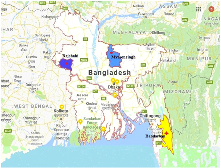
Figure 1: Location of study area inside Bangladesh. Rajshahi, Mymensingh and Bandanban districts share common boundaries with the neighboring country India.

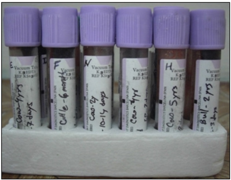
Figure 2: Collected blood sample for hematological and biochemical examination.

Results and Discussion

E. Hematological Study

The concentration of different hematological indices (mean ± SE values) are presented in the Table 1. In this present study, the total number of red blood cells (RBC) count in foot-and-mouth disease cattle at primary, advanced, recovery stage groups (Group B, Group C, Group D) and apparently healthy animals (Group A) were 4.98±1.48 x10⁶/μl, 4.60±0.48** x10⁶/μl, 5.14±1.36 x10⁶/ μl and 5.5±0.29 x10⁶/μl respectively. Similarly, the hemoglobin concentration were 8.85±1.25 g/dl, 8.32±0.78** g/dl, 9.25±2.02 g/dl and 9.57±1.08 g/dl at the primary, advanced, recovery stage groups (Group B, Group C, Group D) and control group (Group A) respectively Table 1. Statistical analysis revealed that the RBCs count and Hb conc. specially at the advanced stage (group C) was significantly (p<0.01) lower than the other groups that might be due to reduction of the process of erythropoesis and hemolysis [20]. Similar findings are also observed by different workers in FMD infection [18,19,21]. On the other hand, FMD infected animals showed significant production of mean corpuscular volume (MCV) (p≤0.05) at advanced (64.27±3.94* fl) stage group as compared to the primary (60.65±2.67 fl) stage, recovery (56.20±2.48 fl) stage and control (51.80±2.86 fl) group Table 1 same findings are recorded by Gokce et al. [19], Ghanem et al. [21], Mohapatra et al. [22], Krupakaran et al. [23] and Gattani et al. [24]. These results could be attributed to endocrinopathy is reported previously by Radostits et al. [20].

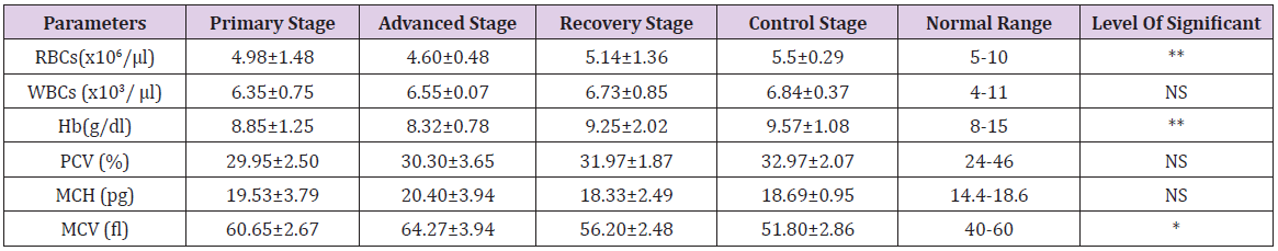
Table 1: Hematological parameters (mean ± SE values) in FMD at primary, advanced, recovered stage group and control group with normal range.

Results are Mean ± SE (Standard Error) in each group. One-way analysis of variance (ANOVA) followed by post hoc Duncan’s test was performed as the test of significance. The difference was considered to be significant when **p<0.01, *p<0.05 compared to FMD control group.
NS = Not significant

Besides these, the total white blood cells (WBC) count, the mean value of packed cell volume (PCV), and the value of mean corpuscular hemoglobin (MCH) in FMD affected animals at primary, advanced, recovery stage groups did not show any significant alteration along with the control group and our findings also are corroborated with the observation made by Mohan et al. [18], Gokce et al. [19] and Al-Rukibat et al. [25].

F. Biochemical Study

The concentration of different biochemical indices (mean ± SE values) are presented in the Table 2. This study demonstrated that the average value of total serum protein were 6.20±0.65 g/dl, 4.32±0.17** g/dl, 5.95±0.57 g/dl and 7.07±0.72 g/dl and albumin concentration were 2.99±0.23 g/dl, 2.17±0.11** g/dl, 3.96±0.18 g/ dl and 4.65±0.18 g/dl in FMD at the primary, advanced, recovery stage groups and control group respectively Table 2. The total serum protein and albumin concentration were significantly (p<0.01) decreased at advanced stage rather than the other groups. Roussel et al. [26] reports that the decrease level of total protein concentration is associated with hepatic and renal damage, starvation, enteropathies that resulting in protein loss and the presence of infection or any lesion in the body is also recorded by Meyer et al. [27]. which was inconsonance with our observation. Low albumin and protein concentrations may also be due to alterations in pancreatic β-cell functions that might have developed during the clinical course of FMD is reported by Barboni et al. [28]. As well as, serum globulin concentration showed a significant decrease (p<0.05) at the advanced stage as compared to primary, recovery stage groups and control group. The serum globulin concentration at the primary, advanced, recovery stage groups and control group were 2.40±0.08 g/dl, 2.14±0.14* g/dl, 2.35±0.30 g/dl and 2.78±0.30 g/dl respectively Table 2, that might be due to hypoglobulinemia in the affected animals, coming in parallel with mention by Gokce et al. [19], Ghanem et al. [21], Mohapatra et al. [22], Krupakaran et al. [23] and Gattani et al. [24].

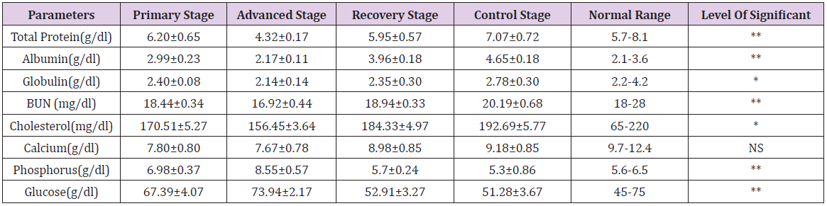
Table 2: Biochemical parameters (mean ± SE values) in FMD at primary, advanced, recovered stage group and control group with normal range.

Results are Mean ± SE (Standard Error) in each group. One-way analysis of variance (ANOVA) followed by post hoc Duncan’s test was performed as the test of significance. The difference was considered to be significant when **p<0.01, *p<0.05 compared to FMD control group.
NS = Not significant

Though the blood urea nitrogen (BUN) concentrations were decreased significantly (p<0.01) at the advanced stage group (18.44±0.34** mg/dl) as compared with the primary (16.92±0.44 mg/dl), recovery (18.94±0.33 mg/dl) and control group (20.19±0.68 mg/dl). It might be due to hypoproteinemia in the affected group. As well as the mean value of cholesterol in FMD at the primary, advanced, recovery stage groups and control group were 170.51±5.27 mg/dl, 156.45±3.64** mg/dl, 184.33±4.97 mg/dl and 192.69±5.77 respectively Table 2, while the normal range is 65- 220 mg/dl. This study was revealed a significant (p<0.01) reduction of cholesterol level at the advanced stage group (Group C) and may be due to dysfunction of pancreatic β-cell is suggested by Gokce et al. [19] and Ghanem et al. [21]. Nevertheless, the mean value of calcium level at the primary, affected, recovered stages and control group was reported very little significant which is also mentioned by Mohapatra et al. [22], Krupakaran et al. [23] and Gattani et al. [24]. This research also revealed a significant (p<0.01) production of phosphorus (P) concentration between the affected and control groups. The mean value of phosphorus (P) in foot-and-mouth disease cattle at primary, advanced, recovery stage groups and control group were 6.98±0.37, 8.55±0.57**, 5.7±0.24 and 5.3±0.86 g/dl respectively. Hyperphosphatemia recorded in our result is also noted by Gokce et al. [19], Ghanem et al. [21], Mohapatra et al. [22], Krupakaran et al. [23] and Gattani et al. [24]. Similarly, Glucose concentration was significantly (p<0.01) increased at the advanced stage (73.94±2.17** mg/dl) group in analogy with the primary (67.39±4.07 mg/dl), recovered (52.91±3.27 mg/dl) stage groups and control group (51.28±3.67 mg/dl) Table 2. An increased concentration of glucose was well documented in cattle affected with FMD is recorded by Elitok et al. [29] and also a common finding in cattle affected by the stress in systemic disease is described by Gokce et al. [19], Paalberg et al. [30] and Yeotikar et al. [31].

Conclusion
From this present study, it can be concluded that the concentration of different hematological constituents revealed a significant reduction (p≤0.01) of red blood cells (RBC), hemoglobin (Hb) and significant production (p≤0.05) of mean corpuscular volume (MCV) especially at the advanced stage (group B) rather than the control groups. Similarly, at 3rd to 7th days of post infection that is in advanced stage, the serum biochemical concentration of total protein, albumin, globulin, blood urea nitrogen, cholesterol are significantly decreased and significant increase (p≤0.01) of glucose and phosphorus in FMD affected animals in comparison to the other respective groups.

For more Articles on : https://biomedres01.blogspot.com/

Recent Therapeutic Options in Cancer Therapy

Recent Therapeutic Options in Cancer Therapy

Introduction
Nanotechnology is already widely applicable in medical field by their various drug delivery systems, in the principle of magnetic resonance in cancer treatments and cellular biology [1-3]. Researchers in nanotechnology have carefully monitored and applied its use in creating the best medical and biological equipment’s and devices so as to be beneficial for patient care [4- 6]. This review briefly details the role of nanoparticles in general medicine and then focusing on its application in treating patients with brain and lung cancers [7-9].

Role of Nanotechnology in Medicine

Nanoparticles are used successfully in the deliverance of basic physical substances such as heat, light, and other substances to the target cell of interest [10,11]. This has been beneficial in using nanotechnology as a treatment option for managing cancers [12]. The nanoparticles enter the target cancer cell and produce a cellular arrest and thereby inhibiting the uncontrolled tumor growth [13]. This further interrupt the proto-oncogenes and tumor suppressor genes causing an abrupt interference of the tumor growth cycle [14]. The highly effective tumor cell penetrance property of the nanoparticles has been proven to be beneficial in combatting the tumor cell biology (Figure 1). The adverse effects in the application of nanotechnology in cancer care treatment is very minimal and hence has shown to be widely applicable[15]. The role of gapmer design in downregulating the cancer cells has also been proven recently [16]. This design model has shown to increase the binding capacity of the nanoparticles to the cancer cells and then downregulates the entire tumor environment [17-19].

The role of nanotechnology in treating brain and lung cancers has been formulated with the LMP in vivo and in vitro levels [20-22]. The LMP formulations reach high levels in these cancer types, hence providing a better chemical and physical stability. These properties further enhance the overhaul performance of nanoparticles effect in brain and lung cancers. The antisense oligonucleotide combination with lipid subunit of nanoparticles produces quaternary aminetertiary cation lipid complex. This complex is very efficient and also cost-effective in cancer therapy [23-25]. Nanoparticles can be combined with various elements, however its combination with polymers and lipid component seems to be the most effective formula [26]. This is mainly attributed to its high bioavailability, compatibility, and also higher safety level. These combinations are used in treating varying cancer types, especially the brain and lung cancers [27].

Figure 1: A schematic representation of the nanoparticle effect. The colloidal nanoparticles are accumulated in tumors due to their higher vascular endothelial penetrance.

Role of Nanoparticles in Brain Cancer Treatment

The role of nanoparticles as a therapeutic option in psychosis is well versed. The anti-psychotic drug, haloperidol develops blocks at neuronal level and thereby ensures slow release of the pharmaceutical substances [28]. This has been beneficial as the relevance of psychosis in modern society has been exponential raising [29]. The dendrimers further alter and induced as nanoparticles. The other popular anti-psychotic medication, risperidone is used effectively in treating schizophrenia [30]. As both these basic and common anti-psychotic drugs are already in treatment plans for psychosis, we now move to the role of nanoparticles in treating brain cancers [31]. Brain tumors are mostly glial based. These are classified as Gliomas by the World Health Organization (WHO). Based on its histological features they are further subtyped into grad 1 to 4. Grade 1 been most mild and grade 4 been most aggressive [32-34]. The application of nanotechnology in treating brain cancers has been limited even though there is abundance of nanoparticles available in the market. This is mainly attributed to the aggressiveness of the brain tumor and also the limitations of nanoparticles in arresting the cell cycle of the tumor. However, when nanoparticle is combined with other chemotherapeutic drugs they produce beneficial effects to the patients by reducing the extent of side effects. This occurs due to the in vivo and in vitro effects of nanoparticles in a tumor environment [35,36].

The application of gold nanoparticles has been effective and productive in brain target drug delivery. The gold electrons are conducted on the metal surface followed by light excitation [37]. The Serine-arginine-leucine (SRL) modified dendrimers produce a high transfusion rate and a low toxicity level. These effects have limitations as they can only be applied in less aggressive forms of brain cancers [38]. In Glioblastoma multiforme, a form of brain tumor, is aggressive and makes it difficult for the procedure to turn effective. Fibrin binding peptide can be induced to treat this case [39]. Studies are still underway in applying polymer, lips, and microbubbles form of nanoparticles to treat brain tumors [40]. These forms have shown lower toxicity compared to other forms of nanoparticles. This application is important as there needs to be an equilibrium between the drug delivery and adverse effects during treatment of aggressive forms of brain cancers [41].

Role of Nanoparticles in Lung Cancer Treatment

Lung cancers are mainly adenocarcinomas. These are followed by squamous cells carcinomas and neuroendocrine cancers [42]. The origin of lung cancer has been cited mainly due to its molecular alterations and environmental-host imbalances [43]. The overexpression of an anti-apoptotic gene, Bcl-2 has shown to be a very important cause for lung cancer origin. The expression of Bcl-2 has been controlled by the antisense oligonucleotides (ASOs) therapy [44]. But this control level has been minimal due to problems in tumor-particle binding ratio, immune nature if oligonucleotide and low in vitro and in vivo nanoparticle concentration. This has led to the demand of lipid nanoparticles which can increase the overall nuclease stability and the circulation time of such oligonucleotides [45,46]. The lipid nanoparticles alter the tumor microenvironment by altering the cell cycle, inhibiting proto-oncogenes, and enhancing cell cycle arrest factors [47]. miR-21 plays an essential role in regulating the propagation of tumor and cancer. QTsome nanoparticles are ideal for inducing strong dosage of the therapy without affecting the sensitivity and increasing the invasion pace [48]. However, extensive research is still required in order to use nanoparticles more commonly in lung cancer treatment.

Conclusion

The role of nanotechnology in patient care has been growing every year. The success of these particles has been attributed to its fast drug deliverance systems and its ability to interrupt the cancer microenvironment. The beneficial effects of nanoparticles in treating psychosis have laid a foundation on which its physical and chemical nature cane be more explored. This holds true for treating cancers especially of the brain and the lungs.

For more Articles on : https://biomedres01.blogspot.com/

RBD and ACE2 Embedded Chitosan Nanoparticles as a Prevention Approach for SARS-COV 2

RBD and ACE2 Embedded Chitosan Nanoparticles as a Prevention Approach for SARS-COV 2

Introduction
A new type of coronavirus-associated persistent pneumonia outbreak called SARS-CoV-2, which causes severe acute respiratory syndrome, was reported in Wuhan, China in Hubei Province in December 2019. In the following weeks, infections spread rapidly to China and other countries around the world [1]. Coronaviruses (CoVs) are known to cause enteric and respiratory diseases in animals and humans, which are positive stranded RNA viruses that are not segmented into large envelopes. Most human CoVs such as hCoV - 229E, OC43, NL63 and HKU1 cause mild respiratory diseases, but two previously unknown CoVs, severe acute respiratory syndrome CoV (SARS - CoV) and Middle East respiratory syndrome CoV (MERS - The worldwide spread of CoV) has drawn global attention to the deadly potential of human CoVs. Genomic analysis shows that SARS - CoV - 2 belongs to the same betacoronavirus family as MERS - CoV and SARS - CoV and shares a sequence that shows a high homology with SARS - CoV. A cellular receptor angiotensin-converting enzyme 2 (ACE2) is mainly mediated by the introduction of SARS - CoV into human host cells. It is expressed in the human respiratory epithelium, lung parenchyma, vascular endothelium, kidney cells and small intestine cells [2].

ACE2 Function as a Double-Edged Sword

ACE2 protein is a carboxypeptidase which has an important function in the conversion of Angiotensin-1 to Angiotensin-(1-9) and generally found in lungs. ACE2’s normal funtion is so important since the ratio of Angiotensin-1/Angiotensin-2 has major effect on sustainability of lung function [3]. Li et al. showed that ACE2 protein also has a role as a major attachment side of Coronaviruses [4]. These two important functions make ACE2 a double edged sword since its function is so crucial for protecting the lungs from lung failure [5]. However, ACE2 involvement in the infection mechanism of SARS-CoV viruses,makes ACE2 an important target of theurapatic approaches. So it becomes more necessary to focus on structural form of ACE2 and its components [6].

ACE2 Distribution along Different Tissues

ACE2 protein which is the main attachment side of SARSCoV has found in many different tissues. The mapping of ACE2 expression in different tissues can potentially identify the possible routes of infection for SARS‐CoV, and possible routes of spread and replication throughout the body. One of the most important of these locations is the lung alveolar epithelial cells which causes the main symptom and signs of COVID19 and it is the responsible from high number of deaths [7]. Numerous variety of studies showed that ACE2 mRNA is highly expressed in renal, cardiovascular, and gastrointestinal tissues [8–10]. Hamming and colleagues study is one of the most important studies which shows the distribution of ACE2 along different types of tissues. The most striking result in this study, addition the previous studies results, is the demonstration of ACE2 expression in the basal layer of the non‐keratinizing squamous epithelium of the nasal and oral mucosa and the nasopharynx [7].

SARS-CoV-2 Cell Entry Mechanism

A distinctive feature of coronaviruses is that they recognize a variety of receptors, including both protein receptors and sugar receptors. Coronaviruses enter cells first by recognizing a host cell surface receptor for viral binding and then creating an endosome. Receptors play an important role not only in viral binding but also in membrane fusion process [11]. In order for a viral infection to begin, a receptor expressed by host cells needs to bind to the virus ligand. ACE2 the host receptor identified by researchers working with SARS-CoV, is now claimed to be a receptor for COVID-19 [12]. One of the most important of these locations is the lung alveolar epithelial cells which causes the main symptom and signs of COVID19 and it is the responsible from high number of deaths [7] . Importantly, the sequence of the COVID-19 spike(S) protein receptor binding domain is similar to that of SARS-CoV, which caused a pandemic in 2003. Mutagenic analyses revealed the domains of SARS and ACE2 receptor which provides efficient interaction between these two. One of these studies is conducted by Xiao et al.and results showed that an independently folded region called Receptor Binding Domain(RBD) which is in between the aminoacids of 318-510 on SARS-Spike(SARS-S) protein, binds to ACE2 receptor with higher affinity than the other remaining parts of SARS-S protein [13]. S protein is the trimeric protein found in metastable inhibition conformation for fusion of viral membrane and host cell membrane. Each of the S1 subunits contains a receptor binding domain (RBD) that mediates receptor recognition. When the RBD is attached to the host cell, the balance of the trimeric structure is disturbed, which causes the S1 subunit to shed and the S2 subunit to change its conformation to take the form of postfusion [14]. The SARS-CoV-2 S1 / S2 region is believed to be cleaved by the cathepsin L, similar to the SARS-CoV [15]. Cell studies carried out by the researchers were predicted that after the spike protein that binds to ACE-2 and then, the virus is introduced into the host cell via cathepsinB /L and TMPRSS2 proteases [16]. Although the newly introduced SARS-CoV-2 entry mechanisms and endocytic pathway are not fully known, it is known to use the ACE2 receptor, which is the same as SARS-CoV for viral entry. The sensitivity of SARS-CoV-2 to the inhibitory effect of chloroquine (CQ) suggests that this new CoV will use the same endocytic pathway for entry into host cells. SARS-CoV, seen as a possible endocytic pathway, enters the cell through autophagy. Autophagy is controlled by a group of proteins encoded by autophagy-related genes (ATG) in several successive stages [17].

SARS-CoV-2 Structure

A coronavirus have four structural proteins, including spike (S), envelope (E), membrane (M), and nucleocapsid (N) protein (Picture 1). S protein displays the most important roles, including viral attachment, fusion and entry [18]. The S protein provides viral particules entry into host cells. Firstly, it engages to a host receptor through the receptor-binding domain (RBD) in the S1 subunit. Subsequently, it fuses the viral and host membranes through the S2 subunit [19]. Therefore, it is critical to define the RBD in SARSCoV-2 S protein for the development of virus attachment inhibitors, neutralizing antibodies, and vaccines [20]. There are more than one study on this subject in the literature. In a study belongs to Lan, J et al, they expressed the SARS-CoV-2 RBD (residues Arg319-Phe541) and the N-terminal peptidase domain of ACE2 (residues Ser19- Asp615) in Hi5 insect cells and established the final model contains residues Thr333 to Gly526 of the SARS-CoV-2 RBD and residues Ser19 to Asp615 of the ACE2 N-terminal peptidase domain [21]. Furthermore, Tai et al. identified the region of SARS-CoV-2 RBD at residues 331 to 524 of S protein. Then, they made recombinant RBD protein using pFUSE-hIgG1-Fc2 expression vector, expressed the protein in mammalian cell 293T. They have demonstrated SARSCoV- 2 RBD bound to sACE2 in a dose dependent manner. Moreover, they have also established , binding between SARS-CoV-2 RBD and sACE2 with 50% effective dose (EC50) was stronger than that between SARS-CoV RBD and sACE2. As a result of study, Tai et al., have suggested that SARS-CoV-2 RBD protein could be developed as an effective therapeutic agent against SARS-CoV-2 infection [20]. Ultimately, based on these studies, in our hypothesis, we decided to use RBD residues to bind to the ACE2 receptors on the host cells (Figure 1).

Chıtosan Nanoparticles

Chitosan is a biocompatible, biodegradable polymer that is considered safe for use in the human diet and approved for dressing applications [22-24]. Chitosan polymer has been used as a transporter in polymeric nanoparticles for drug delivery by different routes of administration [25]. The Chitosan polymer has chemical functional groups that can be modified to achieve certain goals and make it a polymer with a wide range of potential applications in human health and various fields. The chitosan nanoparticles and chitosan derivatives nanoparticles have a positive surface charge and mucoadhesion properties that can stick to mucous membranes and release the drug load in continuous release [26]. Chitosanbased nanoparticles has several applications in non-parenteral drug delivery for cancer, lung diseases, gastrointestinal diseases, administration of the brain and treatment of ocular infections [27]. The Chitosan nanoparticles show low toxicity in both in vitro and some in vivo models.

a. Modified Chitosan Nanoparticles with Antibody

Drug delivery vehicles such as polymeric nanoparticles modified with specific ligands such as antibodies have been widely used for targeted therapy. When designing nanoparticle-antibody conjugates for drug delivery or medical applications, several properties of the structure of the nanoparticles are important. The NPs must be chemically and biologically inert, can be stable in physiological conditions, must move freely in the body, contain a surface that is easily conjugated to the targeting antibody [28]. Here, we design a new type of chitosan nanoparticles for treating the coronavirus disease, for this goal we’ll binded the ACE2 receptor and RBD protein to the surface of chitosan nanoparticles by chemical method.

The Fast and Effective Method: The Usage of Inhaler Spray

Inhaled treatments give easy, stable and effective results in various lung diseases (eg: cystic fibrosis, asthma) [29]. Low doses and low side effects are among the advantages of this form. The most important advantage of inhalation over parenteral doses is that it does not require sterilization and is easy to apply. The drugs reach the bronchial muscles at a higher concentration than other systemic applications by inhalation and their effects are at maximum level. Since the local metabolism of inhaled drugs is slow, its effects last longer (Bizim makale). Another reason that we prefer treatment method through inhalation is the presence of ACE-2 receptors in the nasopharyngeal region [7]. As a result of the literature review, we decided that inhaled therapies for lung and respiratory diseases are more effective at the local level and thus we planned our study accordingly.

Hypothesis: RBD and ACE2 Embedded Chitosan Nanoparticles Prevent to Attach SARS CoV2 to Host Cells

As plasma therapy and other medical treatments are insufficient and vaccination studies take a long time, we planned to develop an prevention therapy so that people can return to their daily life activities. In the literature searches, we established that ACE 2 was located in both the respiratory tract and nasopharyngeal epithelium. Considering the normal physiological function of Ace 2, it is not a matter of discussion of complete inhibition of the receptors in this field. Therefore, we thought it could be enough to provide inhibition during the time people spend outside. In our study to provide an adjustable inhibition, we anticipated the use of chitosan nanoparticles that can be compatible with the body. We assumed that the virus RBD residues should be added to these nanoparticles. Thus, inhibition of host cells ACE2 will prevent the attachment of viruses. We also planned to add ACE2 antibody on nanoparticles. In this way, the nanoparticles will be attached to host cells with the RBD tip, and to viral particles with the ACE2 tip (Figure 2). Extensive studies on biocompatibility of chitosan nanoparticles in literature screening make it possible to test this experiment in a short time on humans. Moreover, it is easy to add antibodies to chitosan therefore we chose these nanoparticles. We anticipated the use of inhaler spray as a more specific and effective method for respiratory and nasopharyngeal receptors (Figure 2).

Figure 1: The Structure of SARS-CoV 2.

Figure 2: Attachment of modified chitosan nanoparticles to the host cell ACE 2 receptor and attachment of viruses to ACE 2 on the nanoparticle.

For more Articles on : https://biomedres01.blogspot.com/

Monday, May 30, 2022

Impact of a Model of Educational Intervention on Intestinal Parasitism in Children of Havana. 2021 Second Post-Evaluation and Intervention Study

Impact of a Model of Educational Intervention on Intestinal Parasitism in Children of Havana. 2021 Second Post-Evaluation and Intervention Study

Introduction
Among infectious diseases, those produced by parasites constitute important health problems for man and at the present time they are a medico-social problem that affects not only the so-called Third World countries, but also those with the highest development. In studies carried out in children from suburban regions of the American continent, at least seven parasites predominate: ascariasis, whipworm, oxyuriosis, amebiosis, hookworms, giardiasis and strongyloidosis [1]. Intestinal parasitism affects all people equally, however, due to their characteristics, the child population has a high level of susceptibility to suffer them, since there is a greater opportunity of contact with these parasites because they spend a large part of the day in schools and carry out activities collectively, which can favor the conditions for the transmission of some parasitic diseases, especially those in which their main transmission mechanism is the oral fecal route, in addition to presenting a lower immunological level [1-2] From a global perspective, intestinal parasitism is a major health problem in underdeveloped countries where it causes significant morbidity and mortality. Thus, the mortality caused by the three most frequent intestinal parasitic infections in the world is not negligible: 60,000 deaths a year from ascarislumbricoides, 65,000 deaths a year from hookworms and necatoramericanus, and 10,000 deaths a year from trichuristrichiura [3].

Symptomatic intestinal parasites can cause some morbidity and mortality, but this is not very significant. However, it represents a risk for groups with poor hygiene, such as nurseries or institutions for the mentally handicapped, or for patients with primary or secondary immunosuppression, in whom the infection can be severe. In addition to immunosuppression, various sociological phenomena make the topic of intestinal parasitism of current interest to the pediatrician. The most important are emigration and the adoption of children from third world countries, but also the phenomenon of globalization, with constant mobility of large masses of the population and frequent trips to underdeveloped countries for tourism or work reasons that expose the indigenous population. to the contagion of endemic parasites in certain areas. The orientation of the problem in these cases must be done taking into account not only the symptoms but also the origin of the population or the place of displacement since certain pathogens will predominate over another [4].

Intestinal parasitic infections by helminths and protozoa are among the most common in man in Latin America [5]. These have been consistently and considerably estimated in their impact on public health; However, in the last decade, its recognition as an important public health problem has increased even more [6]. In Venezuela, various studies indicate variable prevalences, but most of them have been carried out in urban populations. There is great morbidity, among protozoa, especially blastocystis hominis and entamoeba coli, and among helminths, ascaris lumbricoides [3- 4]. Intestinal parasitism represents an alarming health problem in Venezuela due to the large number of people affected and the intense organic disturbances that they can cause. Many times it corresponds to the only morbid process of the patient and sometimes they aggravate other concomitant diseases Due to the aforementioned and taking into account that the mission of the doctor is the promotion and prevention of health, it was decided to carry out an educational intervention study in children of primary education of the Municipality of Marianao Province of Havana , in order to determine the influence of this work on hygienic practices and the prevalence of intestinal parasitism in students, identify the associated symptoms, as well as compare the hygienic practices of the population before and after the educational work.

Materials & Methods

An intervention study was carried out in order to determine the influence of educational work on hygienic practices and the prevalence of intestinal parasitism in students from preschool to sixth grade. 210 children were studied, five for each grade of the six primary schools enrolled in said institution. These schools have already been previously evaluated and intervened, for which the results meter will help us to see the impact of the intervention carried out. The homes were visited to explain the objective of the study to the parents or guardians of the children, their informed consent was collected and a survey was applied to them. The symptoms and clinical signs presented in the children were analyzed, to describe the health education of the family, the quality of the drinking water, the washing of hands before eating food and after defecation, the presence of vectors, the presence of vectors, was taken into account. washing fruits and vegetables, if they are used to walking barefoot, playing with dirt or biting their nails.

Then a stool sample was taken by spontaneous defecation, guiding the parents about the need for non-contamination of it. Each child was given a sterile vial containing 7% formaldehyde solution. The fecal samples were processed in the Microbiology Department of their Polyclinic, where they were subjected to direct examination with Lugol’s solution with 1% eosin and a concentrated examination by the Willis method for enrichment of helminth eggs. A sample was taken from each child in the morning, without previous cleaning, for the diagnosis of Enterobius vermicularis (pinworm) by the Graham method. The clinical evaluation of the parasitized children was carried out, the laboratory results were analyzed and appropriate treatment was applied.

Parasitized Children Received Treatment According to International Standards

The information collected made it possible to identify learning needs and changes in attitudes, for which a study program was designed. They were formed five groups of 20 people each, structuring activities in three sessions of one hour each, with a weekly frequency for each group and develop throughout the month of November 2019. Six months after the educational intervention, the second sample collection and the second application of the survey were indicated, with characteristics similar to the previous one. The percentage was used as a measure for each variable. The research results were presented to summarize the information in tables. The information collected was processed and stored on a Pentium 4 microprocessor, using the Microsoft Office 2003 data processing programs contained in the Windows XP Professional operating system.

Results

The prevalence of intestinal parasitism (Graph 1) was observed that out of a total of 210 schoolchildren, 137 children were parasitized for 65.2 % and only 73 were not parasitized for 34.8 %, so it can be considered that in schoolchildren studied there is a high prevalence of parasites. Parasitic agents in school (Table 1) were more frequently Enterobius vermicularis in 65 children for a 4 7.4 %, and Giardia lamblia and E. Histology and tica 34 and 23 school for a 2 4/8 and 16, 8 % respectively. After the intervention was applied, all the parasitic agents decreased. The most frequent symptoms presented by parasitized children (Table 2) before and after the intervention were anal itching in 44 students, sleep disorders with 31 and irritability in 27 for 32.1%, 22.6% and 9.7%, respectively. After the intervention, the frequency in each of them decreased, although it is worth clarifying that there are symptoms that appear as part of the child’s physiology at school. In the hygienic practices of the population before and after the educational intervention (Table 3), an inadequate hygiene of the population was observed before the intervention, after which all the hygienic habits of the population were positively modified (Table 4). The epidemiological factors present in the home of the schoolchildren were the most prevalent Do not wash foods consumed raw with 28.1% and the consumption of non-potable water with 51%, after the intervention all modifiable epidemiological risks decreased In the variable quality of drinking water (Graph 2) it was shown that 51% of the patients did not boil their drinking water, 63% of them treated the water, with the bottled water trade and the myths that it contains 55% of those who consumed this water were shown to be parasitized After the educational work, those who consumed the water without boiling decreased to 8%, while those who boiled or chlorinated it represented 5% and 11% respectively.

Figure 1: Prevalence of intestinal parasitism in primary school children.

Note: Source: parasitologicalstudy.

Figure 2:Water consumption in the homes of schoolchildren before and after the educational intervention.

Note: Source: sociodemographicregistry.

Table 1: Parasitic diagnosed in school before and after the educational intervention.

Note: 137 Source: primary registry and parasitological study

Table 2: Most frequent symptoms in parasitized children before and after the educational intervention.

Note: N:137 Source: sociodemographicregistry.

Table 3: Hygienic Practices of the population before and after the educational intervention.

Note: N: 210 Source: primarysurvey.

Table 4: Epidemiological factors present in the homes of the schoolchildren before and after the intervention.

Note: Source: sociodemographicsurvey.

Discussion

The Enterobius vermicularis causes the disease called Enterobiasis or pinworm, with a worldwide spread, because this parasite does not require favorable environmental conditions, since the transmission is direct from person to person, without the need for intervention from the floor. This parasitosis occurs in individuals of all ages, but is higher in school age. In a study carried out by Díaz and collaborators, in students of Basic Education of the Municipality of Cacique Mara, in the state of Zulia in Venezuela, a 45.9% prevalence of helminthiasis was demonstrated with a predominance of oxyuriasis, these results coinciding with those of the present investigation. Devera et al., Reported a prevalence for enterobiasis of 48.8%, well above the other parasitosis, results similar to the present study [4-7]. The authors refer that oxyuriosis becomes frequent because the eggs are highly infective from the moment the adult female parasite lays them on the margins of the anus, and there may be self-reinfection by the hands or indirectly by contaminated items such as furniture, desks, bathrooms, or by inhalation of dust in homes and institutions [8-9]. In this study, the most frequent symptoms were (after anal itching and irritability) abdominal pain and loss of appetite, related to enterobiasis or ascariasis, a parasite that was identified in the first and second order in the study. In a study carried out by Kucik and collaborators, they state that among the most frequent symptoms caused by enterobiousvermicularis were irritability and anal itching; while ascarislumbricoides is more associated with diarrhea, abdominal pain, decreased appetite, and weight loss, and trichuristrichiura is associated with anemia, decreased appetite, and weight loss [10- 12].

After the educational intervention was carried out, intestinal parasitism was significantly reduced and hygienic practices increased, indicating that inadequate hygienic practices influenced the presence of parasitism, similar conclusions reported by Medina and González in their study Intestinal Parasitism in Spain [12-15] As it is a comparison of the same subject before and after, it can be affirmed that the educational work together with the treatment helped the success of the group work. Hernández, Rauda and collaborators in a study carried out in San Salvador, on the socioepidemiological factors related to the specific prevalence of intestinal parasitic infections caused by protozoa and helminths in children under 14 years of age from the Atonatl community, metropolitan area of San Salvador, pointed out that the lack of habit of applying chemical or thermal treatment to water, prior to consumption, was significantly related to the parasitism found in children, which coincides with the results of the present work .

Conclusion
The prevalence of intestinal parasites in school children studied was elevated preoperatively, predomin or the Enterobius vermicularis. An obvious decrease in the prevalence of parasitism was evidenced after the educational intervention carried out. The most frequent symptoms in parasitized children were anal itching, irritability, abdominal pain and loss of appetite, which decreased after the intervention. The educational intervention applied was effective, since hygiene practices such as washing hands before eating food or after defecating, walking barefoot, playing with dirt, biting nails and not boiling drinking water, were significantly modified.

For more Articles on : https://biomedres01.blogspot.com/

The Skin Aging Process and Anti-Aging Strategies

  The Skin Aging Process and Anti-Aging Strategies Introduction Appearances play a vital role in society as it is how one communicates to ot...