Showing posts with label Journals on Neuro Imaging Open Access Medical Journal Biomedical Open Access Journals Biomedical Journal Articles Biomedical Journal Impact Factor. Show all posts

Saturday, December 10, 2022

Antioxidant Capacity and Antitumoral Activity of Citrus Paradisi Essential Oil


Antioxidant Capacity and Antitumoral Activity of Citrus Paradisi Essential Oil

Introduction

Phytomedicine has been of increasing interest in recent years. Over 65 % of the world’s population relies on traditional and medical approaches to treating diseases [1]. The return to phytotherapy is more than using an herb to treat an illness or a crude herbal preparation rather than a specific isolated component. Plants are considered essential resources for researchers to prove and develop new drugs. Generally, using plants in the treatment of disease has a long history [2,3]. Thus plants have been primary resources for producing traditional drugs effective in treating cancer and inflammatory diseases [4]. In addition, plants containing essential oils have bioactivity against a host of bacteria, fungi, viruses [5,6]. Currently, essential oils of medicinal plants are increasingly used thanks to their antibacterial, anti-inflammatory, antiseptic, antiviral, antifungal, antioxidant, and antitumor potential [7]. Essential oils are also used in other areas of economic interest, such as cosmetics, perfumes, and aromatherapy [7,8]. Fragrant and aromatic plants such as members of the Asteraceae, Lamiaceae, Rutaceae, and Verbenaceae produce essential oils, which historically have been important in traditional medicines [5]. Citrus species have beneficial pharmacological activities, including antibacterial, vascular protectant, antispasmodic, analgesic, antipyretic, antiinflammatory, and antitumoral effects [9-11].
Citrus peel essential oils have also been searched for their natural antioxidant and antimicrobial properties [12,13]. Furthermore, studies have shown the synergistic effect of two or more ingredients of essential oils against various human pathogens. It is necessary to know more about Citrus paradisi essential oil (CPEo) antioxidant, cytotoxic, and antitumor activity. Therefore, the DPPH, ABTS, FRAP, and NO• techniques studied the antioxidant potential in a chemical system. Furthermore, to check the antitumor activity, MTT was realized using HeLa and MCF-7 tumor cells.

Material and Methods

Samples

In this study, the grapefruit (citrus paradisi) was collected in March 2018 from Sfax, Tunisia. Sfax is one of the biggest coastal cities in Tunisia. It is located in the eastern part of the country. The climate in this area is arid to semiarid with irregular and torrential precipitations [14]. After the collection, it was washed, peeled off and cut into small pieces.

Hydrodistillation

The zest of citrus paradisi was hydro distilled using a Clevengertype apparatus to recuperate the essential oils for 2h to produce the volatile constituents. The distilled essential oils were dried over anhydrous sodium sulfate; then separated from the distillate by liquid-liquid extraction using cyclohexane solvent. The recuperated oils were stored at + 4 °C.

Antioxidant Activity: Chemical System

DPPH Radical Scavenging Assay: Different aliquots from the stock solution (200μl in 1 ml Me OH) of essential oils were mixed with 500μl of 0.2mM diphenyl picryl hydrazine (DPPH) final volume brought to 1mL. The mixtures were vigorously shaken and allowed to stand in the dark for 30min at room temperature. The absorbance was measured by spectrophotometry (LKB BIOCHROM® ULTROSPECPUS 4054 UV/VIS) at 517 nm against a control sample without DPPH. The percentage of radical scavenging activity was calculated using the following equation:

DPPH scavenging effect (%) = (A0- A1)/A0 × 100

A0: The absorbance of the control at 30min A1: The absorbance of the sample at 30min.
ABTS+ Radical Scavenging Effect: The antiradical activity was performed by the ABTS+ free radical decolorization assay as developed by Re, et al. [15]. The 2,2-azino-bis-3- ethylbenzothiazoline-6-sulfonic acid (ABTS) was prepared as an aqueous stock solution (7mM). The ABTS radical cations (ABTS+) were produced by the reaction of the ABTS stock solution with 2.5mM of ammonium persulfate methanolic solution. First, the reaction mixture is incubated in the dark for 16h at room temperature. Then, the solution is diluted to an absorbance of 0.7 ± 0.02 at 734nm to form the working reagent. Next, the reaction mixtures containing 100μl of the sample at different concentrations and 900μL of reagent were incubated at 30 °C for 6min. Finally, the antioxidant power of each sample was expressed as the inhibition percentage calculated according to the following formula:
ABTS+ scavenging effect (%) = (A0- A1)/A0 × 100
A0: the absorbance of the control at 6min
A1: the absorbance of the sample at 6min.
Ferric-Reducing Antioxidant Power (FRAP): This method is based on the plant’s ability to reduce ferric iron (Fe3+) to ferrous iron (Fe2+). The mechanism is known to be a marker of electron donor activity [16]. For 1mL of the sample at different concentrations, 2.5mL of a solution phosphate buffer (0.2M, pH 6.6) and 2.5mL of 1 % K3Fe (CN) 6 potassium ferricyanide solution were added. The mixture is incubated at 50 °C for 20min and then cooled to room temperature. Then, 2.5mL of 10 % trichloroacetic acid (TCA) is added to stop the reaction, and then the tubes are centrifuged at 3000rpm for 10min. 2.5ml of the supernatant are then added to 2.5mL of distilled water and 500μL of a 0.1 % solution of iron trichloride (FeCl3, 6H2O) [16]. The absorbance reading is performed against a blank at 700 nm using a spectrophotometer. Ascorbic acid is used as a positive control. The increase in the absorption capacity of the components indicates the increase in the reduction of iron.
Scavenging Activity of Nitric Oxide (NO·): NO scavenging activity of the essential oils was determined as previously described [17]. Briefly, 0.1mL of the essential oils (0–0.3mg/ml in DMSO) was incubated with 0.5mL of sodium nitrite (0.01mg/mL in 100mM sodium citrate pH 5) at 37 °C for 2h. After incubation, 0.5mL of Griess reagent was added, and the absorbance was read at 540nm using a spectrophotometer (Pharmacia, Uppsala, Sweden). The equation obtained the percentage of RNS scavenging:
NO· Scavenging effect (%) = (A0- A1)/A0 × 100
A0: The absorbance of the control
A1: The absorbance of the sample.

Cytotoxicity Activity

Cell Lines and Culture Conditions: In this study, cancerous cells were used: HeLa and MCF-7. HeLa is a transformed cell line expressing the HPV18 virus (human papillomavirus) [18]. MCF-7 (Michigan Cancer Foundation-7) was isolated from a 69 years old woman with metastatic disease [19]. Hela and MCF-7 cell lines were supplied by ATCC (Manassas, VA, USA). All cells were grown in RPMI 1640 medium (Gibco) supplemented with 10 % (v/v) foetal calf serum (FCS) and 2mM L-glutamine in tissue culture flasks (Nunc). They are incubated at 37 °C, 95 % air and 5 % CO2.
MTT: For the test of cytotoxicity activity, the MTT (3-(4,5-dimethylthiazol-2-yl)-2,5-diphenyltetrazolium bromide) (Sigma-Aldrich, St. Louis, MO, USA) test was done [20]. First, HeLa or MCF-7 cells in 96-well plates were exposed to different concentrations of CPEo (3 wells for each concentration) and incubated for 48h at 37 °C. Then, 20μl of MTT was added to each well. After 4h of incubation at 37 °C, the supernatant was removed, 180μL of DMSO/Methanol (1V/1V) were added to each well to solubilize the formazan crystals. Finally, the plates were shaken 15min at room temperature, and the absorbance was detected at 570nm with a spectrophotometric plate reader.

Statistical Analysis

The statistical studies are carried out using the program SPSS (19.0). The t-Student test carried out the comparison between the averages. The results are represented as mean ± standard deviation (SD).

Results

The Yield of Grapefruit Essential Oils

The yield of the extraction is defined as the ratio between the mass of essential oil obtained and the mass of the plant material.
Y (%) = A/A1 ×100
A: Quantity of extracts recovered in g.
A1: Quantity of dry vegetable matter used for extraction expressed in g.
CPEo yield was 1.32g Eo / 100g dry matter. This result proves that the zest of the fruit was rich in Eo.

Antioxidant Effects

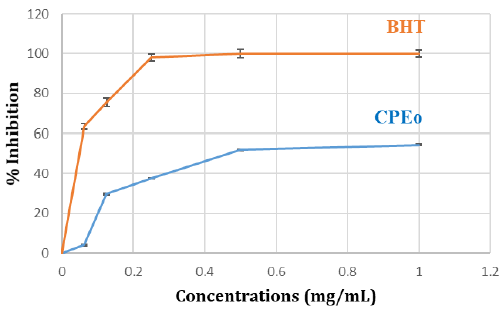
DPPH Radical Scavenging Assay: The curve relating to DPPH radical scavenging shows that the inhibition percentage of the free radical increases with the increase of concentration, either for BHT or CPEo (Figure 1). In effect, Citrus paradisi essential oil has a significant antiradical effect attending 54 % and can be qualified such having an important antioxidant activity.

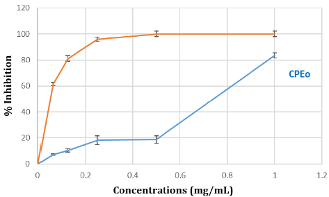
ABTS+ Radical Scavenging Effect: The results illustrated in the curve show that the percentage of inhibition gradually increases with the concentration of grapefruit essential oil. At a concentration of 1mg/mL, the inhibition rate reaches 83 % (Figure 2).

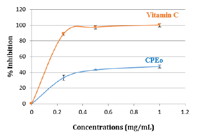
Ferric-Reducing Antioxidant Power (FRAP): The FRAP assay is usually used to measure the capacity of the sample to reduce the ferric complex to the ferrous form. The FRAP assay confirmed the antioxidant activity of CPEo, with values equal to 66 % (Figure 3).
biomedres-openaccess-journal-bjstr

Figure 1: Antiradical activity against the radical DPPH in the percentage of inhibition CPEo. Aliquots of various concentrations of CPEo (0, 0.2, 0.4, 0.6, 0.8, 1mg/mL) and standard BHT were mixed with DPPH and incubated in the dark. The antioxidant activity of CPEo was measured using a spectrophotometer at 517nm. Results were expressed as mean inhibition percentage (%) ± standard deviations (n = 3). BHT was used as the reference standard.
biomedres-openaccess-journal-bjstr

Figure 2: Anti-radical activity against the radical ABTS in percentage of inhibition of CPEo. Various concentrations of CPEo (0 to 1mg/mL) and acid ascorbic were mixed with FRAP reagents. The reduction of ferric ion (Fe3+) to ferrous form (Fe2+) by CPEo produces an intense blue light revealed as a change in absorption at 700nm. Results were expressed as mean inhibition percentage (%) ± standard deviations (n = 3). Ascorbic acid at various concentrations (0 to 1mg/mL) was used as standard.

biomedres-openaccess-journal-bjstr

Figure 3: Inhibition (%) of standard Vit C and CPEo by Ferric Reducing Antioxidant Power (FRAP) assay. Various concentrations of CPEo (0 to 1mg/mL) and acid ascorbic were mixed with FRAP reagents. The reduction of ferric ion (Fe3+) to ferrous form (Fe2+) by CPEo produces an intense blue light revealed as a change in absorption at 700nm. Results were expressed as mean inhibition percentage (%) ± standard deviations (n = 3). Ascorbic acid at various concentrations (0 to 1mg/mL) was used as standard.

NO· Scavenging Assay

The capacity of citrus paradisi essential oils to scavenge NO was also measured. CPEo was checked for its inhibitory effect on nitric oxide production. The inhibition on free radical percentage increases with elevated concentrations of essential oil and Vitamin C (Figure 4).
biomedres-openaccess-journal-bjstr

Figure 4: Inhibition (%) of standard Vit C and CPEo by Ferric Reducing Antioxidant Power (FRAP) assay. Various concentrations of CPEo (0 to 1mg/mL) and acid ascorbic were mixed with FRAP reagents. The reduction of ferric ion (Fe3+) to ferrous form (Fe2+) by CPEo produces an intense blue light revealed as a change in absorption at 700nm. Results were expressed as mean inhibition percentage (%) ± standard deviations (n = 3). Ascorbic acid at various concentrations (0 to 1mg/mL) was used as standard.

Cytotoxicity Activity

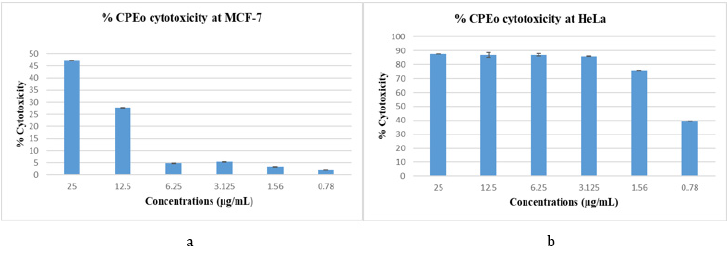
The MTT test studied the cytotoxic effect on HeLa and MCF-7 cells. Cells were cultured in 96-well plates for 48 h, in the presence and absence of the different concentrations of CPEo. The percentage of cytotoxicity was calculated. Our results showed an antitumor potential of the grapefruit essential oils on both cancer cells: Hela and MCF-7 according to the concentrations used: from 0.78 to 25μg / mL (Figure 5).
biomedres-openaccess-journal-bjstr

Figure 5:

(a) Study the cytotoxicity of the essential oil of the grapefruit outer wall in the HeLa
(b) and MCF-7 cell lines.
Cytotoxic activity of citrus paradisi essential oil on cancer cell lines. MTT assessed cell viability. The percent growth reduction was calculated from the extinction difference between treated cell culture and the control. Results are the means of three repetitions.

Discussion

The current study was designed to extract the essential oil from grapefruit. It was obtained by hydro distillation, which is the oldest method and the most used because it is very easy to achieve. Our results agree with studies, which confirmed that Citrus is rich in essential oil, and their yields varied from one plant species to another, ranging from 0.2 to 2.0 % [21]. Other work has shown that yield of Citrus paradisi essential oil collected in September in southern Taiwan was attending 0.37 %, and others were collected in October from Iran were 0.85 % [22,23]. So the difference in EOs yields may be related to the plant’s origin, the environmental conditions, time of collection, and extraction method [24]. The CPEo analyzed in this work showed potent radical scavenging activity. We have demonstrated that CPEo has an interesting antioxidant activity highlighted by the DPPH, ABTS+, FRAP, and NO. Our results agree with the literature, which showed that citrus EO possesses an anti-free radical activity DPPH of 17.7 to 64 % [7]. Other studies on grapefruit seed oil showed a high antioxidant potential (61 %) [25]. Furthermore, the effect of citrus paradisi essential oils on ABTS free radical was determined and exhibited a higher radical scavenging activity. Our results corroborate with other studies showing a significant antioxidant effect of C. paradisi EO against ABTS radicals [22]. In addition, the CPEo had a significant FRAP value, which was in accord with the results obtained by Jang et al. that confirmed the higher FRAP values in the peel of grapefruit [26]. The superoxide anion assay is commonly used to evaluate the superoxide anion radical-scavenging ability of plant extract. Therefore, CPEo was checked for their inhibitory effect on nitric oxide production in the present study, attending 47 %. Our results agree with studies that confirmed that the superoxide values of grapefruits varied from 36.83 % to 50.31 % in peels [27]. The antitumor potential of CPEo was tested on HeLa and MCF-7 tumor cells. Our results showed a considerable antitumor activity dependent on essential oil concentration. This result agrees with the work of Zu et al., which shows that essential oil from grapefruit exhibited an antiproliferative effect on MCF-7 cells [28]. Furthermore, Monajemi et al. studied the impact of different concentrations of essential oils of other species of the citrus family as Citrus limon, Citrus medica, and Citrus sinensis on MCF-7 and Hela [29]. They proved a significant decrease in viability in a dose-dependent manner in both tumor cells [29].

Conclusion

Citrus paradisi essential oils are well known for their flavor and fragrance properties and numerous aromatherapeutic and medicinal applications. Accordingly, the essential oils of Citrus species exhibited intense antioxidant activity. In addition, the essential oil of Citrus paradisi showed an antitumor effect against both cancers cells, HeLa and MCF-7.


For more Articles on: https://biomedres01.blogspot.com/

Wednesday, September 28, 2022

Remote Monitoring of the Health Status of Pregnant Women in the COVID-19 Pandemic

Remote Monitoring of the Health Status of Pregnant Women in the COVID-19 Pandemic

The Role of Remote Technologies in the Quality Management System and Safety of Medical Care

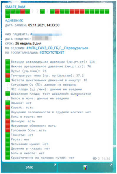
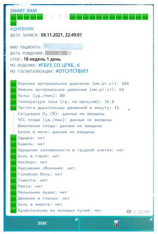
On April 26, 2021, Deputy Chairman of the State Duma Irina Yarovaya at a meeting of the Presidium of the Council of Legislators of the Russian Federation under the Federal Assembly of the Russian Federation called for simplifying the exchange of data between medical institutions and patients. In the Sverdlovsk region, an automated information system of mobile notifications «AIST_SMART» for pregnant patients and doctors began to operate. Using a smartphone or, say, a tablet, pregnant patients in their personal account get the opportunity to keep an electronic diary of self-control of their health. The diary has the functions of automatic interpretation of the results and the formation of signal information for the obstetrician-gynecologist. Now pregnant women do not need to fill out paper diaries of self-control, call their doctor or the reception of the antenatal clinic or wait for a doctor’s call in order to report the results - the process is fully automated. The women’s consultation received an IT tool for remote interaction with pregnant women and women in child child. The introduction of «AIST_SMART» technologies made it possible to replace paper diaries with electronic ones. Medical data of the patient are collected in a single database and allow you to track the dynamics of the patient’s health around the clock. The results of electronic diaries are automatically processed by the system and if no abnormalities are detected, the data is simply recorded in the system and does not disturb the doctor (Figure 1).

In case of detection of deviations in the patient’s state of health, the system marks the identified deviations and sends a notification to the doctor about the current state (Figure 2). Mobile notifications instantly convey accurate and detailed information about the patient’s state of health and thus contribute to the timely decision on hospitalization in case of detection of criteria for weighting the course of NCVI. All notifications in case of deviations are automatically sent to the attending physician and the doctor in the Obstetric Remote Consultation Center (hereinafter referred to as the ADCC) for the routing of the patient 24/7. Remote health monitoring functions as follows.

biomedres-openaccess-journal-bjstr

Figure 1: The data of the expanded diary of self-control at the NKVI, all indicators are normal.

biomedres-openaccess-journal-bjstr

Figure 2: The data of the expanded diary of self-control at COVID-19 with deviations in the state of health.

Registration in the System «AIST_SMART»

To register the patient in the personal account at the initial appointment of a pregnant patient, a consent-instruction [1] is issued to connect to the mobile service «AIST_SMART» with an individual QR code. At home, the patient reads the QR code using the camera of her smartphone or tablet and, according to the instructions, undergoes the registration procedure, forming a digital four-digit PIN-code. From now on, it is guaranteed 24/7 technical support. The QR code serves as the patient’s identifier and the link between her electronic medical record (EHR) in the AIST «RAM» and the personal account in the «AIST_SMART» system. To register a doctor in your personal account, you must log in to the medical information system - AIST «RAM», in which all medical personnel of the obstetric service in the region work. Open the «Personal Account» tab and register by scanning an individual QR code. So, in order to access electronic self-control diaries, the doctor and the patient connect to the AIST_SMART service, and after registering in the system, notifications about the results of remote health monitoring will be received on their mobile device. The doctor does not need to call on the phone to find out how she feels, what her temperature is, the symptoms of SARS, etc.

How the Mobile Alert System Works

Formation of Notification of the Result of Self-Control Diaries

This process is fully automated. AIST_SMART performs the role of an intellectual assistant to the obstetrician-gynecologist/ midwife. The patient fills in the diary data, and the doctor receives ready-made results with automatic interpretation. Now the patient will not forget to call the antenatal clinic, and the doctor will be able to make decisions on the tactics of conducting comprehensively, taking into account the results of the patient’s home self-control and his obstetric status.

Patients with COVID-19 are Asymptomatic/Mild and Receiving Care on an Outpatient Basis (at home)

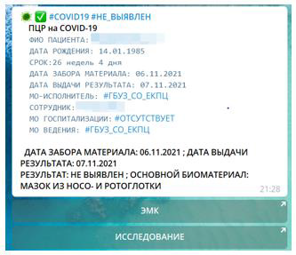
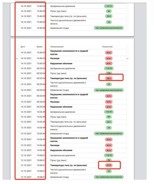
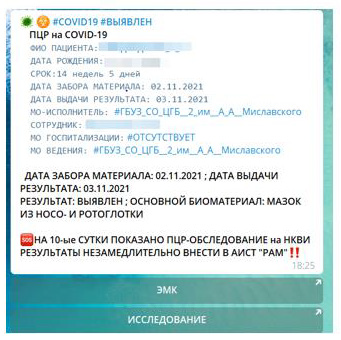
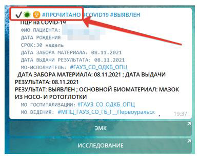
Upon receipt of the results of testing in a pregnant woman / maternity for COVID-19, the data are entered by medical personnel in the AIST «RAM». Notifications about the results are automatically generated in the personal account «AIST_SMART» (Figures 3 & 4). These notifications are automatically sent to both the patient and the doctors. With what there is control that the patient is also informed about the result (Figure 5). If a positive result is detected on the COVID-19, the patient receives notifications 2 times a day about the need to fill out a self-control diary, which is also informed by the doctor - full feedback (Figure 6). The doctor of the ADC, based on the results of the self-control diary (Figure 7) and obstetric status according to the data in the electronic medical record (hereinafter referred to as the EHR) in the AIST «RAM», where there is information about all the results of the examination, the course of pregnancy and diagnoses, decides on further management tactics: to continue outpatient treatment or hospitalization in a covid hospital. The ADCC doctor fixes his decision in the EHR, making out a remote consultation for the attending physician of the antenatal clinic or obstetric hospital (if the patient is in the hospital at the time of detection of the COVID-19).

biomedres-openaccess-journal-bjstr

Figure 3: Mobile NOTIFICATION of PCR result for COVID-19: not detected.

biomedres-openaccess-journal-bjstr

Figure 4: Mobile NOTIFICATION of PCR result for COVID-19: DETECTED.

biomedres-openaccess-journal-bjstr

Figure 5: Marking about the patient’s reading of the results of the examination.

biomedres-openaccess-journal-bjstr

Figure 6: Mobile notification that a reminder has been sent to the patient to complete a self-monitoring diary.

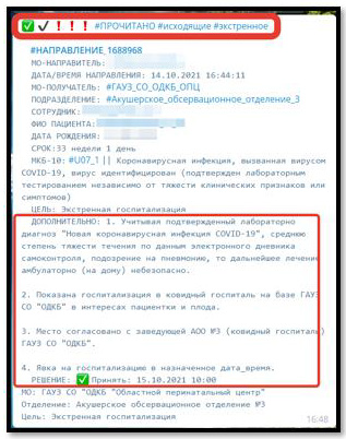
If a decision is made on the need for hospitalization, the doctor of the ADC through a confidential «working» chat in AIST_SMART can contact the patient and clarify her consent to hospitalization and the possibility of transportation by personal transport. If consent is obtained (Figure 8), the ADC doctor makes an additional referral for (re-) hospitalization to a particular covid hospital for pregnant women and women in childcare, taking into account available places. The patient receives a notification about the referred referral indicating the covid hospital, the date and time of hospitalization (Figure 9). If it is necessary to organize transportation, the doctor of the ADC has resources through communication with the medical organization where the patient is on the dispensary register and agreeing on the method and time of transportation by the NSR team in compliance with epidemiological rules (Figure 10).

biomedres-openaccess-journal-bjstr

Figure 7: Dynamics of the state of health according to the electronic diary of self-control.

biomedres-openaccess-journal-bjstr

Figure 8: Communication with the patient through confidential “working” chat in AIST_SMART.

You do not Need to Receive a Paper Direction

If necessary, you can print the direction at the place of treatment of the patient, using a single information space of the regional obstetric monitoring of AIST «RAM». All the directions that a woman received during pregnancy are reflected in her personal account in the «My directions» section. The patient can open any document, even if the connection with the internet has disappeared.

biomedres-openaccess-journal-bjstr

Figure 9: Notification of referral to a Covid hospital and labeling of reading by a patient.

Advantages of Remote Monitoring of Health

The transition to electronic diaries of self-control allows you to identify the weighting of the course of ARVI / ARI in the case of outpatient treatment (at home) with COVID-19, and timely send the patient to hospitalization to prevent adverse events, which is from the main directions of the quality management system and safety of medical care. AIST_SMART allows you to create constant feedback [2] with the patient and thereby form a patient-centric model of care as one of the priority areas for the development of modern medicine and healthcare in general. All of the above increases the compliance of doctor-patient interaction and directly affects the quality and safety of medical care in the difficult conditions of the NCVI pandemic, which meets the modern needs of society and solves the tasks set by the Government of the Russian Federation in the field of digitalization of healthcare [3-8].

biomedres-openaccess-journal-bjstr

Figure 10: Finding out the possibilities of transportation and hospitalization to the covid hospital through a confidential “working” chat in AIST_SMART.

For more Articles on: https://biomedres01.blogspot.com/
Tuesday, June 28, 2022

Cobalt Doped TiO2/rGO Nanocomposites as Highly Efficient Photocatalyst for Water Purification


Cobalt Doped TiO2/rGO Nanocomposites as Highly Efficient Photocatalyst for Water Purification


Introduction

Photocatalysis is a crucial research filed, which solves the problem of energy and environmental pollution in the world in an economical and sustainable way [1]. Titanium dioxide (TiO2), as the most common candidate among various semiconductor photocatalysts, has been widely utilized in the environmental filed because of its high activity, long-term stability and low toxicity [2-6]. However, because of its wide band gap (Ebg≈3.2eV for anatase type TiO2) and high recombination rate of electron-hole pairs, TiO2 can solely adsorb the UV light which is merely 3~5% of solar spectrum, resulting in low utilization of the majority of the solar energy [7-9]. In order to overcome these drawbacks, various improvement methods have been explored including heterogenous composition [10,11], element doping [12,13], surface modification and the like. Among them, the element doping of TiO2 photocatalysts has been considered as a feasible method to improve the interfacial charge-transfer efficiency, narrow the band gap and delay the recombination of carriers. Up to now, transition metal such as Co [14,15], Pt [16], Sn [17] and Fe [18] has been reported to be successfully doped into TiO2, and the light response wavelength of the obtained materials showed significant red-shift. According to literatures, transition metals cobalt is considered as one of the best candidates to reduce the electron-hole recombination rate and transfer the adsorption edge to the visible light region [19- 21]. The cobalt oxide-loaded TiO2 (TiO2-CoO) support with reduced graphene oxide (rGO) was fabricated by sol-gel method and utilized to remove 2-chlorophenol (2-CP).

The removal efficiency of 2-CP was 98.2% with the ternary nanocomposite in the visible region [22]. The ternary rGOTiO 2/Co3O4 nanocomposites were successfully prepared by coprecipitation method, and exhibited the highest degradation performance of methylene blue (MB) and crystal violet (CV) dye under visible light [23]. As a result, cobalt doped TiO2 photocatalysts have shown superior performance in degrading various organic pollutants. Graphene oxide (GO), due to its excellent electrical conductivity, large surface area and chemical stability, has attracted wide attention as a substrate for promoting the uniform distribution of heterojunction materials and enhancing the photocatalytic activity [24-28]. Due to the conjugated structure of GO, the nanocomposite of modified TiO2 supported with graphene oxide were the perfect combination to enhance the charge separation during the electron-transfer processes. Therefore, the coupling of graphene oxide with some semiconductors has received particular attention in recent years [6]. In this paper, Cobalt doped TiO2/rGO composites was successfully fabricated through hydrothermal method for MB degradation. The results demonstrated that the Co- TiO2/rGO composites remarkably enhanced the MB degradation efficiency. Furthermore, recycling degradation experiments revealed excellent stability of the fabricated Co-TiO2/rGO nanocomposites for treatment of target contaminant.

Materials and Methods

Materials

Cobalt nitrate hexahydrate (Co(NO3)2·6H2O, 98%), Tetrabutyl titanate (C16H36O4Ti), glacial acetic acid (C2H4O2), ethanol (C2H5OH) and macrogol 400 (HO (CH2CH2O) n H) were obtained from He Dong Hong Yan reagent factory of Tian Jin. Natural flake graphite (≥99.85%) was purchased from Sinopharm Chemical Reagent Co. Ltd.

Catalysts Synthesis

Co-TiO2 catalysts were prepared by one-step hydrothermal method. In a typical synthesis procedure, 10mL tetrabutly titanate was dissolved in ethanol (20mL) to form homogenous solution “A”, whereas 0.02g Co (NO3)2·6H2O dissolved in a solution of ethyl alcohol, glacial acetic acid, macrogol 400 and deionized water to form solution “B”. Subsequently, solution “A” was introduced into solution “B”, and the obtained dispersion was heated at 180 °C for 5h. In following step, the prepared catalysts were washed by centrifugation with ethanol and dried at 80 °C. The obtained composites were light yellow particle and calcined in a muffle furnace at 500 °C for 3h, and the obtained sample was named as Co- TiO2. The GO was synthesized by modified Hummers method. 20mg GO powder was dispersed in a solution of deionized water (40mL) and ethanol (20mL) through 30min ultrasonic, and then 200mg Co-TiO2 was introduced to the GO suspension under vigorous stirring. Subsequently, the solution was heated at 140 °C for 5h. The resulting precipitate was washed with deionized water and dried at 80 °C. The final composites powders were labelled as Co-TiO2/rGO- 2. For comparison, the samples prepared by adding 10mg, 30mg GO powder were denoted as Co-TiO2/rGO-1 and Co-TiO2/rGO-3, respectively.

Characterization Methods

The purity and crystallinity of the prepared samples were collected by Bruker D8 Advance X-ray diffraction (Germany) with Cu Kα radiation. The morphology of the photocatalysts was characterized via Scanning electron microscope (Hitachi SU-4800). The Ultraviolet-Visible (UV-Vis) diffuse reflectance spectra (DRS) were implemented by using UV-3600 Plus. X-ray photoelectron spectroscopy (XPS) were obtained by ESCALAB 250XI (ThermoFischer Electron Corporation, USA). Electrochemical measurements were carried out on CHI 660E electrochemical workstation.

Photocatalytic Degradation

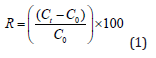
The photocatalytic efficiency of Co-TiO2/rGO samples was investigated with MB degradation under visible light. The visible light source was Perfect 300W Xe-lamp (with a 420nm cut-off filter). In each experiment, 20mg of Co-TiO2/rGO composite were added to 150mL MB solution (20mg/L). The suspension was stirred in the dark for 60min to ensure the attainment of adsorption-desorption equilibrium. 5mL sample solution was extracted at predetermined time and analyzed by UV-3600 plus. The removal efficiency (R) of MB was calculated by Eq. (1).

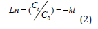
It was expected that the degradation of the MB obeyed the pseudo-first-order reaction kinetics as follows:

where C0 (mg/L) was the initial concentration of MB, Ct (mg/L) was the concentration of MB at time t, k was the kinetic constant.

Results and Discussion

Structure Characterization

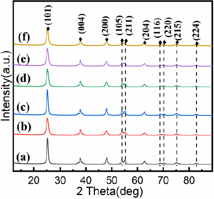
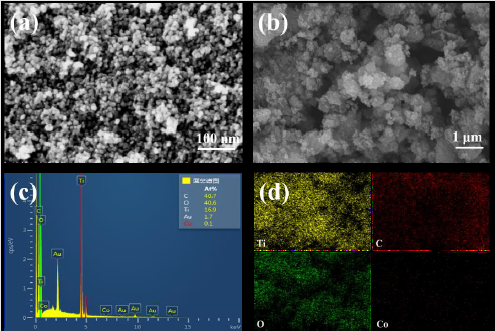
Figure 1 were the XRD patterns of the as-prepared nanocomposites. It was clear that all samples exhibit similar diffraction peaks. The peaks located at 2θ = 25.34°, 37.85°, 47.99°, 54.04°, 62.67°, 68.79°, 70.31°, 75.05° and 82.49°, which could be indexed to (1 0 1), (0 0 4), (2 0 0), (1 0 5), (2 0 4), (1 1 6), (2 2 0), (2 1 5) and (2 2 4) planes of anatase TiO2, demonstrating the high purity and good crystallinity of the samples [29]. The diffraction peaks of Co were not observed, which might be owing to the low content of Co (NO3)2· 6H2O or the cobalt ions were uniformly dispersed into the anatase crystallites. It was noteworthy that the peak intensity corresponding to the (2 1 1) crystal plane in the cobalt-doped nanocomposites varied, indicating that the presence of Co2+ ions around Ti4+ [30]. No significant diffraction peaks were noticed for XRD patterns of Co-TiO2/rGO nanocomposites when compared with Co-TiO2 nanoparticles, which was described the low rGO content in the composite, or of the TiO2 loading on the rGO surface [31,32]. Surface morphology of the as-prepared composites was assayed through SEM analyses. It could be seen from Figure 2a that the Co- TiO2 particles were subsphaeroidal and well-dispersed. Figure 2b showed that the agglomeration occurred when subsphaeroidal Co- TiO2 particles were combined with graphene sheets. The element composition of the Co-TiO2/rGO-2 nanocomposites were confirmed by EDS analysis. In the element mapping images (Figures 2c & 2d), C, Ti, O and Co disperse uniformly in the selected area of Co-TiO2/ rGO-2, suggesting that cobalt atoms were successfully doped into the composites. According to these images, the cobalt atoms were evenly distributed in TiO2 particles, indicating that the interaction between cobalt and TiO2 particles was excellent in the hydrothermal synthesis procedure [15].

Figure 1: XRD patterns of

a) TiO2;

b) Co-TiO2;

c) TiO2/rGO;

d) Co-TiO2/rGO-1;

e) Co-TiO2/rGO-2;

f) Co-TiO2/rGO-3

Figure 2: SEM images of

a) TiO2;

b) Co-TiO2/rGO-2;

c) C and

d) EDS analysis of Co-TiO2/rGO-2.

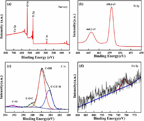
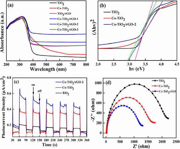
The chemical oxidation state of Co-TiO2/rGO-2 nanocomposites were measured by XPS analysis (Figure 3). As shown in Figure 3a, the XPS survey spectrum of the Co-TiO2/rGO-2 presented that C, O, Ti and Co elements could be revealed, which could consistent well with the result of EDS element mapping. The spectrum of Ti 2p (Figure 3b) exhibited two main peaks at 464.2 and 458.4eV, which were assigned to the Ti 2p1/2 and Ti 2p3/2 [33]. The C 1s spectrum of Co-TiO2/rGO-2 composite was fitted into four peaks at 292.3eV, 288.1eV, 285.9eV and 284.3eV, which were signed to C= O, C= O= C, C= OH and C= C/C= H, respectively [34,35]. In the Co 2p core level of the Co-TiO2/rGO-2 nanocomposites (Figure 3d), the peak appearing at 781.2 corresponded to Co (II) ions [36,37]. The optical property of TiO2 and Co-TiO2/rGO was inspected by UV-Vis adsorption spectra, as displayed in Figures 4a & 4b. Pure TiO2, with equal to 3.18eV and adsorption edge at 390nm, showed almost no visible light adsorption. Compared with the adsorption edge of pure TiO2, a strong light adsorption intensity at approximately 430nm was observed for the Co-TiO2/rGO-2 composites, which was associated to the formation of Ti-O-C bonds, resulting in reduced excited photons energies and hence low band gap energy [38]. As a result, the visible light adsorption efficiency of Co-TiO2/rGO-2 can be effectively enhanced due to the cobalt cations and rGO, which is beneficial to improving the photocatalytic degradation activity.

In order to reveal the behaviors of charge transfer and separation in the prepared photocatalysts, the photocurrent response and electrochemical impedance spectroscopy (EIS) were recorded [39]. Figure 4c showed the transient photocurrent responses of TiO2, Co-TiO2 and Co-TiO2/rGO-2 composites. It could be found that the photocurrent densities of Co-TiO2/rGO- 2 composites were significantly higher than that those of other samples, implying the efficient separation efficiency of electronhole pairs. Figure 4d exhibited EIS changes of TiO2, Co-TiO2 and Co- TiO2/rGO-2 composites. It was clearly observed that the Co-TiO2/ rGO-2 possessed much smaller arc radius relative to TiO2 and Co- TiO2, indicating that Co-TiO2/rGO-2 had lower resistance and faster separation of electron-hole in the charge transfer processes, which could well correspond to the photocurrent results.

Figure 3:

a) Full XPS spectrum and high-resolution spectrum of

b) Ti 2p

c) C1s and

d) Co 2p

Figure 4:

a) UV-Vis diffuse reflectance spectra of TiO2, Co-TiO2, TiO2/rGO and Co-TiO2/rGO;

b) Plot of Kubelka-Munk function versus band gap energy of TiO2, Co-TiO2 and Co-TiO2/rGO-2;

c) The transient photocurrent density of TiO2, Co-TiO2 and Co-TiO2/rGO-2;

d) Electrochemical impedance spectra of Nyquist plots of TiO2, Co-TiO2 and Co-TiO2/rGO-2.

Photocatalytic Performances

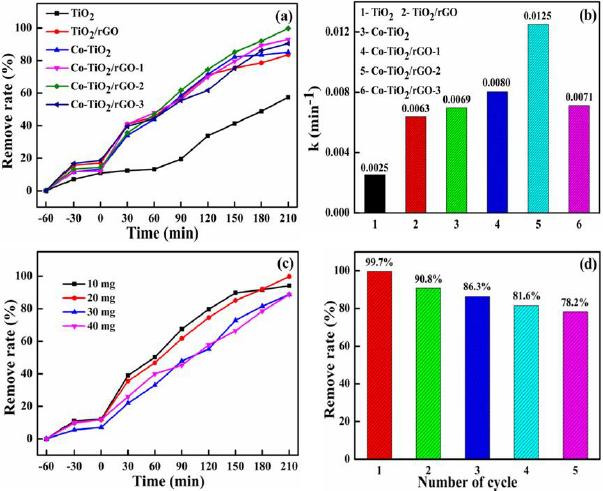
The photocatalytic performances of the TiO2, Co-TiO2, TiO2/rGO and Co-TiO2/rGO composites were evaluated by degradation MB. As exhibited in Figure 5a, the Co-TiO2/rGO-2 nanocomposites had the highest photocatalytic performance. For pure TiO2 nanoparticles, only 57.4 % of the MB was removed following 210 min under visible light irradiation. Nonetheless, the removal percentage of MB by TiO2/rGO and Co-TiO2/rGO-2 nanocomposites was 83.5% and 99.7%, respectively. The enhanced activity of the Co-TiO2/rGO-2 nanocomposites might have been attributed to the introduction of Co ions and rGO. Figure 5b manifested the kinetic constant (k) of the as-prepared photocatalytic. The k value of pure TiO2 and TiO2/ rGO were 0.0025 and 0.0063 min−1, respectively. While the Co-TiO2/ rGO-2 nanocomposites exhibited the highest MB photodegradation rate (0.0125 min-1), which was almost 5 and 1.98 times faster than those of the TiO2 and TiO2/rGO, respectively. To identify the optimum dosage of the photocatalyst, a series of experiments were carried out by varying the concentration of catalyst from 10mg to 40mg in 150mL of MB (20mg/L) (Figure 5c). It was realized that the removal efficiency of MB increased from 69.3% to 99.7% with the Co-TiO2/rGO-2 nanocomposites increased from 10mg to 20mg, which was ascribed to the availability of enough active sites on the catalyst surface. Whereas the remove efficiency decreased with a further increase in the Co-TiO2/rGO-2 dosage, which was ascribed to the agglomeration of the photocatalyst. Based on the above results, the optimal dosage of Co-TiO2/rGO-2 nanocomposites for MB degradation was to be 20mg. The stability and recyclability of the photocatalyst exerts great impact on the operating cost of wastewater treatment. Therefore, the stability of the photocatalysts was evaluated for the Co-TiO2/rGO-2 nanocomposites and the results were showed in Figure 5c. The study indicated that the removal efficiency of MB was still 78.2% after five recycling runs, indicating the activity of the recovered Co-TiO2/rGO-2 was stable enough for recycling. Therefore, Co-TiO2/rGO-2 nanocomposites were expected to be promising in environmental remediation because of their excellent photocatalytic activity and stability.

Figure 5:

a) Photodegradation of MB under simulated solar irradiation over the as-prepared photocatalytic;

b) The kinetic constants of the as-prepared photocatalytic for the MB photodegradation;

c) Efficient of the dosage of the MB photodegradation by Co-TiO2/rGO-2;

d) Effect of cycling times on photocatalytic efficiency on Co-TiO2/rGO-2

Proposed Mechanism for Photocatalytic Degradation of MB

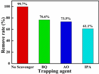
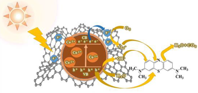
To determine the active species (such as •OH or h+ or •O2 - radicals) and further explore the photodegradation mechanism, isopropyl alcohol (IPA), ammonium oxalate (AO) and 1,4-benzoquinone (BQ) were used as the radical scavengers [40,41]. The experiment data revealed that the photocatalytic activity of Co-TiO2/rGO-2 was decreased by adding the radical scavengers but to different degrees (Figure 6), indicating that all the above active radical species were responsible for the MB degradation. Notably, the photocatalytic performance dropped sharply to 61.1% with the addition of IPA, demonstrating that h+ radical was the main active species in the MB degradation process. Based on the above characterization and photocatalytic activity results, a plausible mechanism of Co-TiO2/ rGO-2 for MB degradation has been proposed and shown in Figure 7. The improvement of TiO2 photocatalytic performance could be explained as follows:

1) The doping of optimal Co2+ into the lattice of TiO2 nanosheet could efficiently reduce the band gap width of TiO2 and increase the adsorption of visible light [42,43].

2) The specific surface area of the composite increased due to adding rGO, and more active sites could be provided for photocatalytic activity [44].

3) Under the excitation of visible light, the electrons generated by the conduction band of TiO2 were captured and transferred by the graphene layer, which improved the electron-holes separation efficiency [45,46].

The electrons subsequently react with the oxygen molecules adsorbed on the surface of the catalyst to generate •O2 - to degrade MB. At the same time, the residual h+ within the TiO2 VB can be directly or through water oxidation to generate ·OH radicals, and in turn photo oxidize of MB [15]. In summary, the addition of Co metals to TiO2/reduced graphene oxide composite have demonstrated to be beneficial for degrading MB, which was consistent with the electrochemical measurements. The synergy effects of Cobalt doped TiO2 and rGO was conducive to the formation of the active sites and the facilitation of the high photocatalytic performance. Therefore, Co-TiO2/rGO composite offered an excellent combination of high activity and long-term performance durability.

Figure 6: Effects of radical scavengers on the degradation of MB over Co-TiO2/rGO-2 nanocomposite.

Figure 7: The probable photocatalytic degradation mechanism for MB by the Co-TiO2/rGO nanocomposites.

Conclusion

In conclusion, an efficient Cobalt doped TiO2/rGO photocatalyst was successfully prepared, and the properties of Co-TiO2/rGO nanocomposites were investigated. It was noticed that the Co-TiO2/ rGO-2 revealed an excellent photocatalytic performance in the MB degradation process. Compared to TiO2, MB degradation percentage was increased from 57.4% to 99.7% in the existence of Co-TiO2/rGO- 2. This phenomenon could be explained as the special properties of reduced graphene oxide components and cobalt dopant, which facilitate the separation of photo-generated carries and extend the adsorption spectrum of TiO2 into visible region. Furthermore, the degradation percentage of MB was still obtained to 78.2% after five cycles. Therefore, Co-TiO2/rGO nanocomposites have promising applications in degradation of the organic compounds in the coloring, petroleum and leather industries.

For more Articles on : https://biomedres01.blogspot.com/


Monday, May 2, 2022

Pyometra in a Cat: A Clinical Case Report


Pyometra in a Cat: A Clinical Case Report


Introduction

Pyometra is an acute or chronic suppurative inflammation of the uterus. It is characterized by endometrial hyperplasia with cystic dilation of endometrial glands and accumulation of a neutrophil-rich exudate in the uterine lumen. The incidence of feline pyometra is still not well documented and probably underestimated because queens often don’t present with clinical signs [1]. No prevalence data for pyometra have so far been described in cats, but observations of most veterinarians are that the disease is observed less commonly than in dogs. The most common clinical finding in case of 75% of pyometra cases is mucopurrulent to hemorrhagic vaginal discharge [2]. The clinical presentation of pyometra is similar in cats and dogs. In ‘open-cervix pyometra’ a blood stained; purulent vaginal discharge may be the only clinical sign. Animals with ‘closed-cervix pyometra’ may not show any vaginal discharge and are more commonly systemically ill because resorption of bacterial toxins from the uterine lumen into the circulation can result in endotoxaemia. Bacteremia may also occur. Non-specific clinical signs such as anorexia, vomiting, lethargy, loss of weight and unkempt appearance can also be observed [3]. Polyuria and polydypsia do not occur as often as in dogs. They were reported only in 9% of the cases [4].
Abdominal ultrasound is the most important diagnostic tool in a pyometra case. The uterine horns typically appear distended with hypo-/ to hyperechoic fluid with or without flocculation. The uterine wall often appears thickened with irregular edges and small hypoechoic areas consistent with cystic changes of the endometrial glands. The pyometra can be diffuse or segmental. Cytology of the uterine or vaginal discharge is likely to reveal degenerative neutrophils and phagocytized bacteria. Leukopenia can be present in around 5% of the cases [4]. Treatment includes correction of fluid deficits, proper administration of antibiotics against bacterial organisms and removal of infected uterine contents. The other management includes surgical removal of ovary and uterus (ovariohysterectomy) or use of by PGF2α [3]. The decision to try medical or surgical therapy is based on the physical status and breeding capacity of the queen.
However, some complications may develop after ovariohysterectomy (OHE), such as ovarian remnant syndrome (ORS). This syndrome may develop because of the failure to totally remove both ovaries (most commonly the right ovary) at OHE, or the presence of a partial or complete separation of a portion of normal ovary (the fragment may be located near the ovary or in the broad ligament) that is not detected at OHE. In some cases, uterine stump pyometra may occur because of ovarian remnants and this situation may be fatal in affected queens. Worldwide, fatal complications occur as a result of surgical errors in routine OHE. In this article, we report and discuss the procedure and importance of ORS in a queen.

Materials and Methods

History and Clinical Examination

An eleven years old local breed cat was admitted to Teaching and Training Pet Hospital and Research Centre, Chittagong Veterinary and Animal Sciences University, Bangladesh, with history of anorexia, chronic emaciation. At first, general physical examination was done, then special examination was done. On physical examination, body temperature found 1010C, heart rate 174 beats per minute and respiratory rate 42 breaths per minute.

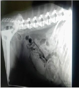
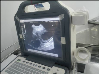
On abdominal ballottement the uterus felt harder and enlarged than normal. Lateral radiograph revealed multiple tubular, radioopaque fluid filled structures from caudal to mid abdomen (Figure 1). The structures appeared distinct and separate from the intestinal loops. Ultrasonography was performed using a B mode real-time 5MHz linear transducer. The finding of abdominal ultrasound was found multiple anechoic fluid filled area without foculation (Figure 2). Then blood sample was collected for doing routine examination and serum analysis.

Figure 1: Multiple tubular, radio-opaque fluid filled structures

Figure 2: Multiple anechoic fluid filled pocket.

Surgical Management

Restraining and Anesthesia: Firstly, the cat was being held on its side with its back against the handler, while the handler grasps the front and back legs, with a forearm across the cat neck. As premedication agent atropine sulphate was administered (Injection Atropine®, Techno drug, Bangladesh, 0.04mg/kg body weight intramuscularly) and as muscle relaxant xylazine hydrochloride (Injection xylazine®, Indian Immunologicals Ltd, India, 1mg/ kg BW intramuscularly) administered. Again, as a general anesthesia ketamine hydrochloride (G-ketamine®, Gonoshasthaya Pharmaceuticals Ltd., Bangladesh, 15 mg/Kg body weight intravenously) was administered. The maintenance anesthetic dose was given half of the initial dose during the surgery. Preparation of surgical area was carried out after shaving and removing hairs. 70% alcohol scrubbed onto the skin around the surgery area, the area is then covered until surgery, since nothing must touch it once it is cleaned.
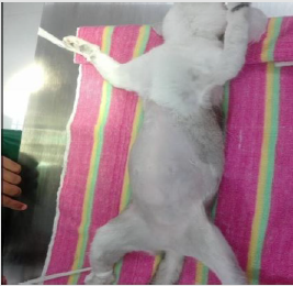
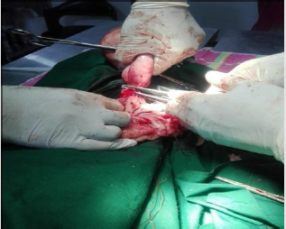
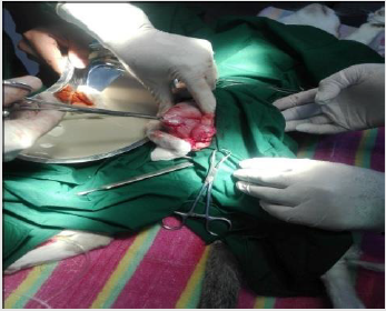
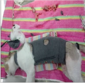
Surgical Procedure: The cat was being laid on her back (Figure 3) and a sterile drapper was placed over her. Close monitoring of temperature, blood pressure, heart rate, gum color, pulse strength and depth of anesthesia was done. An incision was made in the middle of the underside along the length of the abdomen. After exposing the abdomen by laparotomy, the uterine and ovarian blood vessels were properly secured and the ovaries, uterine horns and uterus were completely removed. The abdominal wall was closed with catgut (size: 1-0). The skin was then closed with cross-mattress suture pattern using silk. The sutured wound was covered with the benzoin seal. During the entire operative period, 5% dextrose saline was intravenously infused.

Figure 3: Lying the cat at dorsal recumbent position.

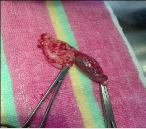
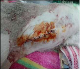
Post-Operative Care: After surgery, antibiotic ceftriaxone @20 mg/Kg body weight (Injection Triject vet 1gm®, SK+F Pharmaceuticals, Bangladesh) was administered intramuscularly daily for 7 days. Antihistaminic chlorpheneramine maleate @1mg/ Kg body weight (Injection Astavet®, Acme Laboratories Ltd., Bangladesh) was administered intramuscularly daily for 7 days. Analgesic (Injection meloxicam @40 mg/Kg body weight and Injection Melvet®, Acme Laboratories Ltd., Bangladesh) was administered subcutaneously daily for 5 days for pain management. The patient was kept in clean squeeze cage and observed for 7 days. No complication was noted, and the bitch recovered uneventfully. On the 14th day, the suture was removed, and it was noticed that the surgical site was healed completely (Figures 4-9).

Figure 4: Ligating the uterus at the base of the uterine body.

Figure 5: Removal of ovaries.

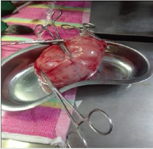
Figure 6: Removal of uterus.

Figure 7: Release of pus from uterus.

Figure 8: Application of benzoin seal.

Figure 9: Cat after surgery.

Results and Discussion

Pyometra is a uterine inflammatory disorder characterized by cystic endometrial hyperplasia [5]. Potter et al. Potter et al. [6] concluded that the prevalence of pyometra in cats increases with age in sexually intact female cats and mainly after parturition, while Agudelo, [7] suggested that the disease is common in queens older than three years and in other queens older than five years with no relationship to the number of parturitions, these close to findings were reported in this case. Hagman et al. [8] found comparatively higher prevalence of pyometra in Bengal cat which is almost similar to this study. Pyometra is a disease of the middle-aged or older animal which was also stated by Brady et al. It could be speculated whether this increase is related to degenerative changes in the uterus or other conditions such as ovarian pathologies or uterine neoplasia that more often affect older animals and may predispose for developing pyometra. But it has been described also in younger cats [9-11].

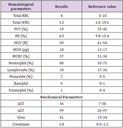
Present study before treatment the hemoglobin level of cat was decreased indicating anemia which agrees with the previous reports [12,13]. This might be due to loss of red blood cells by diapedesis into uterine lumen apart from depressed feed intake and impaired erythropoiesis under toxemic condition in severely affected cases [14]. The PCV level was decreased in the bitches indicating a mild normocytic, normochromic philia might be due to and regenerative type of anemia [15]. According to Greene et al. [16] total erythrocyte count before treatment was decreased in the bitches affected with pyometra indicating anemia which is similar to this study. It might be associated with the toxic depression of the bone marrow whereas severe non-regenerative, microcytic, hypochromic anemia accompanied by extremely high white blood cell levels might be indicative of a concurrent blood loss possibly by diapedesis into luminal pus and due to shortened life span of circulating erythrocytes associated with iron deficiency [17]. Different degree of leucocytosis was observed in bitches affected with pyometra which is consistent to this study. It might be due to severity of the inflammation varying between animals.

In the present study, absolute neutrophilia, lymphopenia, monocytosis with normal eosinophil count was the most consistent finding among the bitches affected with pyometra. Neutrophilia with regenerative shift to the left might be due to retention of purulent exudates in the uterus which exerts a chemotactic effect on neutrophils resulting into accelerated granulopoiesis and lymphopenia might be due to severe stress and elevated monocyte count might be due to chronic suppurative process [12]. Neutrophilia is a typical feature in hematology of bitches affected with pyometra [18] which might be due to influence of toxins in pyometra [19]. The ovariohysterectomy is the useful treatment of pyometra. Potter et al. [6] recorded that 61% of affected cats were spayed or died because of complications relating to reproductive tract disease. The case fatality for pyometra overall was 5.6%. In dogs it is reported to be 3% to 4% [20]. The reason for the higher fatality rate in cats is not known, but one theory could be that this species is less sensitive to endotoxin, or not as prone to show clinical signs unless they develop sepsis [21].

Pyometra can cause liver and kidney function changes (Nak et al. 2001). Occasionally, in this study ALT level moderately increased which support the finding of Nak [22]. Because of septicemia hepatocellular damage were happened resulting diminished hepatic circulation and cellular hypoxia in the dehydrated cats. In this study, decreased ALT result can explain by a process of inhibition of liver enzyme synthesis or possible hepatic membrane damage. Renal dysfunction may develop secondary related to bacterial endotoxin to pyometra. In this case, blood urea nitrogen and creatinine concentration increased it might be due to dehydration [23-27]. A high creatinine concentration was determined in 12% of a group of cats with pyometra Kenney et al. [28].

In this study, ovariohysterectomy was performed under general anesthesia using xylazine hydrochloride and ketamine hydrochloride which is almost similar to study of Deniz et al. [28]. The causes of postoperative wound dehiscence include rough handling and tearing of tissues during surgery, improper selection of the suture material, inefficient suturing, infection, hematoma or seroma formation, failure to obliterate dead space and training of the animal. In such cases, debriment and fresh coaptation of the wound is indicated [29].

Table 1: Hematological and biochemical analysis of pyometric cat.

For more Articles on : https://biomedres01.blogspot.com/ 
 

Faba Bean Varieties Response to Phosphorus Application on Yield and Yield Components at Kulumsa Area of Tiyo District, Arsi Zone, South-Eastern Ethiopia

  Faba Bean Varieties Response to Phosphorus Application on Yield and Yield Components at Kulumsa Area of Tiyo District, Arsi Zone, South-Ea...