Showing posts with label Journals on Medical Sciences. Show all posts

Tuesday, August 4, 2020

Current Recommendations on Treatment of Acute Diverticulitis – Mini Review

Current Recommendations on Treatment of Acute Diverticulitis – Mini Review


Introduction

Diverticulosis is a common disease in Western world and is associated with a Western lifestyle with low fiber diet, smoking, constipation. It is quite rare in the East, however it’s incidence is growing also in the Eastern countries. Diverticula are a small outpouching of the colonic wall and are harmless, unless they became inflammed or cause other symptoms. Colonic diverticula can occur anywhere in the colon or rectum, but they most often occur in the descending and sigmoid colon [1]. The majority of individuals (80-85%) with colonic diverticula remain asymptomatic, 10-15% of people with colonic diverticulosis develop abdominal symptoms, such as abdominal pain, bloating or changes in bowel habits. Those symptoms, without macroscopical changes within diverticula, we describe as a symptomatic uncomplicated diverticular disease (SUDD). The other complications to diverticular disease are acute inflammation of the one or more diverticula (acute diverticulitis), bleeding, fistulas with other organs, intestinal obstruction. The lifetime risk of developing acute diverticulitis in people with colonic diverticulosis is about 4% [2]. This mini-review article summarizes some latest guidelines and suggestions regarding treatment of acute divericulitis, which were proposed mainly by the World Society of Emergency Surgeons (WSES) [3].

Mini Review

Diverticulum is the protrusion of the inner layer of the intestinal wall (mucosa and submucosa) through the weak points in the muscular layer of the intestinal wall, forming small pouches (diverticula) that bulge out the large bowel. Inflammation of diverticula is defined as diverticulitis and is most sommon in the sigmoid colon [4]. Acute diverticulitis can be simply classified as uncomplicated and complicated. In uncomplicated acute diverticulitis the inflammation is limited to the bowel wall and does not spread beyond the visceral peritoneum. Complicated diverticulitis is, when the inflammation spreads beyond the bowel wall with formation of pericolic or distant abscesses or perforation in to the abdominal cavity with consequent diffuse purulent or stercoral peritonitis [5]. The most simple and understandable classification of complicated diverticulitis was proposed by Hinchey, which classifies complicated diverticulitis in to four stages. Stage 1 is a acute diverticulitis with a pericolic abscess, stage 2 is acute diverticulitis with a distant intra-abdominal abscess, pelvic abscess or retroperitoneal abscess. Hinchey stage 3 describes complicated acute diverticulitis with diffuse purulent peritonitis and stage 4 with diffuse stercoral peritonitis. Hinchey classification helps us to decide, whether to treat patients conservatively or more invasively with percutaneous drainage or surgery.
Uncomplicated acute diverticulitis can be managed conservatively with antibiotics. Some authors believe, that mild cases of uncomplicated acute diverticulitis are self-limited and dont need antibiotic therapy. Mild cases of uncomplicated acute diverticulitis dont need admission to hospital but need to be carefully followed-up [6]. Approximately 15-20% of patients have complicated acute diverticulitis and have an abscess on CT scan, when they are admitted to hospital. Patients with smaller diverticular abscesses (less than 4-5 cm) may be treated by antibiotics alone. Patients with larger abscesses (more than 4-5 cm) can best be treated by percutaneous drainage combined with antibiotic therapy. Whenever percutaneous drainage of the abscess is not feasible or not available, based on the clinical conditions patients with large abscesses can be initially treated by antibiotic therapy alone. However, careful clinical monitoring is mandatory [3]. In Hinchey stage 3 and 4 acute diverticulitis surgical management is mandatory. Some authors recommend minimally invasive procedure with only laparoscopic lavage and drainage of the abdominal cavity in Hinchey stage 3, when there is present purulent peritonitis without evident hole in the colon.
However, further studies need to be done to elucidate the role of minimally invasive surgery in complicated diverticulitis. The other option, which is currently the gold standard for treatment of a perforated acute diverticulitis with diffuse peritonitis, is standard open surgery with removal of the diseased colon and formation of terminal colostomy (Hartmanns procedure), irrigation and drainage of the abdominal cavity [3,4,6]. The colostomy can be temporary or permanent, depending on the patients age, general condition, comorbidities and also on patients desire to reconstruct the large bowel. In young and healthy patients with acute perforated diverticulitis some authors recommend one stage procedure with primary anastomosis, however there is a risk of anastomotic dehiscence and another operation with formation of the terminal colostomy [3]. Even in young and healthy patients with massive peritoneal contamination it is the best option to perform two-stage procedure with Hartmanns procedure first and terminal colostomy closure later [3-6].

Conclusion


Acute diverticulitis is a complication of diverticulosis. It can be uncomplicated or complicated. Uncomplicated forms of acute diverticulitis can usually be managed conservatively. Complicated acute diverticulitis can also be managed concervatively with antibiotics or in combination with percutaneous drainage. Surgical management must be employed for advanced forms of complicated acute diverticulitis with perforation and peritonitis.

How the Cure for Cancer was Discovered-https://biomedres01.blogspot.com/2020/08/how-cure-for-cancer-was-discovered.html

More BJSTR Articles : https://biomedres01.blogspot.com

How the Cure for Cancer was Discovered

How the Cure for Cancer was Discovered


Opinion

In this account, I must keep places and identities secret but, if you need to know the details, I shall be pleased to tell you in confidence because great emotion is attached to cancer. Everyone expects to die. A man I had known for many years told me that he had prostate cancer and asked if CellSonic VIPP (very intense pressure pulses) could save him. I told him there was a chance. We had a report from a European university that cancer cells were hit with the pressure pulses of the CellSonic type and the replication of mutant cells stopped immediately. There was also a hypothesis from China that pressure kills cancer cells. The authors did not know how to apply the pressure internally so they were only theorising but I knew because the pressure exerted by CellSonic is the same as that used in lithotripsy to fracture kidney stones. The most important consideration was safety. Under no circumstances should the patient be at risk of a cancer being caused or an existing cancer spreading.
The technology in CellSonic goes back forty years when the first non-invasive surgery was invented to break kidney stones. Instead of surgically opening the kidneys, the stones were hit by a lithotripter generating sudden pressure pulses aimed into the body to break the stones into sand small enough to pass through the bladder. Over time, millions of patients had been treated in hospitals in all countries of the world and there were no reports of causing or spreading cancer. I was involved with the first lithotripter in Britain in 1987 and from that had developed CellSonic. On the basis of bad news travelling faster than good news, and there had been no bad news, I was confident that it was safe to treat the cancer patient and the patient, who was in the medical business, agreed. He had the first treatment on a Monday morning. The protocol was 300 pulses per tumour so it took two minutes. No anaesthetic. No drugs. On Wednesday morning he had the second treatment and I telephoned to enquire on progress.

The answer was that the patient was alright. It was unbelievable. He had been dying a few days before. I was angry with them for making a joke in a serious situation so the phone was passed to the patient and his reply was, “I am better now.” And he was. His immune system told him that the cancer had gone. Blood tests the following week confirmed it. More patients were treated and again it was on the third day that the patient declared themselves cured. Exactly how the cancer was cured was not understood until a meeting at Bradford University in England a few months later where they divulged their discovery of the different permittivity values for cancer tissues and healthy tissues. In other words, cancer is an electrical fault. That made sense. The CellSonic pulses lasting less than a nanosecond caused by a 25,000-volt arc over a onemillimetre gap were forming a short duration electromagnetic field. In the words of Dr Steve Haltiwanger, we had created a nonsurgical form of irreversible electroporation using a combination of sound waves PLUS a high-powered electric field. This combination had never been done before and Dr Haltiwanger complemented me by saying, “Noble prize work if you can live to collect it. This is a paradigm-breaking disruptive technology. WOW!” We now have a good working theory to explain how CellSonic VIPP works in cancer. It is the combined effect of sound and electric field that produces the unique effects of VIPP. Take away the short duration electrical field and just use sound waves or a slower rise time electrical field you will not see the same effects - which is why CellSonic VIPP technology is different from competitors.

Therapy of Hyponatremia: End of Era or Minority Report?-https://biomedres01.blogspot.com/2020/08/therapy-of-hyponatremia-end-of-era-or.html

More BJSTR Articles : https://biomedres01.blogspot.com

Therapy of Hyponatremia: End of Era or Minority Report?

Therapy of Hyponatremia: End of Era or Minority Report?


Introduction

I congratulate the authors on their success in investigating and treating this case of severe hyponatremia (HN) [BMJ 2008;337: a2377]. I admire the impressive style and delightfully stimulating “Endgame” analysis. The report highlighted hormonal pathways that play vital role in the patho-physiology and replacement/ maintenance therapy of secondary HN of Sheehan Syndrome (SS). The case report seems impeccable but appraisal/criticism using scientific reasoning analysis illustrates the multidimensional complexity of HN puzzle. It demonstrates how the primary insult causing SS and its secondary HN remains obscure that may be a different primary cause of HN. In sport spirit, may I contribute thoughts to this intellectual “Endgame” analysis? I have spent my career life seriously studying, analysing and resolving the puzzle of HN starting with that of the transurethral resection of the prostate (TURP) syndrome [1] and beyond [2]. The first reported case of HN as the TURP syndrome [3] was made possible after serum sodium estimation became available in clinical practice- as based on previous original animal research work [4].
Brainstorming and anecdotal therapy of HN was first aired at the BMJ and Lancet when I had nothing but observations, ideas and questions. Now I have nothing but answers believed to be worth more than singing about in my kitchen! The points mentioned below are all relevant to the diagnosis, primary insult, pathoaetiology, clinical presentation and the correct therapy of HN.
I. The report mentioned the diagnosis based on timely detection of HN Nadir of 116mmol/l. It occurred postoperatively- 2 days before acute presentation with cerebral MASKs that may range from confusion to coma [5] while its earlier hyper-acute presentation MASK is mainly circulatory shock [6]. Other hyper-acute presentation MASKs include severe cardiac [7], respiratory [8] and renal failure or sudden arrest and death.
II. The condition presenting with shock MASK to urologists, gynecologist, surgeons and anesthetists in theatre and ICU earlier, is the same as that presenting, with coma MASK to physicians later. Its main serum marker is the HN Nadir, if detected.
III. The original event of difficult labour, ruptured uterus, blood loss (of 1.5L in this case), and caesarean section surgery are important predisposing factors for the insult inducing brain and pituitary edema, infarction and later atrophy.
IV. The pituitary oedema is part of a general oedema affecting brain cells among other vital organs but the brain is contained within the skull. The pituitary is perhaps more vulnerable to vascular necrosis being contained within its closed Selal Turcica. This is a compartmental compression syndrome that compromises its blood supply causing infarction, so it may be the only affected but other cerebral areas and cranial nerves have been reported.
V. The use of vasopressin or ergometrine recommended for control of uterine bleeding [8] in the developed countries and, at times, incorrectly used to induce labour in developing countries, plus the endogenous vasopressin or anti-diuretic hormone (ADH) release are also important predisposing factors in the etiology of primary HN causing brain/pituitary oedema and SS.
VI. The quantity of 5% Dextrose solution given during labour and surgery should be mentioned, as it augments the primary dilution HN causing brain cell oedema and/or pituitary oedema that cause SS and its secondary HN. In lethal cases, 6-7l of 5% Dextrose were reported, by a great author and authority on HN [9] with a correct hypothesis of cellular hypoxia-anoxia [9], despite normal arterial oxygen saturation.
VII. The mentioned hypotension “episode” is consistent primary insult of both the TURP syndrome [6] and SS, that is usually but incorrectly attributed to a known type of shock such as hemorrhagic, hypovolemic or septic shocks.
VIII. The quantity and type of fluids used for combating this hypotension “episode” of shock, to induce volume expansion and to correct hypotension are relevant to the pathogenesis of the brain/pituitary oedema. Please compare the quantity of fluid used to correct a hypotension episode with a 1.5l of blood loss.
IX. Based on volume and osmotic milieu regulations, therapeutic fluids used in clinical practice are of two types: Sodium-free fluids (type 1) and sodium-based fluids (type 2) that induce volumetric overload (VO) with respect to time (t) of the types; VO1 and VO2, , respectively [1].
X. Such volume/time (VO/) concept is directly responsible yet took years to unravel as it is rarely reported or thought relevant as it conflicts with received wisdom.
XI. The classification and type of HN of both primary and secondary to SS, in relation to clinical and biochemical severity grade, require 2 more grades worse than severe in prognosis to represent cases of “critical” and “close” to death cases [5-12].
XII. The induction rate, period to presentation and correction rate signify time importance.
XIII. Grading of severity should match the HN nadir, clinical presentation and the VO/t gain.
XIV. The timely affirmed serum hypo-osmolality and HN nadir are detectable before therapy only, and relate to severity [1].
XV. The associated high sodium loss in hyperosmolar urine was documented in animal research [13] not in the post-TURP clinical research as bladder irrigation did not allow it. XVI. The hydrocortisone replacement for SS HN covers its mineralocorticoid effect that prevents sodium loss contributing to the hyperosmolar urine.
XVII. Hydrocortisone is recommended for shock therapy- when used for combating the primary hypotension “episode” of the hyper-acute TURP syndrome HN shock it minimizes such sodium loss in urine. This partially explains the high sodium loss in hyper-osmolar urine during severe HN and hypoosmolality of the TUR syndrome.
XVIII. Excess sodium loss in urine despite serum hypoosmolality was detected in experimental setting by a great most prolific researcher on the TURP syndrome [13]. I thank the authors for resolving this conflicting issue.
XIX. The main cause of sodium loss in hyperosmolar urine despite serum hypo-osmolality is the excess ADH on the kidney. It implies predominance of volume over osmotic regulation by a responsive kidney. This may cause renal shut down or acute renal failure (ARF). So, VO/t may cause ARF as it does hypotension Shock!
XX. The rejuvenated hypertonic sodium therapy (HST) is the correct life-saving therapy of severe HN of “Critical” and “Close” to death Grades. It requires immediate rapid infusion of 5% NaCl or 8.4% NaCO3- the later is readily available and equally effective.
XXI. Hypertonic Sodium is a potent ADH suppressor that induces massive dieresis with low sodium hypo-osmolar urinewhen high doses of loop diuretics fail [1]. By the end of 10-20 minutes infusion, recovery from coma is witnessed. It corrects the vascular hypotension shock and ARF- inducing massive dieresis shedding the excess VO. This dries the patient out of an internal drowning state, so please do not re-infuse similar volume again!
XXII. The hypertonic sodium therapy is no longer contraindicated, and was not an error when rejuvenated as anecdotal. It is the current therapy recommended by authorities on HN [14]. Its success has been proved by old reliable research [3,4] as was rejuvenated and statistically proved in a recent prospective study [1]. It does miracle if timely given early before brain oedema causes permanent brain damage of coma and vegetative state [9-12], pontine myelinolysis [12] or other cerebral focal damage such as SS.
XXIII. Excess volumetric expansion by any infused VO2 fluid may mask or erase HN Nadir, cause dysnatraemia, hyperosmolarity gap and irreversible shock. Delay in therapy also causes permanent brain damage or death [8-12].
XXIV. Cranial nerves may be focally affected and total bilateral permanent blindness has been reported after the TURP procedure and brain oedema- with and without HN. XXV. Even if all known causes of secondary HN are excluded, the prevalence of primary dilution HN in current hospital practice remains high (1% of all postoperative patients in USA [9]).
XXVI. The HN incidence of severe morbidity and mortality is also high. However, because it wears many presentation MASKs, rarely diagnosed at the hyper-acute setting, its serum HN Nadir can be easily erased or masked without being corrected on infusing VO2 fluid. Also because HN or the TURP syndrome is not listed as cause of death in any mortality register, the cause of death may be recorded under any name of its MASKs such as circulatory failure, cardiac, respiratory or cerebral infarction causes of death [5-12]. Thus the TURP syndrome is thought rare, not seen or does not exist.
XXVII. The TURP syndrome HN has apparently “vanished” from urology just by replacing VO1 (1.5% Glycine) with VO2 (Saline) as irrigating fluid for the TURP procedure! Or, is it reincarnating?
XXVIII. The incidence of HN in SS is 35% in postpartum women [15,16] and 16.8% in post trauma brain injury [17]. All cases have partial or complete empty Sella Turcica on MRI that, no doubt, will increasingly prove valuable tool in early diagnosis of VO/t insult and shock.
XXIX. The secondary HN of SS affects both women and men [15,16]. The TURP syndrome HN affects men and women after the TUR procedure, and affects men, women and children after 5% Glucose infusions [8-12]. The incidence of postoperative HN is 1% in USA [10].
XXX. Most VO/t goes missing from the overloaded circulation as it cannot be accommodated within the vascular system with maximum capacity of 7 litres and the excess far exceeds any loss by normally functioning kidneys before ARF occurs. Where does it all go?
XXXI. The intra-cellular part of the missing VO of both VO/t types causes primary brain oedema and/or pituitary infarctionthe exact pathological mechanism require verification.
XXXII. There are paradoxes here that are inconsistent with received wisdom and current recommendations based on accumulated errors. Examples: Hypotension is not synonymous with hypovolemia. Both types of VO/t cause hypotension shock and ARF, but how and why? This require longer explanation and introduction of new evidence [1,2], but a short answer is:

XXXIII. A new phenomenon called magnetic hydrodynamic mixer discovered in the porous orifice (G) tube [2] may prove powerful contender hypothesis for replacing the erroneous physiological low on the capillary-interstitial fluid exchange making the correct foundation for resolving HN and other puzzles of clinical syndromes caused by VO/t. The evidence on volumetric overload shocks (VOS) [18-20] and the replacement for Starling’s law have been reported [21].

Advancement of Bio inks in three Dimensional Bioprinting-https://biomedres01.blogspot.com/2020/08/advancement-of-bio-inks-in-three.html

More BJSTR Articles : https://biomedres01.blogspot.com

Monday, August 3, 2020

Advancement of Bio inks in three Dimensional Bioprinting

Advancement of Bio inks in three Dimensional Bioprinting


Introduction

Three dimensional bioprinting is the modern technique extensively used in regenerative medicine and tissue engineering for the development of multifaceted biological structures such as complex tissues and organs. An American inventor named Chuck Hull coined the concept of three dimensional printing in early 80’s. Three dimensional bioprinting works by depositing cell-laden biomaterial in a predesigned architecture to engineer functional organs and tissues. Computer aided design (CAD) is being used to design complicated three dimensional structures by processing the data obtained from MRI, X-ray and other imaging techniques. In this way a more personalized, highly precise and patient specific structures could be created efficiently within minimum time and resources. After designing the desired structure, next step of bioprinting involves the deposition of “bio inks” in the designed shapes. These bio inks or biomaterials are enriched with living cells and medium supporting and nourishing these cells [1-2].
Rheological behavior, gelation kinetics, swelling properties and surface tension of bio inks are the key characteristics which influence printability. But the core aspect here is bio fabrication which mainly rely on bio ink dispensing. Thus it is necessary to choose appropriate dispensing technique for relevant bio ink. Three main methodologies vastly used for bioprinting includes; inkjet bioprinting, extrusion based bioprinting and laser assisted bioprinting [3]. Inkjet bioprinting is based on two techniques i.e. thermal inkjet bioprinting and piezoelectric bioprinting. Former vaporizes the bio ink using a heating element and creates pulses of pressure which expels the droplets out. While in piezoelectric bioprinting, acoustic waves are produced by piezoelectric crystals which forces the fluid out of the nozzle head. Droplet size of inkjet bioprinter is 10-50μm and its formation is mainly dependent on the surface tension which arises due to the cohesive forces in the molecules of the liquid. Inkjet bioprinters are vastly used for bioprinting owing to its precision and compatibility with various bio inks but on the other hand, nozzle blockage due to early gelation of bio ink and loss of viability of living cells due shear and heat stress reduces the efficiency of inkjet bioprinters [4].
Unlike inkjet bioprinter, extrusion based bioprinter (EBB) dispenses filaments of hydrogel which are 150-300 micrometer in diameter. Bio ink is fed to plastic syringes and it is extruded out via piston (pneumatic) or screw-driven (mechanical) assembly. Pneumatic bioprinting provides better control of flow rate of bio ink whereas mechanical assembly aids the spatial control and printing of viscous bio inks. Major drawback of this methodology is poor resolution and like inkjet bioprinting; compromised cell viabity and nozzle clogging also occurs [5-6]. Another methodology used for bioprinting is LIFT (Laser induced forward transfer) which is orifice free bioprinting technique. The assembly of LIFT consists of three layers i.e. donor substrate, an absorbing layer made up of gold or titanium and bio ink layer. Laser beam is focused on the absorbing plate which creates a pressure bubble due to local evaporation thus impelling a small quantity of hydrogel towards the collector platform by leaving the donor layer although a loose connection remains. This methodology addresses the nozzle clogging, shear and heat stress [7].

Bio inks for 3D Bioprinting

Raw material used for the manufacturing of 3D printed complex biological structures are considered as bio inks which the substances are mainly comprised of living viable cells or tissues and the matrix which holds these cells together and provide nutrition and support to the cells. Bio inks imitates extracellular matrix which allows adhering, proliferating and differentiating of bio printable cells. Selection of bio inks for 3D printing of the biomaterials is an imperative task. There is a pool of physical and chemical aspects of bio inks to be considered for bioprinting. Basic requirements for bio inks include bio printability, in situ gelation, viscoelasticity, biocompatibility with live cells, tissue regeneration, permeability of oxygen, nutrients and metabolic wastes, biodegradation and sheer thinning [8]. Recent studies have reported various biomaterials can be manipulated as bio inks. An overview of these materials will be discussed in following section. These biomaterials are categorized into two main classes; natural biomaterials and synthetic biomaterials. Both have their own distinct properties such as natural biomaterials show high degree of biomimicking and ability of self-assembling whereas stability, controllability and photo crosslinking are the key features of synthetic polymers.

Agarose Based Bio inks

Agarose is a polysaccharide which is derived from marine algae and has diverse application in biomedical field due its property of gel formation [9]. Agarose based bio inks imparts stability to the 3D structures and it can be used in conjunction with other biomaterials to increase the efficiency. A study has manipulated alginate, agarose and carboxymethyl-chitosan to develop functional neurons from neural cells derived from humans and printed the stable structures by encapsulating the cells [10]. A study has made a comparison between agarose and other hydrogels based on biocompatibility and printability by loading the gels with mesenchymal cells and 3D printed cartilage to examine the differentiation of cartilage to fibrocartilage. All the gels showed viability of around 80% while agarose and alginate showed higher cell differentiation [11,12]. Rapid gel formation, high degree of biocompatibility and rheological properties make it highly preferable for bioprinting. However, owing to its viscous nature it is not commonly used in inkjet bioprinters as it causes nozzle clogging [7].

Collagen-Based Bio inks

Collagen is a protein which is obtained naturally from the extra cellular matrix of many mammals and it is extensively used as bio ink for tissue regeneration, tumor modeling and other tissue engineering techniques. Owing to integrin-binding domains of collagen it facilitates cell adhesion, attachment and proliferation. It is most suitable for extrusion based bioprinting but due to the presence of high degrees of fibrous micro-architecture its use in inkjet bioprinter is limited. Other limitations of collagen based bio inks include, slow gelation rates, instability and formation of fibrous structure at high temperature [13]. A study has reported that cell attachment and cell proliferation of chondrocytes was enhanced by using collagen in conjunction with sodium alginate moreover it has also suppressed the differentiation of the incorporated cells and it is also suggested that the combination has also increased the mechanical strength of the structure thus it is preferred choice for the tissue engineering of the cartilage [14]. In another study has reported that viability and differentiation of human hepatocytes stem cells is improved when collagen was used in varying concentrations in lieu of alginate [15]. Engineered structures have shown enhanced biological and physical properties and a greater number of cells remained after printing. Moreover, engineered tissues and complex models of tumors were also developed by using the transglutaminase-crosslinked gelatin [16].

Hyaluronic Acid Based Bionks

Hyaluronic acid (HA) naturally occurs in all connective tissues and extra cellular matrix of cartilage of all mammals and shows the characteristics like collagen type I. HA is vastly used for tissue engineering as it is highly biocompatible and imparts flexibility to the hydrogels. Chemical modifications of HA increase its rheological properties and photo-cross linking. As HA plays a key role in embryonic development, thus it is favorable to the living cells in the hydrogels and structures bio printed using hyaluronic acid are more stable and mechanically controllable. However, owing to its slow gelation ability it is not suitable for extrusion based bioprinting, but it can be used efficiently by blending it with other hydrogels [1]. The adhesion property of hyaluronic acid based hydrogels was increased by the adding the oligopeptides having cell-adhesive properties while mechanical properties were not affected by this addition [17]. Moreover, Ruthenium-based complexes with visible light also polymerizes the hyaluronic acid-gelatin based bio inks. Owing to this ability cell differentiation and viability of adipose stem cells has been improved [18]. Additionally, hyaluronic acidcarboxymethyl cellulose increases the viability and stability of bioprinter structures, moreover the mechanical properties were also enhanced by modifying the HA based gels with methylcellulose [19].

Fibrin-Based Bio inks

Fibrin is the major clotting protein and can be transformed into hydrogel by the enzymatic reaction of thrombin and fibrinogen. It is highly biocompatible and possess biodegradation, but the structures formed using fibrin hydrogels are mechanically weak. It supports cell proliferation and growth. Due to its property of filaments formation the structures thus produced show high amount of deformation without breakage. It can be used with inkjet bioprinting, but it can cause nozzle blockage due to filament formation. In-vitro properties of fibrin has been investigated by developing a three dimensional structure of urethra [20]. Moreover, the combined effect of fibrin and hyaluronic acid by encapsulating the Schwan cells to investigate nerve regeneration [21].

Matrigel TM

Matrigel is a synthetic material and a commercial product mainly composed of gelatinous extra cellular material which promotes vascularization and outgrowth of cells from various tissue fragments, and promotes the differentiation of cultured cells, additionally complex cellular behavior is also observed when cells are cultured on Matrigel [22-23]. Matrigel imparts mechanical strength, stability and high survival rates to the 3D bioprinted structures as compared to alginate and agarose [24]. It is not a favorable choice as bio ink for extrusion based and droplet based bioprinters [25-26] but due to its optimal viscosity and thermal crosslinking ability it is widely used in laser based bioprinters [27]. Matrigel has been used as a vital instrument for the construction of rodent tumor xenograft models to develop innovative cancer treatments [28]. Moreover, Matrigel was used as a bio ink to print three dimensional biostructure using human osteosarcoma [29].

Pluronic® F-12

Pluronic® F-12 is a synthetic polymer having surfactant properties [30]. It is mostly used in combination with other bio ink materials e.g. PEG to deliver and slowed release of drugs [31] and with methacrylate hyaluronic acid to engineer biostructures with increased mechanical strength [32]. Complex structures can be engineered owing to the reversible properties of Pluronic® F-12. Moreover perusable channel like structures can also be constructed using this material [33]. This material is acceptable for bioprinting for extrusion based bioprinter and but not favorable for droplet based bioprinting due to high viscosity and thermosensitive nature. A study reported that interaction ability of mesenchymal stem cells derived from bone marrow was increased when bioprinted with Pluronic®.

Conclusion


3D bioprinting technology is a futuristic approach for tissue engineering and synthesis of other biological structures with greater feasibility. By improving the quality of bio inks and scaling up the commercialization of 3D products can open new horizons. It can facilitate the patients who are in dire need of specific organs and dependent on the organ donor moreover it can also address the emergency medical needs of patients. Various bio inks with their advantage and disadvantages and their suitable bioprinters for the development of complex 3D biostructures are discussed in this review which will improve the commercial application of 3D printing technology. Natural and synthetic bio inks such as agarose and fibrin based inks and other extracellular matrix based bio inks are showing promising results but there is need to address the problems arising in the printing process. Moreover it is required to modify these bio inks so that single bio ink should have the capability to be used with various bioprinters. 3D bioprinting is a modern technology which has the potential to form such complex biostructures which will help to overcome the hurdles in all medical fields.

A Hierarchical Reduced Markov Model for Reliability Evaluation of Phased-Mission Systems-https://biomedres01.blogspot.com/2020/08/a-hierarchical-reduced-markov-model-for.html

More BJSTR Articles : https://biomedres01.blogspot.com
A Hierarchical Reduced Markov Model for Reliability Evaluation of Phased-Mission Systems

A Hierarchical Reduced Markov Model for Reliability Evaluation of Phased-Mission Systems


Introduction

Many practical systems, such as those encountered in electronic, aerospace and nuclear power have multiple phases of operations during their mission and are generally referred to as Phased-Mission System (PMS) [1]. Compared with general system, the reliability evaluation of PMS is much more complicated because of the components dynamic behavior as well as their dependence across the phases. Considerable efforts have been made in the analysis of PMS. Generally, existing methods can be roughly classified into two groups: Analytical methods and simulation methods [2]. The analytical methods are further classified into combinatorial approaches, state-based approaches, hierarchical approaches and modularization approaches. The combinatorial approaches, especially the binary decision diagram-based algorithm [3,4], exploit the Boolean algebra to reduce the model scale, which are effective in evaluating non-repairable PMS. The state-based approaches are mainly based on Markov model [5,6], which suffers from the state-explosion problem when is applied to large-scale systems. The simulation methods typically offer greater generality in system representation, but they suffer from computational inefficiency and great computer memory consumption [7].
The above-mentioned methods mainly apply to small-scale PMSs due to the high computational complexity. This paper proposed a reduced Markov reliability model by using hierarchical reduction techniques. Compared with the traditional Markov model, the proposed method can efficiently reduce the number of system states, and avoid the well-known state-explosion problem. The remainder of the paper is organized as follows. Section 2 describes the hierarchical reduced Markov model. Section 3 illustrates our method with a specific example. Lastly, section 4 concludes the study and discusses future works.

The Hierarchical Reduced Markov Model

It is common that the real engineering systems provide hierarchical feature which can be used to advantage in the PMS reliability analysis. For example, thrust subsystem for an aircraft frequently effect all phases of a mission, maybe in different quantities, at each stage of flight [8]. Furthermore, the top-level subsystem can be decomposed into lower level components such as compressor and turbine runner. Such structure is common in many other engineering systems, not just those relating to aircraft. The hierarchical architecture is also a usual style in software reliability estimation, in which systems are decomposed into different layers according to the components’ calling relations [9]. And it is also used to describe the interpreter relations of different layers in communication software. Accounting for hierarchical structure in the phase failure logic will enable a more reduced Markov model to be developed. One way of taking advantage of these features is using hierarchical reduction method which can reduce the state space of Markov model efficiently by subsystems replacement. The main idea of this method is generating a hypothetical component to replace the top-level subsystem. The components of subsystems are considered to be all in series or in parallel. Thus, the failure rate and repair rate of the hypothetical component can be derived by manipulation of the corresponding components’ parameters in the subsystem. Also, it is important that the subsystems replaced need to be totally independent of all other sections of the mission failure fault tree.
The detailed process of the hierarchical reduction method is discussed taking a n components subsystem for example. Let λi and μi be the failure rates and repair rates of component Ci . Also, λs and μs are the failure rate and repair rate of hypothetical component Csrepresenting the subsystem.
1) if the n components are series, λs can be given as
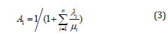
Let As denote availability of the subsystem, where is defined by
A series system is working when all of the components do not fail. If state 1 represents that all components are operational and 1 A is the steady-state availability of system in state 1, then A1 can be computed as follows.
Since state 1 is the only working state in series system, it can be derived that. As = A1 Moreover, according to Eq. (2) and (3), we have
From Eq. (1) and (4), we can use the hypothetical component Cs with failure rate λs and repair rate μs to replace the series subsystem.
2) if the n components are parallel, μs can be given as
For parallel system, the formula of availability As is presented in [19] as follows.
in which Ai = μi/λ +μi is the availability of component ci . Hence, using Eq. (2), (5) and (6), we obtain
From Eq. (5) and (7), we can use the hypothetical component cS with failure rate λS and repair rate μs to replace the parallel subsystem.

Case Study

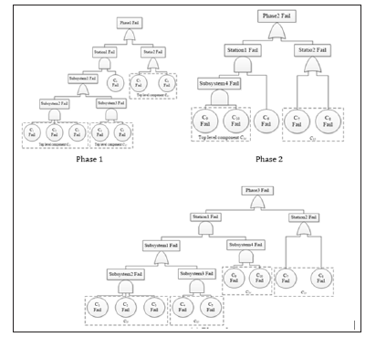
In this section we describe a case study to verify the proposed reduced Markov model for PMS reliability analysis. The mission reliability of the PMS has been calculated by the traditional Markov method. The example PMS is a space flight Telemetry, Tracking and Control (TT&C) system. The failure fault trees of the example TT&C mission are shown in Figure 1, in which station1, station2, and station3 are composed of c1~C6, C7~C8, and C9~c10 respectively. Assume that the failure rates and repair rates of components c1~c10 are unchanged across phases. The relevant parameters are detailed as follows: failure rates μi=3.33×10⁻³ ; repair rates μi=3.33×10⁻²; phase transition time t1 = 20s , t2 = 120s and t3 = 85s .
Figure 1: The failure fault tree of the three basic phases.
biomedres-openaccess-journal-bjstr
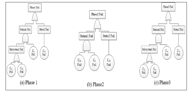
By analyzing fault trees of the three basic phases, there are four subsystems can be reduce using hierarchical reduction method. These are S1with components c1,c2 and c3 , S2 with components c4 and c5 , S3 with components c7 and c8 , and S4 with components c9 and c10 . Therefore, the top level subsystems S1 , S2 , S3 and S4 can be replaced by four hypothetical components cS1 , cS2 , cS3 and cS4 respectively. For the series subsystem, failure rate and repair rate of the top-level components can be computed by using Eq. (1) and (4). Similarly, Eq. (5) and (7) can be used for the parallel subsystem. The computed parameters of the top-level hypothetical components are presented in Table 1. After hierarchical reduction, the new PMS phases configuration is shown in Figure 2.
Table 1: Computed parameters of the hypothetical components (unit: min).
biomedres-openaccess-journal-bjstr
Figure 2: New phases configuration of the PMS after hierarchical reduction.
biomedres-openaccess-journal-bjstr
From Figure 2, we can derive that the number of reduced system states of the three phases are 16, 8, and 16 respectively. For the original PMS, these are 256, 32, and 512 respectively. Because the number of components is decreased efficiently by using hypothetical components to replace the top-level subsystems, and it will slow down the increase in transition rate matrix scale. The PMS reliability and computation time of the traditional Markov method, and the hierarchical reduction method are presented in Table 2. The computing errors of each phase between traditional method and the hierarchical method are shown in Figure 3. Results in Table 2 indicate that there is an excellent match between the proposed method to the traditional Markov method, with relative error less than 1.19×10⁻⁵. Furthermore, the computation time of our method is less than the traditional Markov method. This is mainly because that the number of system states of simplified Markov model is less than that of traditional models. However, from Figure 3 we can see that the computing errors increase dramatically with the mission time. Thus, the hierarchical reduced Markov model is applicable for short duration PMS.

Conclusion

This paper presents a reduced Markov model using hierarchical reduction approach to evaluate the PMS reliability. The traditional Markov model suffers from the problem of huge transition rate matrix. Our approach takes advantage of PMS provide hierarchical feature, and an simplify the original Markov model of each phase by hierarchical reduction. In comparison to the traditional Markov approach, the proposed method achieves less time cost, also has good computation accuracy. However, the hierarchical process may cause computation errors. In future work, more attention will be addressed on error analysis, and efforts should be made to improve current method to achieve more accuracy and efficiency.

Respiratory Muscle In Post-Polio Syndrome: Highlights-https://biomedres01.blogspot.com/2020/08/respiratory-muscle-in-post-polio.html

More BJSTR Articles : https://biomedres01.blogspot.com

Faba Bean Varieties Response to Phosphorus Application on Yield and Yield Components at Kulumsa Area of Tiyo District, Arsi Zone, South-Eastern Ethiopia

  Faba Bean Varieties Response to Phosphorus Application on Yield and Yield Components at Kulumsa Area of Tiyo District, Arsi Zone, South-Ea...