Showing posts with label Journal of biomedical research and reviews. Show all posts

Friday, May 6, 2022

Recurrent ADEM Mimicking Young Stroke

Recurrent ADEM Mimicking Young Stroke

Introduction
ADEM, also known as post-infectious encephalomyelitis, is a demyelinating CNS disorder that usually follows the occurrence of infection, or more infrequently, after the administration of a vaccination [1-3]. ADEM should be suspected when one or more of the following features are present such as a multifocal and polysymptomatic initial presentation, the presence of signs and symptoms suggestive of meningoencephalitis, encephalopathy, bilateral optic neuritis, cerebrospinal fluid (CSF) fluid pleocytosis along with the typical magnetic resonance imaging (MRI) picture [4,5]. It affects children more than adults but can affect anyone. Although it is monophasic by definition, relapsing forms of ADEM have also been recognized.

Case Report

A 30-year male, right-handed, presented with a history of fever for 7 days, holocranial episodic acute severe headache for 3 days, which was associated with nausea, photophobia, and phonophobia. Headache was short-lived but was recurrent, it persisted for around 2-3 hours and get relieved. No triggering factor for this exacerbation. No associated neck pain. The patient also had 1 episode of involuntary body movements, frothing from the mouth, urinary incontinence with loss of consciousness for 1 day. H/O altered sensorium with the irrelevant talk was present. A few hours later, the Patient complaint of Weakness of the left upper and lower limb with facial deviation to the right side. Weakness was to the extent he was unable to lift hand and legs above the bed. On examination, patient was conscious but altered sensorium, left UMN facial nerve palsy, left hemiparesis (Grade- 3/5), brisk deep tendon reflexes, bilateral Extensor plantar reflex, no meningeal signs, no involvement of bowel/bladder, and the sensory deficit was there. Systemic examinations were normal. Blood picture and biochemical investigations were within normal limits. Magnetic resonance imaging (MRI) brain showed focal hyperintense lesions (FLAIR and T2) in the subcortical white matter of right basal ganglia, right corona radiate, left parietal- temporal- occipital region [(Figure 1)-MRI July 2018], and bilateral cerebellar hemisphere and no restriction in DWI &meningeal enhancement in a gadoliniumbased MRI contrast study.

Figure 1: Magnetic resonance imaging brain showing focal hyperintense lesion ( FLAIR& T2) in cerebral cortex and subcortical white matter indicating demyelination of brain in the first episode (July 2018).

Cerebrospinal fluid (CSF) study was done which shows Sugar 45mg/dl, protein 248mg/dl, cell count 25/cmm (99% lymphocytes), negative for Gram-stain or Ziehl–Neelsenstain, and no growth in culture, viral encephalitis panel, and TB PCR was negative. Keeping diagnosis of ADEM, he put on intravenous immunoglobulin (IVIG) for 5 days then oral prednisolone and antiepileptic, and was discharged in stable condition. After 3 months, in November 2018, he was again admitted with a similar severe headache, left hemiparesis grade 2/5, left UMN type facial palsy with left focal seizure and secondary generalization, and global aphasia. MRI brain showed focal hyperintense, demyelinating lesions [(Figure 2)-Nov 2018], in the same brain territory as in the previous episode [(Figure 1)-MRI July 2018]. Immunological markers: Antinuclear antibody, antineutrophil cytoplasmic antibody, anticardiolipin antibody, lupus anticoagulant were negative. Serum Lactic acid and PBF for Sickle cell were normal. MR Venography and CT angiography brain plus neck vessel were normal. His treatment was IVIG for 5 days, followed by oral Prednisolone in the tapering dose and antiepileptics and the patient showed improvement. After follow-up on 1 month, clinically/MRI brain was normal (Figure 3).

Figure 2: Magnetic resonance imaging brain showing focal hyperintense lesion ( DWI& T2) in cerebral cortex and subcortical white matter indicating demyelination of brain in the second episode (Nov 2018).

Figure 3: MRI brain after 1 month showed disappearance of lesions.

Discussion

The index case presented with recurrent episodes of headache, seizures, encephalopathy, a focal neurological deficit in form of hemiparesis and aphasia. The differential diagnosis of recurrent CNS lesions is considered [6]:
1) Recurrent Meningo Encephalitis
2) Reversible cerebral vasoconstriction syndrome(RCVS)
3) MELAS
4) Primary CNS vasculitis/Angiitis
5) Recurrent CNS inflammatory demyelinating disorder like NMO, MS, ADEM
Of the above, Systemic diseases like MELAS were excluded as, S. Lactic acid was normal, no family history, and no multiorgan manifestations like myopathy, hearing impairment, short stature, dementia, and Recurrent meningoencephalitis was excluded as there was no stiff neck or signs of meningismus and CSF HSV PCR was negative. Multiple sclerosis (MS) and ADEM, the two major CNS inflammatory demyelinating diseases, are difficult to differentiate in the initial episode. MS is a continuous demyelinating disease with a characteristically relapsing-remitting course [6]. Although recurrence is characteristic of MS, a second ADEM is described. Furthermore, there are no definite guidelines to differentiate MS and ADEM [6].
Recurrent ADEM (RADEM) is defined as the occurrence of a new episode with a recurrence of the first symptoms and signs, 3 or more months after the first ADEM event and after at least 1 month completing therapy, without a new central nervous system (CNS) lesion (clinical or neuroimaging). The incidence of the second episode has occurred in 10–18% of cases [7]. The category of recurrent ADEM was eliminated in the 2013 criteria, and replaced by the term multiphasic disseminated encephalomyelitis (MDEM), describing 2 episodes consistent with ADEM, separated by at least 3 months [8]. In our patient, 2 episodes of headache, seizures, encephalopathy, a focal neurological deficit in form of hemiparesis and aphasia separated by a period of 3 months, elevated CSF protein, an absent oligoclonal band in CSF, same territory MRI lesions, and complete clinic neuroradiological recovery clinch to the diagnosis RADEM. In previous studies, there are only a few adult RADEM have been reported [8-10]. However, more than two ADEM should be suspicious for MS. Neuropsychiatric features may be the main presentation of a relapse. Since recurrent ADEM is a corticosteroid-responsive condition, awareness and early diagnosis are mandatory [10].

Conclusion
RADEM can be diagnosed clinically, initiated treatment at the earliest because it is most important for the outcome.

For more Articles on : https://biomedres01.blogspot.com/

Wednesday, December 8, 2021

Biostatistical Analysis on Medicinal Plant Toxicity

Biostatistical Analysis on Medicinal Plant Toxicity

Introduction
The human brain is the body’s control center, receiving and sending signals to other organs through the nervous system and through secreted hormones [1]. It is responsible for our thoughts, feelings, memory storage and general perception of the world. The stomach secretes acid and enzymes that digest food. Ridges of muscle tissue called rugae line the stomach. The stomach muscles contract periodically, churning food to enhance digestion. The pyloric sphincter is a muscular valve that opens to allow food to pass from the stomach to the small intestine [2]. The spleen is the largest organ in the lymphatic system. It is an important organ for keeping bodily fluids balanced, but it is possible to live without it [3].

The spleen is located under the ribcage and above the stomach in the left upper quadrant of the abdomen [4]. A spleen is soft and generally looks purple. It is made up of two different types of tissue. The red pulp tissue filters the blood and gets rid of old or damaged red blood cells. The white pulp tissue consists of immune cells (T cells and B cells) and helps the immune system fight infection. The degree of the toxic effect is not the same in all organs [5]. Usually there are one or two organs which show the major toxic effect. These are referred as target organs of toxicity of the particular substance. Terminalia Chebula tree is about 50-80 feet tall in height [6]. It has round crown and spreading branches. The bark is dark brown with some longitudinal cracks. Leaves are ovate and elliptical, with two large glands at the top of the petiole. The flowers are monoecious, dull white to yellow, with a strong unpleasant odour, borne in terminal spikes or short panicles. The fruit is mild laxative, stomachic, tonic, alterative, antispasmodic [7,8].

It is useful in opthalmia, hemorrhoids, dental caries, bleeding gums, ulcered oral cavity. Its paste with water is found to be antiinflammatory, analgesic and having purifying and healing capacity for wounds. Its decoction is used as gargle in oral ulcers, sore throat. Its powder is a good astringent dentifrice in loose gums, bleeding and ulceration in gums [9]. It is good to increase appetite, digestive aid, liver stimulant, stomachic, gastrointestinal prokinetic agent, and mild laxative. The powder of T. chebula fruits has been used in chronic diarrhea. It is used in nervous weakness, nervous irritability. It promotes the receiving power of five senses. It is adjuvant in hemorrhages due to its astringent nature and good for chronic cough, chorizo, sore throat as well as asthma. Also it is useful in renal calculi, dysurea, retention of urine and skin disorders with discharges like allergies, urticaria and other erythematous disorders. Because of the wide use of this plant, theis study is aim at evaluating the sub-acute toxic effect of the ethanol leaf extract of Terminalia Chebula in Wister rats [7-9].

Materials and Method

Animals

Male and female Wister rats were obtained from College of Veterinary Medicine, Northwest A&F University, Yangling, Shaanxi, China. They were maintained on standard animal pellets and given water ad libitum. Permission and approval for animal studies were obtained from the College of Veterinary Medicine, Northwest A&F University.

Plant Collection

Fresh leaves of Terminalia Chebula were collected from its natural habitat from nearby Yunnan province, China. The plant was authenticated from College of Veterinary Medicine, Northwest A&F University.

Plant Extraction

The leaves were shadow dried for two weeks. The dried plant material was further reduced into small pieces and pulverized. The powdered material was macerated in 70% ethanol. The liquid filtrates were concentrated and evaporated to dryness at 40°C in vacuum using rotary evaporator. The ethanol extract was stored at -4°C until used.

Animal Study

Twenty-four (24) rats of either sex (125-300g) were selected and randomized into four groups of six rats per group. Group 1 served as the control and received normal saline (10ml/kg) while the rats in groups 2, 3 and 4 were giving 200, 400, and 800 mg/kg of extract respectively. The weights of the rats were recorded at the beginning of the experiment and at weekly intervals. The first day of dosing was taken as D0 while the day of sacrifice was designated as D29.

Haematological Analysis

The rats were sacrificed on the 29th day of experiment. Blood samples were collected via cardiac puncture. The blood was collected into sample bottles containing EDTA for hematological analysis such as Hemoglobin concentration, White Blood Cell counts (WBC), differentials (neutrophils, eosinophils, basophils, lymphocyte and monocyte), Red Blood Cell count (RBC), platelets and Hemoglobin (Hb) concentration using automated Haematology machine (Cell-Dyn, Abbott, USA).

Food and Water Consumption

The amounts of feed and water consumed were measured daily as the difference between the quantity of feed and water supplied each day and the amount remaining after 24hours.The rats were sacrificed on the 29th day of experiment organs were harvested for further gross histo-pathological analysis.

Statistical Analysis

Data were expressed as the Mean ±Standard Error of the Mean (SEM). Data were analyzed statistically using one-way Analysis of Variance (ANOVA) followed by Dunnett’s post hoc test for multiple comparisons between the control and treated groups. Values of P≤ 0.05 were considered significant.

Results

Effect of 28 days Oral Administration of Terminalia chebula on Hematological Parameters in Rats

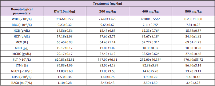
Terminalia Chebula caused significant (p<0.05) decrease in the level of red blood cell, hemoglobin, platelet etc. and significantly (p<0.05) caused an increase in mean corpuscular hemoglobin concentration in the rats at the dose level of 400 mg/kg compared to the control. The level of basophiles, neutrophiles, eosinophils and lymphocytes were however not significantly (p<0.05) affected by mean corpuscular hemoglobin concentration (Table 1) (Figures 1-6).

Table 1: Effect of 28 days oral administration of ethanol leaf extract of Ocimum canum on hematological parameters in wistar rats.

Note: Data presented as Mean ± SEM: n = 6, One-way ANOVA, followed by Dunnett’s post hoc for multiple comparison *significantly different from the Distilled Water (DW) control at p<0.05. DW = distilled water.

(WBC = White Blood Cells; RBC = Red Blood Cells; HGB = Hemoglobin; HCT = Hematocrit; MCV = Mean Corpuscular Volume; MCH = Mean Corpuscular Hemoglobin; MCHC = Mean Corpuscular Hemoglobin Concentration; PLT = Platelet, LYM = Lymphocyte; NEUT = Neutrophils; EOSI = Eosinophils; BASO = Basophils).

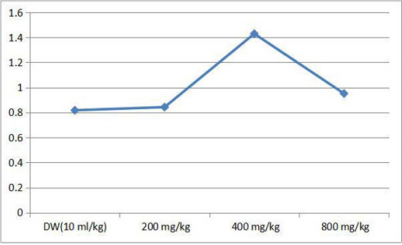
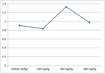
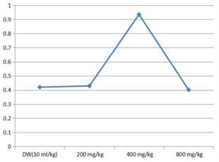
Figure 1: Showing effect of Terminalia Chebula on the brain body weight ratio.

Figure 2: Effect of Terminalia Chebula on rat stomach body weight rate ratio.

Figure 3: Effect of Terminalia Chebula on spleen body weight ratio in rat.

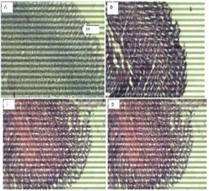
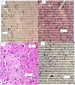
Figure 4: The brain (Hematoxylin and eosin. H and E ×100). (a) Control group shows normal neurons (N). (b) 200 mg/kg. (c) 400 mg/kg.(d)800 mg/kg of ethanol stem extract of Terminalia Chebula.

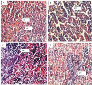
Figure 5: The Stomach (Hematoxylin and eosin. H and E ×100). (a) Control group shows normal red (R) and white (W) pulp. (b) 200 mg/kg (c) 400 mg/kg, (d) 800 mg/kg of ethanol leaf extract of Terminalia Chebula.

Figure 6: Spleen (Hematoxylin and eosin. H and E ×100). (a) Control group shows normal stomach mucosa (M). (b) 200mg/kg, shows normal features. (c) 400mg/kg, shows normal features. (d) 800mg/kg, shows normal features.

Effect of 28 days Oral Administration of Ocimum canum on Body Weight (g) in Rats

In the 1st, 3rd and 4th week significant (p<0.05) increase was observed at 200 mg/kg dose level by ethanol leaf extract of Terminalia Chebula, while in the 2nd week there was increase in body weight by the extract but not significant when compared with the control (Table 2).

Table 2: Effect of 28 days oral administration of Ocimum canum on body weight (g) in rats.

Note: *Significantly different from the Distilled Water (DW) control at p<0.05. DW = distilled water.

Effect of 28 days Oral Administration of Terminalia chebula on Relative Organ to Body Weight Ratio in Rats

The brain, spleen and stomach were observed to be significantly (p<0.05) increased in rats with dose level of 200 mg/kg of the ethanol plant extract. At higher doses of 400 and 800 mg/kg there was no significant (p<0.05) change (Table 3).

Table 3: Effect of 28 days oral administration of Ocimum canum on relative organ to body weight ratio in rats.

Note: *Significantly different from the Distilled Water (DW) control at p<0.05. DW = distilled water.

Effect of Oral Administration of Ethanol Leaf Extract of Terminalia Chebula on Histology of Brain, Lungs, Spleen and Stomach of Rats

Histopathological examination of the brain showed normal features at all doses and slight vacuolation at 400 mg/kg dose of the extract. The lungs showed normal features at all doses respectively. The spleen showed Slight lymphocyte hyperplasia at all doses and normal features at the control (10 ml/kg). The stomach showed normal features at all doses of the extract administered.

Discussion
Herbal medicine is becoming popular particularly in developing counties and there is an increased interest in green medicine simply because it is considered safe relative to conventional medicines [10-12]. Traditionally, plants and plant extracts were used to cure many diseases and disorders. However, before usage it is of utmost important to ensure its safety [13,14]. The extract may be therapeutically very efficient but if its toxicity assessment is not worked out, it will not be accepted. Hence, toxicity assessment of plants with proven therapeutic use is of utmost important13. Toxicity data are required to predict the safety associated before the use of medical products [13-15].

Hematological parameters are useful indices that can be employed to assess the toxic potentials of plant extracts in living systems [16,17]. They can also be used to explain blood relating functions of chemical compound/plant extract. Present result showed that ethanol leaf extract of Terminalia Chebula caused a reduction in the level of red blood cells, hemoglobin, hematocrit, mean corpuscular volume, mean corpuscular hemoglobin and mean corpuscular hemoglobin concentration which means that it can significantly reduce oxygen carrying capacity of the blood and thus cause anemia. Anemia is a condition where the blood has insufficient red blood cells to carry oxygen from the lungs to the rest of the body or not enough hemoglobin [18-20], the iron-rich protein that carries oxygen inside the red blood cells and gives blood its red color [21-24]. Anemia takes several forms and may vary in severity and duration [22,24]. Also reductions in Packed Cell Volume (PCV) and Red Blood Cell (RBC) were also observed in rats administered with the extract. This implies that Terminalia Chebula could cause disturbances in osmoregulatory system of the blood cells and/or oxidative injury to the cell membrane. The extract could suppress the haemopoietic system. The reduction may have also occurred due to lysis of blood cells. Sule et al, 201225 also observed decrease in RBC, PCV, hemoglobin and lymphocytes in rats fed with extracts of Acalypha wilkesiana.

The major functions of the white blood cell and its differentials are to fight infections, defend the body by phagocytocis against invasion by foreign organisms and to produce or at least transport and distribute antibodies in immune response [26]. The extract had no effect on white blood cell parameters, suggesting that it does not disturb or improve immune system. In this study, there was significant increase in the mean body weight ratio of brain, stomach and spleen of Wister rats administered with ethanol leaf extract of Terminalia Chebula at 400 mg/kg dose below and beyond which there was no effect. The probable reason could be that the plant extract has some toxicity effects on these organs which tend to increase their sizes. This may also suggest that the plant may have a biphasic tendency. That is, at a particular dose it has an oxidant effect, while at higher dose it may be reversed or act as an antioxidant. In addition, reports indicated that tannins, saponins, volatile oils, saponin glycosides and alkaloids were detected in fresh and dried samples of Terminalia Chebula [19,26- 28]. Histology observation also agrees with other parameters that the plant extract may be deleterious at some doses while at other doses, the plant may be safe.

Conclusion
Though, biochemical analysis was not carried out in this work, result suggests the need for caution while consuming the plant. It is also important to establish the right dose that will be beneficial for the purpose intended. Further work that will include biochemical study may be done to give a better and broader insight on the possible activity of Terminalia Chebula in the body.

For more Articles on : https://biomedres01.blogspot.com/
Wednesday, May 26, 2021

Future Prospects of Soybean in Pakistan

Future Prospects of Soybean in Pakistan

Case Report
Pakistan is an agriculture country. Majority of Pakistan’s population is dependent upon Agriculture and agricultural products. Pakistan is spending a lot of money on the edible oil import. The import bill of edible oil was Rs.284.546 billion (US$ 2.710 billion) [1]. Globally soybean is grown on an area of 120.48 million hectares but in Pakistan total area under soybean cultivation is comparatively less [2]. Farmers are getting low crops production due to these reasons like non-availability of good quality seeds, lack of advanced agricultural mechanization techniques and unavailability of modern methodologies which are applied in commercial crop production to save time and enhancement yield. Value addition is good techniques in which raw materials is converted into usable products e.g. Potato is value added in different products like processed Chips, finger chips etc. But our farmers are unaware about the value addition of products to get maximum profit [3]. Soybean is an important oil seed crop. It contains 20% oil content and 40% protein. It is considered as world healthiest food ever. Increased protein intake such as soybean meal has always been associated with suppression of appetite.

Soybean has been historically called as “meat of the field” or “meat without bones” due to these reasons it is highly rich in protein content. Genistein (an isoflavone phytonutrient in soybean) is used for cancer-prevention. One cup of soybean (172 g) contains 298 calories and copper (78 %) manganese (71%) phosphorus (60 %), iron 49 % omega-3 fats (43%) fiber (41 %), magnesium (37 %), vitamin K (37%) ,potassium (25%) [4]. It is a leguminous crop that is grown to fix nitrogen in soil to increase the fertility of soil through Rhizobacteria. Due to its low production it is grown on a very low area of Pakistan. Value added products of soybean include soymilk, soy flour, soy protein and tofu etc. Early planting of soybean is a critical factor in maximizing yield potential, if crop production technology is followed. Early planting of soybean crop can help to develop a large canopy, which may lead to an increase in photosynthesis, resulting in extension of the reproductive growth stage, ultimately increasing the yield potential [5]. Now days we are facing the problem of climate change throughout the world. Due to this factor environmental temperature has been changed. It is affecting badly the production of all crops due to shortage of water. So, it is need to grow such alternate crops that can survive on low water availability.

So, soybean is a crop which can thrive in all conditions. It has deep root system and can survive under less water availability. It is therefore high time to realize the fact that Pakistan is spending a lot of foreign exchange for the import of edible oil to meet the local demands. We should look for some alternate sources of edible oil from our own sources. We have fortunately good fertile lands and hardworking farming community which can be engaged for growing crops having good potential of edible oil like soybean. Government should provide good quality seed of soybean varieties at affordable prices to the farmers so that they can plant this seed in their more productive areas rather than marginal lands. Government should also fix support price to this crop and arrange to purchase the farmers produce at such price so that maximum farmers can be attracted towards its cultivation. If these suggestions are followed it is hoped that we can bridge the gap between our edible oil demand and supply

More BJSTR Articles: https://biomedres01.blogspot.com

Tuesday, April 6, 2021

Cell Renewal and Regeneration

Cell Renewal and Regeneration 

Introduction

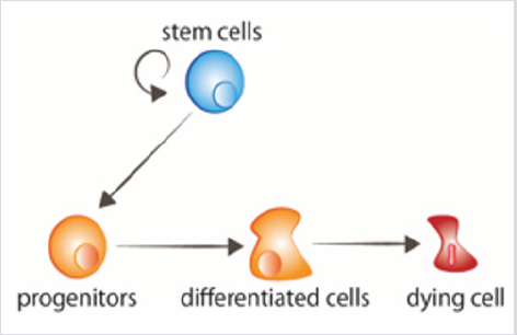
Cell or tissue renewal and regeneration are the two main developmental requirements of adult organisms. Both processes have as starting point a population of stem cells, normally located in a specific environment called the “niche” [1], which provides them the required signals to maintain the stemness properties, or to differentiate to the required different cell types (Figure 1). Stem cell proliferation and differentiation must be coordinated with the death of the cells that need to be replaced. In addition, processes such as cell migration, epigenetics, and cellular communication, are also necessary for proper cell renewal [2,3]. Fast renewal tissues can be recognized by a higher mitotic activity. Conversely, slow renewal tissues contain less mitosis, and may not be easily recognized from non-renewing areas which may also present some mitosis [2]. The fate decisions of stem cells during proliferation directly influence tissue renewal and homeostasis. Therefore, understanding the regulatory mechanisms that sustain a balanced cell division and differentiation is critical. Extracellular signals (e.g., tissue microenvironment, intracellular ROS, and cytokines) as well as intracellular factors (e.g., epigenetic machineries, transcription factors and DNA damage response) are responsible for the regulation of stem cell division.

figure 1: Cell turnover Stem cells proliferate, giving rise to progenitors that thereafter receive the signals to differentiate. Aged cells receive signals to die.

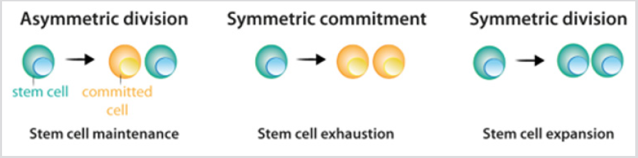
Stem cells show three possible options of division: [1] asymmetric division, in which one stem cell and one committed daughter cell are originated; [2] symmetric commitment, which yields two committed daughter cells; and [3] symmetric division, which yields two daughter cells that maintain stem cell properties [4] Figure 2. Although it could be predicted that asymmetric division is the only mechanism that enables the maintenance of a stable population of stem cells, the current data from lineagetracing experiments demonstrated that in most tissues, the balance between stem cell proliferation and the generation of differentiated offspring is achieved at the level of the whole stem cell population. The loss of stem cells due to differentiation or cell damage, induces symmetric division to fill this gap [5]. After stem cell division, the cells that follow the differentiation process pass through different stages that are defined by a combination of transcription factors that control the activity of the appropriate repertoire of genes, and allow their commitment and terminal differentiation. For each cell lineage the end product of the sequence of decisions is a specific differentiated cell type [6] Figure 3a. Under most circumstances, cellular identity - the product of normal differentiation - is stable within tissues, and its maintenance is crucial for normal tissue function. Such stability is achieved through epigenetic regulation - e.g. histone demethylation and acetylation - that results in heritable patterns of tissue-specific gene expression [3,7].

figure 2: Division pattern of stem cells.

a) During asymmetric division, stem cells give rise to one stem cell, which maintains the stem cell population, and one cell that becomes committed to differentiation.

b) During symmetric commitment, stem cell division gives rise to two daughter cells that became committed to differentiation.

c) During symmetric division, stem cell division gives rise to two stem cells. As explained in the text, current experimental data indicates that the three modes of division can occur while maintaining the stem cell population.

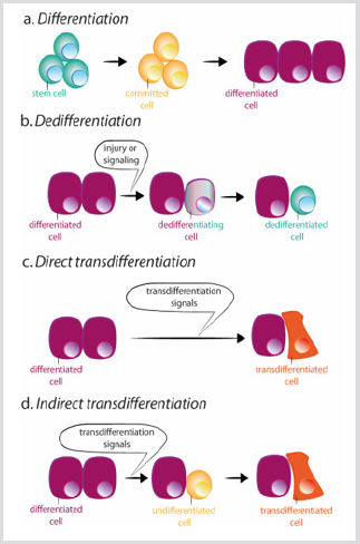
However, loss of cell identity can occur. Indeed, cells from Drosophila imaginal disc are able to transdetermine and acquire a new adult fate following transplantation [8]. In this situation, extracellular cues seem to reprogram some precursor or differentiated cells to acquire characteristics of either a more stem state or a new differentiated state. There are two mechanisms by which a cell can change its identity: dedifferentiation, and transdifferentiation. Dedifferentiation refers to the process by which a differentiated or committed cell acquires characteristics of a less mature cell [9] Figure 3b. The most dramatic example of dedifferentiation is the in vitro conversion of terminally differentiated cells into pluripotent cells (induced pluripotent stem cells, iPSCs), by the overexpression of a limited number of transcription factors [10]. Transdifferentiation, by contrast, occurs when a differentiated cell changes it transcriptional program and converts into another differentiated cell type. The process can occur through an intermediated step of dedifferentiation towards a less mature stage before the conversion into the new differentiated cell, or directly, without the intermediated stage [9] Figure 3c-d. The direct conversion of fibroblasts into myoblasts by the ectopic expression of MyoD is an example of the second process [11].

figure 3: Schemes of differentiation, dedifferentiation and trans differentiation.

a) During normal differentiation, stem cells give rise to committed cells which in turn differentiate in different cell types.

b) Dedifferentiation consists of the acquisition of stem cell properties by a differentiated cell. Trans differentiation can occur in a direct or indirect way.

c) During direct trans differentiation, a differentiated cell acquires the transcriptional program of another cell type, usually closely related, as for example exocrine to endocrine pancreatic cells, becoming a different differentiated cell.

d) During indirect trans differentiation, a differentiated cell dedifferentiates before adopting the new transcriptional program of the other cell type.

Dedifferentiation and transdifferentiation also occur in a natural way in response to an injury, or tissue loss [12]. Dedifferentiation, for example, occurs naturally during limb regeneration in the urodele amphibians. After limb amputation, cells adjacent to the wound dedifferentiate, forming a blastema that consists of undifferentiated cells that proliferate and eventually, redifferentiate into the same cell type to create all the components of the lost limb[12]. Natural transdifferentiation occurs indirectly: first, the cell dedifferentiates; and then the natural developmental program is activated, allowing the cell to differentiate into the new lineage[12]. Tsonis and collaborators described a natural mechanism of transdifferentiation in a newt. They found that when lenses are removed, pigmented epithelial cells from the dorsal iris transdifferentiate, and regenerate the missing tissue. To achieve this, pigmented epithelia cells must first dedifferentiate and proliferate to create new lens cells, and then differentiate into the mature cells of the lens [13]. In both situations – dedifferentiation and redifferentiation into the same cell type or transdifferentiate to a new cell type – a complex network of signaling pathways may control the transcriptional program acquired by each cell in the perfect time-point.

hus, the spatiotemporal control of gene expression is continuously required during animal homeostasis, and during a regenerative process. However, during regeneration, cells must re-adjust to the new situation, which requires making more profound decisions at a cellular level, often including processes of dedifferentiation and transdifferentiation that during homeostasis are scarce.

More BJSTR Articles :  https://biomedres01.blogspot.com/
Thursday, April 1, 2021

How freckles correlate with normal body temperature?

How freckles correlate with normal body temperature?

Introduction

Normal body temperature also called normothermia or euthermia. the thing that keeps the organisms at optimum operating temperature is called the temperature control mechanism. The rate of chemical reaction that affects the homeostatic mechanism [1]. By the time of the day, normal temperature varies from person to person. a range of normal temperature is the measurement of normal temperature. The 37 is the range of normal body temperature. The temperature changes in a day many times like a healthy person have o.5. a person is hungry sleepy sick or cold his temperature changes every moment of his body [2]. Normal temperature is sensitive to many hormones. Temperature also varies with the change of season during each year. This is called a circannual rhythm. everyone has a different normal body temperature. For an adult, normal body temperature can be 97. 99 [3]. When the temperature of the body is higher this is also called fever. For children, fever is more complicated. Between 3 months or 3 years and has a rectal temperature over 102. Your temperature based on where your body you measure it. Freckles are small brown spots on the skin. overproduction of melanin causes the freckle which is responsible for hair colour and skin [4]. There are two types of a freckle. Epihalides or lengths. Epihalide is most common in the skins of people. But lignite’s in the more production of melanin during pregnancy causes a dark spot on the skin. This includes agin sunspot or freckles [5].

Material and Method

We check the body temperature by a different method. We also check by the different method like a thermometer. The thermometer also has different types like automated or by mercury. First, we take a thermometer or check their normal body temperature. 98 is the normal temperature. put the thermometer into your mouth below the tongue. If you have a fever it shows on the thermometer. note that reading [6].

Project Design

We check the normal body temperature of the body. And last we noted their temperature. All the student was related to our university.

Statistical and Analysis

we use MS Excel software to find the statistical analysis of normal body temperature. P<0.1 is a significant value.

Result and Discussion

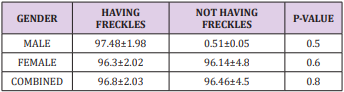
A relationship between freckles and normal body temperature (Table 1). The result is significant. They introduce that there is a strong relationship between body temperature and freckles [7,8].

Table 1:

Conclusion

We concluded this discussion in these words that there is a strong connection between freckles and normal body temperature when we talk about the overall population.

More BJSTR Articles : https://biomedres01.blogspot.com/
Wednesday, March 24, 2021

Assessment of Diaphragmatic Function by Ultrasonography: A Device to Record Breathing Signals on an Ultrasound Machine

Assessment of Diaphragmatic Function by Ultrasonography: A Device to Record Breathing Signals on an Ultrasound Machine

Introduction

In recent years, several works have supported the significance of ultrasonography in the assessment of diaphragmatic function [1,2]. The excursions of both hemidiaphragms can be measured by M-mode ultrasonography (US). Normal values of diaphragmatic motion have been reported [3] and M-mode US is able to detect hemi diaphragmatic paralysis or hypokinesis [4-6]. In patients suffering from neuromuscular disease, the measurement of diaphragmatic excursion during forced breathing can predict an impairment in vital capacity [7]. Two-dimensional ultrasonography (B-mode US) makes it possible to measure diaphragmatic thickness at various breathing time such as the end of normal expiration (functional residual capacity), at the end of normal inspiration and at total pulmonary capacity [1]. B-mode US has demonstrated its importance in the diagnosis of diaphragm paralysis [8] and of diaphragm dysfunction in patients suffering from myopathy [9]. Some studies have suggested that speckle tracking ultrasound (STI) was superior to conventional ultrasound techniques [10].

It has been reported that the diaphragmatic parameters measured by STI were related to the measurements performed by M-mode US and B-mode US [11,12]. STI might provide more clarification via the analysis of the longitudinal muscle deformation [13]. Unfortunately, no ultrasound machine has been developed to study diaphragmatic strain. The software used in these circumstances is the software designed to track 2D speckle motion in echocardiograms. Consequently, during the STI analysis of the diaphragm, the tracking time is related to a cardiac cycle time period and not to the breathing cycle. To study the maximum deformation of the diaphragm it would be important to analyses the diaphragm during the entire inspiratory phase. The aim of this work was to develop a system enabling the recording of the respiratory signals on the ultrasound machine.

Methods

The specifications of the device were initially determined: To improve the quality of the analysis of the diaphragmatic function by ultrasonography, it would be useful to record the beginning of inspiration and expiration on the screen of the ultrasound machine and to measure simultaneously the gas volume and the ultrasonographic parameters (hemi diaphragmatic excursion or muscle thickness). Lastly, the device should be usable with any commercial ultrasound machine.

Results

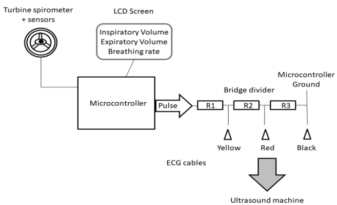
Figure 1 reports the principle of the device developed to record breathing signals on ultrasound machines. Monitoring of the breathing rate and gas volume was performed using a commercially available turbine spirometer (price about €1.6 - $1.9, Flow Mir, Medical International Research, Rome, Italy). The turbine support was built using a three-dimensional printer. This support incorporated 3 sensors (1 infrared led and 2 infrared phototransistors, price: €10 - $11.6) to detect breathing time and assess gas volume. The number of turbine rotations was counted by the sensors to assess the gas volume. The direction of the rotation made it possible to distinguish inspiration and expiration. A microcontroller (Nano, Arduino, price: €25 - $29) was used to acquire and interpret the sensor signals.

Figure 1: Principle of the device developed to record breathing signals on the ultrasound machines.

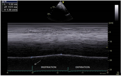
The breathing parameters (inspiratory and expiratory volumes, breathing rate) were displayed on an LCD screen (price: €33 - $38). Furthermore, electronic signals were sent to the ultrasound machine via ECG cables. To record the beginning of inspiration and expiration, a voltage pulse was sent now of the reversion in the rotational direction of the turbine. The duration of the voltage pulse was adjustable (from 500 to 1200 microseconds) and transmitted by the microcontroller to a bridge voltage divider. This bridge divider made it possible to decrease the voltage to 50mV to the first electrode and to 25mV to the second electrode. Consequently, the signal amplitude recorded on the screen of the ultrasound machine differed according to the breathing time i.e. inspiration and expiration (Figure 2). The system was powered via the USB connection of the ultrasound machine.

Figure 2: Measurement of the diaphragmatic excursion (1.52cm) during quiet breathing, by M mode ultrasonography: the voltage pulse reports the beginning of both inspiration (arrow: larger voltage) and expiration (image produced using a Vivid E90 and a M5S transducer, GE Healthcare, Little Chalfont, United Kingdom).

Abbreviations: L: diaphragmatic excursion in cm, dT: duration of the diaphragmatic contraction in ms, V: velocity of contraction in cm/s.

Discussion

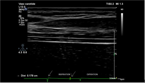
The advantage of the device was to record simultaneously the breathing cycle via the ECG cables and the diaphragmatic images by ultrasonography. The device developed by our team is of interest for several supplementary reasons. The cost of manufacture is low, in total about €70 - $82, including the turbine spirometer, the sensors, the microcontroller and a screen. No change to the ultrasound machine is needed since the transfer of the signals is transmitted via ECG cables. Consequently, the device is suitable for application with any commercially available ultrasound machines. M-mode ultrasonography enables the diaphragmatic motion to be measured whereas the two dimensional mode can measure the diaphragmatic thickness in the area of apposition of the diaphragm to the rib cage. The breathing signals on the screen of the ultrasound machine is a guarantee that the measurement is performed at the right breathing time (Figure 3).

Figure 3: Measurement of diaphragm thickness (d= 0.178cm) in the area of apposition during inspiration (first arrow= beginning of inspiration, second arrow= time of measurement) image produced using a Phillips Affinity 70 Ultrasound system - vascular transducer (L12-3), Koninklijke Philips N.V, Amsterdam, The Netherlands).

In patients suffering from hemidiaphragm paralysis, the signal of the beginning of inspiration can be useful to demonstrate the paradoxical displacement of the paralyzed diaphragm during quiet breathing or deep breathing [6]. The simultaneous measurement of the diaphragmatic motion and the gas volume of the corresponding breathing cycle is also interesting. Indeed, it has been reported on one hand, that the relation between inspired volume and hemi diaphragmatic movement was linear [14] and on the other hand, that diaphragmatic excursions of both sides were quite similar in healthy subjects [3,15]. In patients suffering from hemidiaphragm dysfunction, the right-to-left ratio of maximal excursion on deep breathing can be outside the range of 0.5 to 1.6. In such circumstances, it would be useful to control that the inspired gas volumes corresponding to the diaphragmatic excursions are similar. Furthermore, the work of breathing is the energy expended to inhale and exhale a breathing gas. It can be calculated by the product of the change in thoracic pressure and the change in pulmonary volume.

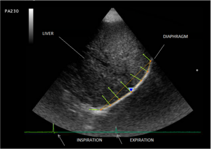
It has been reported that the measurement of the thickening fraction [the ratio (thickness at inspiration-thickness at expiration)/ thickness at expiration] was related to the transdiaphragmatic pressure–time product per breath [16]. Consequently, the simultaneous recording of the gas volume and the thickening fraction should be useful in assessing the work of breathing. The significance of the study of diaphragmatic function by STI has been recently underlined [13]. The device developed by our team can detect the beginning of inspiration and expiration. Since the information is transmitted to the ultrasound machine via ECG cables, the deformation study by the software can be triggered by the breathing cycle and not by the cardiac cycle. Using this procedure it is possible to perform the analysis of the diaphragm during the entire inspiratory phase (Figure 4).

Figure 4: Study of the diaphragmatic deformation triggered by the breathing cycle (first arrow = beginning of inspirationlarger voltage, second arrow= beginning of expiration) - image produced using a Mylab 25 Esaote, Genoa, Italy.

Conclusion

The combination of the recording of the breathing signals and gas volume with the corresponding diaphragmatic thickening or excursion should be useful for a better analysis of the diaphragmatic function.

More BJSTR Articles : https://biomedres01.blogspot.com/
Monday, March 22, 2021

Correlation between Parasitic Infections in HIV Patients Parasitic Infections in HIV Patients

Correlation between Parasitic Infections in HIV Patients Parasitic Infections in HIV Patients

Introduction

Human Immunodeficiency Virus (HIV) is a retrovirus of the Retroviridae family and the etiologic agent of Acquired Immunodeficiency Syndrome (AIDS). When first described in the United States, in 1970, this virus was observed mainly in male homosexuals, hemophiliacs and intravenous drug users [1,2]. Currently, other groups are infected with HIV, including the elderly and wives of men who have sex lives outside of marriage [3]. The genetic material of the HIV is RNA, double-stranded and enveloped. It has the ability to attach and invade mainly CD4+ lymphocytes because of CD4 receptor presence [4]. Other cells also may be invaded by HIV due to its ability to recognize other types of cellular receptors such as the CCR5 receptor present in dendritic cells, macrophages and T cells [5,6]. HIV transmission occurs through unprotected heterosexual and homosexual intercourse. Vertical transmission occurs when the virus breaks through the placental barrier during birth, or by breastfeeding [6].

Another method of transmission is the sharing of needles by drug users, the use of some cutting or perforating utensils containing HIV infected blood [7]. Patients with HIV/AIDS experience an asymptomatic phase followed by the appearance of symptoms. The symptomatic phase is characterized by severe expressions of immunodeficiency and general clinical complications. Significantly, at this point, the patient may develop some opportunistic infection [8]. Parasitic infections are responsible for significant morbidity as well as mortality worldwide [9]. In developing countries, they are considered a serious public health problem due to inadequate sanitation and lack of information, which makes the perpetuation of the life cycle of the parasites possible [10]. In these patients, parasitic infections, which are usually asymptomatic, become symptomatic and progress to serious disease, and causing death [11]. Helminths and protozoan, which affect humans and cause many symptoms, are usually associated with the gastrointestinal tract. These diseases are related to demographic, socioeconomic, physiological and immunological factors.

Thus, immunocompromised patients and/or those undergoing immunosuppressive therapy are more susceptible to contracting infections by these parasites and often with a higher degree of severity. Among the frequent intestinal parasites and related diarrhea in patients with HIV are: Entamoeba histolytica, Giardia lamblia, Ancylostomatidae, Ascaris lumbricoides, Strongyloides stercoralis and others [12,13]. People infected with HIV, enteric infections occur more frequently. Some cases are more likely to have severe recurrence and persistence. In most cases the microorganisms are not identified, although there are potential causes of diarrhea whose sources can be some form of parasitic infection [14]. Overall, these facts motivated the present research which aimed to determine the prevalence of parasitic infections in infected HIV/AIDS patients using medical records of patients who had been or were being assisted by the Joint Health Unit in Taguatinga, DF, during the period from 2005 to 2015.

Methodology

A program of transversal, quantitative and exploratory field research was carried out. The information was collected from medical records of patients with HIV/AIDS. Data were obtained from the Archival System of the Joint Health Unit of Taguatinga, DF. The data included follow-up patients and those who had been referred to be treated and monitored in other hospitals of the regional cities of Brasília, DF. The Ethics Committee of the Foundation of Teaching and Research in Health Sciences (FEPECS) approved the project (Process: 05163312.3.0000.5553 of 04/02/2013).

Target Population

Patients included those who had been diagnosed with HIV/ AIDS and who were or had been monitored and/or who were or had been in treatment at the Joint Health Unit, Taguatinga, DF, from January 2005 to July 2015. The survey was conducted at the Joint Health Unit in Taguatinga, DF, Brazil, and analyzed 502 medical records of these patients.

Criteria for Inclusion and Exclusion of Patients in the Sample

The records examined contained information regarding socioeconomic factors, gender, age, origin, profession, sexual preference, diagnostic tests performed, and medications administered, among other data. Records of patients who had not been diagnosed with HIV/AIDS, who were outside the established period or contained insufficient data to complete the questions, were excluded.

Variable Selection

Data were collected from patient charts, available in Archival System of the Joint Health Unit Taguatinga, DF. The variables chosen for the study were selected from the data included in the charts.

Biosecurity Standards

The study involved no invasive methods. For this reason, the Biosecurity Standards did not apply. The research was, however, in compliance with the recommendations of Resolution No. 196/96 of the National Health Council, maintaining the anonymity of the information that could identify patients with HIV/AIDS.

Sharing of Results

The survey results were passed on to the local group of professionals in this research area. It is very important to identify and differentiate the microorganisms that cause opportunistic infections in immunocompromised patients. For these patients the manifestation of diseases is usually more severe. Furthermore, to bring the situation under control, information and awareness are necessary. The health professionals, who deal with these issues, have the opportunity to inform patients about the necessary precautions to avoid contamination with opportunistic pathogens such as bacteria, fungi, protozoa, helminths and other pathogens.

Statistical Analysis

Data were organized in tables and graphs. The statistical analyses were conducted using Chi Square (X2 ) with the INSTAT 3 statistical program. Correlations were considered statistically significant at p <.0.5

Results

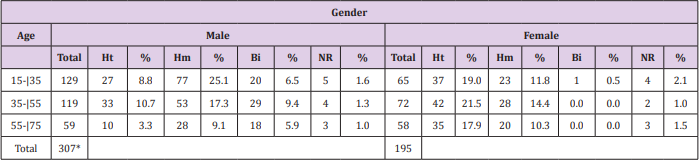
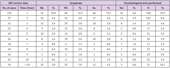
The distribution of the HIV-positive patients of the sample, by age group, gender and sexual orientation, is shown in Table 1. Most patients had been subjected to more than one test to confirm HIV. During the period from 1999 to 2004, three patients had undergone Elisa, 10 had been tested using Western blotting, two had been diagnosed using indirect immunofluorescence, three had had rapid tests and six had been examined by antibody Screening Anti-HIV 1 and 2. The distribution of HIV-positive patients, compared by the year of diagnosis and confirmatory tests performed, as well as the CD4+ cell counts, are presented in Table 2. Correlations between the diagnosis of HIV infection based on symptoms such as eosinophilia and parasitological test results of the patients of the sample, as well as the common symptoms reported that could be indicative of infection by parasites, helminths and protozoa, are shown in Table 3.

Table 1: Distribution of HIV-positive patients by age group, gender and sexual orientation, treated at the Joint Health Unit Taguatinga, Federal District, during the period 2005-2015.

Note: M: Male; F: Female; Ht: Heterosexual; Hm: Homosexual; Bi: Bisexual; NR: not reported; %: Percentage; * Significative difference.

Table 2: Distribution of HIV-positive patients at the Joint Health Unit Taguatinga-DF, by year of diagnosis, confirmatory tests and CD4+ cell count, Federal District, during the period 2005-2015.

Note: El: Elisa; Wb: Western blotting; II: Indirect Imunofluorescence; Rt: Rapid test; Sa: Screening antibodies Anti-HIV 1 and 2.

Table 3: Correlations between the diagnosis of infection with HIV, time period of clinical symptoms and parasitological examination results of patients at the Joint Health Unit Taguatinga, Federal District, during the period 2005-2015.

Note: No: Number; %: Percentage; Dh: Diarrhea; Wl; Weight loss; Eo: Eosiniphilia.

A total of 149 patients were diagnosed with diarrhea, 230 had weight loss and 140 were eosinophilia. Of the patients with diarrhea, 37 tested positive for intestinal parasites or commensals: 19 for Entamoeba coli, nine for E. histolytica, three for S. stercoralis, one for G. lamblia and five for A. lumbricoides. Of the patients who had weight loss, 11 were positive for intestinal parasites or commensals: five for E. coli, two for E. histolytica, one for S. stercoralis, and three for A. lumbricoides. In addition, of those with eosinophilia: 34 were positive for enteroparasites or commensals: 18 for E. coli, eight for E. histolytica, two for S. stercoralis, one for G. lamblia and five for A. lumbricoides. The frequency of parasites and/or commensals diagnosed in HIV-positive patients using parasitological and immunological methods is shown in Table 4. Of the total of 502 patients, 83 had undergone parasitological tests and of these 42 were positive. The most common commensal was E. coli with 22 cases. Of 483 patients who underwent immunological tests, 193 cases were positive for toxoplasmosis, and 11were positive for Chagas disease.

Table 4: Parasitological and immunological test results and species of parasites and commensals found in HIV-positive patients, the Joint Health Unit Taguatinga, Federal District, during the period 2005-2015.

Note: N: Negative; P: Positive: NR: Not performed*; %: Percentage: Ec: Entamoeba coli; Eh: Entamoeba histolytica; Ss: Strongyloides stercoralis; Gl: Giardia lamblia; Al: Ascaris lumbricoides; Tg: Toxoplasma gondii; Tc: Trypanosoma cruzi

Discussion

The age group most affected by HIV a few years ago was 20 to 40 years and men were more prevalent than women [15]. The results found in the present study corroborate these findings. The most affected patients were men (61.2%), then women (38.8%). The most infected age group was between 15-35 years of age for males. Women were most affected between 35-55 years of age. These data show that women are contracting the virus at a younger age and the number of infected women has grown over the years [16,17]. HIV/AIDS primarily affects homosexuals and drug users [1,18]. In asymptomatic homosexuals, an increase of cytotoxic T lymphocytes and suppressor T lymphocytes was observed. In terms of sexual behavior, more men aged 15-35 years were affected by HIV. Of these, 8.8% were heterosexual, 25.1% homosexual, 6.5% bisexual and 1.6% did not report sexual preference. Among women, the most prevalent age group affected by HIV was between 35-55 years, with 21.5% heterosexual, 14.4% homosexual, no cases of bisexuals and 2% did not report sexual preference. The findings confirm results reported in the literature: the male age group with the highest rates of HIV is the homosexuals and among women it is the heterosexuals [19].

The first reported cases of HIV described clinical changes related to the gastrointestinal tract. Most cases noted diarrheal conditions associated with parasitosis [19]. In a study conducted at the University Hospital of Rio de Janeiro, the parasite most often observed was A. lumbricoides [20]. In another study conducted in Rio Grande do Sul, the most common intestinal parasites were T. trichiura, G. lamblia and A. lumbricoides [21]. In the present study, intestinal parasites and commensals diagnosed in parasitological stool tests were: E. coli (most frequent, with 22 cases), E. histolytica, S. stercoralis, G. lamblia and A. lumbricoides. E. histolytica causes amoebiasis. Protozoan infections usually occur in regions where sanitary conditions are poor and typically spread by fecal-oral contamination [22]. In patients with HIV/AIDS, E. histolytica deserves special attention because it can cause fulminant amoebic dysentery [23,24].

G. lamblia is common both worldwide and in the Brazilian population [24]. It infects individuals of all ages, genders, races and social conditions [12]. The frequency of giardiasis in the present study (three cases) was low compared to other research [20,25]. This may be related to the age factor, since all individuals examined were adults and possibly the personal hygiene conditions were adequate, or because the water used by the population had been treated or filtered. It may also be related to the small number of samples analyzed. E. coli is a commensal, usually acquired by ingestion of contaminated food [26], as reported in patients with HIV [16]. The amount of E. coli found (22 cases) in the present study was relatively low, but it corroborate with the literature that show the frequency this commensals in patients HIV positive [27]. A. lumbricoides is the most common intestinal parasite in the world [28]. This disease can affect physical and mental well-being, hinder learning in children and adolescents and is even more complicated for patients with serious diseases such as HIV/AIDS [29]. The frequency of A. lumbricoides (six cases) in the present study was similar to the studies reported in the literature [21,27].

Strongyloidiasis is a parasitic disease caused by S. stercoralis and occurs in tropical and subtropical countries [12,30]. One possible means of transmission is by homosexual relationships. The biological cycle is made possible by internal and external autoinfection, by which large amounts of infective larvae are produced and can complete the autoinfection cycle and spread in the body, causing death in patients with HIV/AIDS [31]. Thus, strongyloidiasis can be considered an opportunistic infection with an AIDS-related agent [32]. The depression of cellular immunity is the conditioning factor for spread of strongyloidiasis [33,34]. Results of the present study were similar to previous studies, with low prevalence of S. stercoralis (five cases) [20,21]. Among the patients who had had immunological tests, 193 cases tested positive for reagents of T. gondii (toxoplasmosis). T. gondii is the causative agent of toxoplasmosis, it has a complex life cycle with two hosts, and several modes of transmission including blood transfusions, and transplants [35]. In Brazil, toxoplasmosis infection among those with the human immunodeficiency virus is of great importance since it is the major infectious disease found in pregnant women [36]. Toxoplasmosis is, therefore, of great medical importance as a frequent cause of death, especially in infants [37,38].

In the present study, T. gondii was the most frequent protozoan (193 cases) diagnosed by the immunological tests. Eleven patients of the study were seropositive for T. cruzi infection, or Chagas’ disease. The correlation of the disease with HIV is worrisome because the literature has shown that the reactivation of Chagas disease in immunosuppressed patients is high and associated with many cases of death [39]. Infection with T. cruzi occurs mainly through diet and blood transfusion [40]. Opportunistic infections are frequent characteristics of HIV patients and often occur due to the depletion of CD4+ T cells in number and function. But these infections can also infect and alter macrophages, Langerhans cells, dendritic cells, and CD8+ lymphocytes. It is for this reason that HIV patients are more susceptible to secondary infections [16]. In a study conducted in São Paulo, 53.6% of patients with HIV had CD4+ cell counts of less than 200/mm3 [27]. In the present study, the CD4+ cell counts of less than 200/mm were found among 8.2% of the patients studied from 1999 to 2004; from 2004 to 2009 it increased to 11.7% and from 2009 to 2015 it was 20.5 %.

Conclusion

In conclusion, it is very important to monitor the evolution of AIDS in patients in Brasília since parasitic infections caused by helminths, protozoa are frequent, and many deaths can be avoided if these pathogens are correctly diagnosed and treated. However, for such treatments to be administered in time it is necessary that serological and parasitological examinations become routine for these patients. This procedure is totally dependent on the request for the examinations by the health care professionals who provide care to these individuals.

More BJSTR Articles : https://biomedres01.blogspot.com/
The Effect of Interface Design and Perceived Risk of Customers’ Satisfaction and Loyalty on Online Booking Application

  The Effect of Interface Design and Perceived Risk of Customers’ Satisfaction and Loyalty on Online Booking Application Introduction The ra...