Usefulness of Comprehensive High Resolution Ultrasound Imaging in Dermatologic Field: Epidermal Cyst
Introduction
Dermatologic ultrasound imaging has been rapidly growing in recently years because of the development of high-resolution multifrequency transducers and multichannel color Doppler machines [1]. The international working group, called DERMUS (Dermatologic Ultrasound) was formed and provided the guidelines for performing dermatologic ultrasound examinations [1] and proposed for an assessment training program [2]. The minimum frequency recommended for performing dermatologic examinations was 15 MHz [2]. Epidermal Cyst is a common slow-growing dermal or subcutaneous epithelial cyst, which contains keratin and is lined by the epidermis [3]. It has been reported that epidermal cyst shows 2 types as pseudotestis and nonpseudotestis types on gray-scale US [4]. Epidermal cysts classically show a round-to-oval shape with posterior echo enhancement and lateral shadow on gray-scale US [5]. As the unruptured type of epidermal cyst represented no substantial blood flow signals, the ruptured type shows pericystic and peripheral vascularity [6] and prominent vascularity in the periphery [7] on color Doppler US. Real-time sonoelastography is an ultrasound-based technique using the property that the tissue strain due to compression is lower in hard tissues [8]. RTE is widely used for diagnosis of breast [9] and thyroid lesions [10] on tissue elasticity. There have been reported that the TRE is still uncommon in the dermatologic field. Patel et al. [11] reported that most epidermoid tumors show a predominant blue color, which represents their hard nature. Park HJ et al. [11,12] suggested that superficial epidermoid tumor exhibits a softer nature than dose malignant tumor but does not have a different strain elastography (SE) pattern from other benign tumors [12]. It has been reported that utility of sonoelastography in differentiating ruptured from unruptured epidermal cyst [5], suggesting the differences in tissue elasticity.
Dermatologic Ultrasound
Dermatologic ultrasound imaging has been rapidly growing in recently years because of the development of high-resolution multifrequency transducers and multichannel color Doppler machines [1]. An international working group, called DERMUS proposed that the contents for level 1 include basic ultrasound knowledge, normal dermatologic ultrasound anatomy, and common pathologic condition [2]. The minimum frequency recommended for performing dermatologic examinations was 15 MHz. We usually perform US examinations for dermatologic diseases with a high-resolution, broad-band (5MHz-18MHz) linear transducer (Nobulus Hitachi, Ltd. Tokyo, Japan). Until now, we have reported some studies in relation to the skin and subcutaneous disease [13- 17]. Epidermal cyst is considered as level 1 content of the training program in dermatologic ultrasound by the international working group, called DERMUS [2].
Epidermal Cyst
Epidermal Cyst is a common slow-growing dermal or subcutaneous epithelial cyst, which contains keratin and is lined by the epidermis [3]. As the rate of malignant transformation into squamous cell carcinoma has been reported to range from 0.01% to 2.00% [4,12,18], accurate diagnosis of epidermal cyst is very important for the establishment of the treatment plan. It is also difficult to distinguish an epidermal cyst from other benign skin tumors, such as pilomatricoma, lipoma, steatocytoma, dermatofibroma and intradermal nevus. The cosmetic outcome is less favorable in cases of inflamed or ruptured cysts because these cysts have indistinct margins, more friable cyst walls, and difficulty in complete removal owing to inflammation and fibrosis of the surrounding tissue. Therefore, preoperative diagnosis is important, and different treatment options should be planned for ruptured and unruptured cysts [5].
Gray Scale Ultrasonographic Features
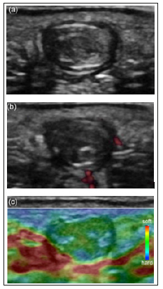
It has been reported epidermal cyst shows 2 types as pseudotesis and nonpseudotestis [4] and the report of the classification of 5 patterns [3]. Pseudotestis type is mostly accompanied with filiform anechoic areas consistent with packed keratin lamellae, echogenic reflectors consistent with cholesterol, sebaceous foci, or calcifications. Nonpseudotestis type is mostly depicted as concentric ring sign which is consistent with alternating layers of laminated keratin and squamous cells [4]. (Figure 1a) showed an ovoid nodule with a laminated, concentric ring pattern with hypoechoic rim, called nonpsendotestis type of epidermal cyst pathologically confirmed. Other features are dermal attachment, focal dermal protrusion [4].
Color Doppler Ultrasonographic Features
Unruptured type shows usually no substantial blood flow. On the other hand, pericystic changes are demonstrated in ruptured type on gray-scale US and color Doppler US shows pericystic and/ or peripheral vascularity. A few peripheral blood flow signals were depicted on power Doppler US (Figure 1b). It has been reported that peripheral low echoic rim, consistent with the capsule was detected in 67% on gray-scale US [19]. It has been suggested that thin and smooth rim enhancement is shown in unruptured type, thick and irregular rim enhancement is represented in ruptured type on enhanced MRI [20]. The rim enhancement might be due to fibrosis and foreign body reaction around the cyst wall remnants and keratinous material [21].
Figure 1: Nonpseudotestis type of epidermal cyst pathologically confirmed in the plantar in a 16-year-old female.
(a) On gray-scale US showed an ovoid nodule with a laminated, concentric ring pattern with hypoechoic rim.
(b) A few peripheral blood flow signals were depicted on power Doppler US.
(c) Real-time tissue elastography showed more green than blue.

Real Time Tissue Elastography Features
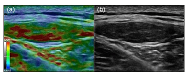
Recently, it has been reported that the usefulness of elastography is described in dermatologic field [22]. RTE is widely used for diagnosis of breast. Rago T et al. [9,10] suggested that low elasticity of thyroid nodules on ultrasound elastography is correlated with malignancy, degree of fibrosis. Park HJ et al. described that the strain elastography grades was from one to four as follows; Score 1 shows very soft nature, high elasticity. Score 2 represents moderate soft nature, moderate high elasticity. Score 3 is consistent with moderately hard nature, moderately low elasticity. Score 4 shows very hard nature, low elasticity. On RTE, they concluded that superficial epidermoid tumor shows a softer nature than dose malignant tumor; however, it does not have a different SE pattern from other benign tumors. (Figure 1c) shows relatively soft nature on RTE as previously reported [12]. The lipoma is hyperechoic compared with the subjacent muscle but isoechoic as compared with the adjacent subcutaneous adipose tissue. Gently curved echogenic lines were showed within the lipoma [23]. Lipoma represented green to orange color, showing grade 1 as previously reported [12]. This case was comprehensively diagnosed as a lipoma using high-resolution ultrasound imaging. Lipoma represent green color with a lot of orange color, suggesting soft nature on RTE (Figure 2a). Gently curved echogenic lines were noted on gray-scale US (Figure 2b). Ruptured type is softer nature than unruptured type on RTE. Park J et al. reported that RTE is able to detect differences in tissue elasticity between ruptured and unruptured epidermal cysts. Park et al. evaluated that US features of superficial epidermoid tumor with a focus on SE features that will help in the differential diagnosis of epidermoid tumor from other benign and malignant soft-tissue tumors. They concluded that superficial epidermoid tumor shows a softer nature than dose malignant tumor; however, it does not have a different SE pattern from other benign tumors [11].
Figure 2: (a) This case was comprehensively diagnosed as a lipoma using high-resolution ultrasound imaging. Lipoma showed green color with a lot of orange color, suggesting softer nature than epidermal cyst on RTE.
(b) Gently curved echogenic lines were obtained in the mass on gray-scale US.

Conclusion
Pseudotestis and nonpseudotestis types on gray scale US and pericystic and peripheral vascularity in ruptured type on color Doppler US were represented. A softer nature than malignant tumor and the differences in tissue elasticity between ruptured and unruptured epidermal cysts on RTE were shown. This comprehensive tools using high-resolution ultrasound including gray-scale US, color Doppler US and RTE are useful for the accurate diagnosis of epidermal cyst.
The Impact of Fencing on Regeneration, Tree Growth and Carbon Stock in Desa Forest, Tigray, Ethiopia-https://biomedres01.blogspot.com/2020/09/the-impact-of-fencing-on-regeneration.html
More BJSTR Articles : https://biomedres01.blogspot.com


No comments:
Post a Comment
Note: Only a member of this blog may post a comment.