The Influence of Pyramid Structure on Cells
Introduction
Numerous biological effects of pyramid structure (PS) have been reported in recent years. For example, Indian scholars found that the seed germination rate and radicle length of plants under the PS were significantly higher than those of the control group [1-3], and other reported effects included accelerated would healing of mice, improved preservation time of food [4-7], and reduction in the occurrence of tumor in mice accompanied by the improvement in liver enzyme activity along with some other biochemical and physiological indicators, which could explain the retardation of the tumor cell growth [8].Despite the accumulating amount of evidence of biological effects reported, there is very little research investigating the underlying biological mechanisms for the observed phenomena. The PS has been proposed to generate a torsion field [9], which has been extensively studied for its biosocial effects. As a portal to receive, process and integrate information in cells, mitochondria play a critical role when cells were exposed to stimuli including external Qi from a Qigong master [10], Chinese texts with different meaning [11], and torsion field [12]. Our group has previously found that the exposure of cultured mammalian cells to the above stimuli can induce changes in numerous parameters related to the mitochondrial functions, such as the production of ATP, ROS, MMP and mtDNAcn, as well as the cellular oxidant capacity reflected by the reduced GSH.In this study, we used 293T cells to examine any potential responses in the mitochondria for cells grown under PS, with the goal of examining potential underlying biochemical mechanisms of the effects generated by a pyramid. We examined parameters including the growth and vitality of the cells, mitochondrial functions including the production ATP, ROS, MMP, the cellular oxidant capacity reflected by the reduced GSH, as well as cell senescence related parameter such as the telomere length.
Materials and Methods
Cell Culture
The 293T cells was obtained from National Infrastructure of Cell Line Resource (Beijing, China, http://www.cellresource.cn) as visceral cells which grow rapidly. The culture conditions and inoculation process of these cells can refer to our previous articles [12].
Pyramid Structure Model
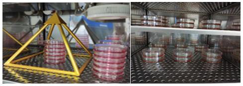
The pyramid model used in the study is made of copper (some studies believe that the effect of copper is more obvious than other materials [3]) according to the true proportion and angle of ancient Egyptian pyramids. The copper pyramid model with a square of length 280 mm and height 280 mm (Figure 1).
Experimental Design
Two groups of 293T cells culture were used, including a treatment group (cultured in the pyramid model and named as ‘PS’), and a control group, named CK (away from the pyramid model). 293T cells were seeded at a density of 5~6×10⁵ cells/plate into a 10cm plate, and after a 3h attachment period, plates of the ‘PS’ group were placed in the pyramid model for 24h treatment. The plates for the ‘CK’ group were placed under normal growth conditions in another room about 50 meters away. The cells were collected and subjected to different assays at 4h and 24h respectively during incubation. All experiments were performed in triplicate (Figure 2).
Biochemical Analysis
In the experiment, the detection methods of various biochemical indexes of 293T cells, such as ATP, ROS, MMP, protein, cell viability, SOD and GSH, were unified in our research group [12].
Gene Expression Analysis
Because of using the same research vector (293T cells), the parameters of gene expression analysis such as transcriptome sequencing and mitochondrial DNA quantification can refer to the previous work [12].
Statistical Analysis
Values of different measurements were normalized to a respective mean control value from untreated samples and expressed as percent control. All data are expressed as mean ± standard deviation (SD). They were analyzed by analysis of variance (ANOVA) and Least Significant Difference (LSD) using GraphPad InStat software, where P <0.05 was considered statistically significant.
Results
Effects of Pyramid Structure on Cell Growth
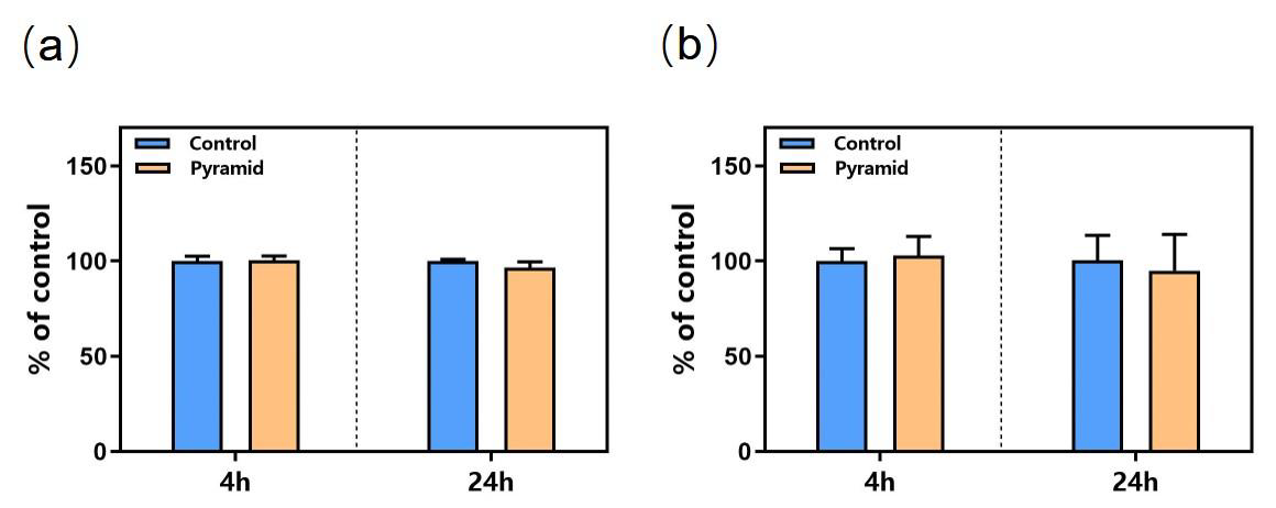
Compared to the control cells, PS did not affect the cell growth and cell viability at 4 hours post treatment and 24 hours (Figure 3), indicating that PS treatment for short period did not affect cell growth evidently (Figure 3).
Effects of Pyramid Structure on Mitochondrion
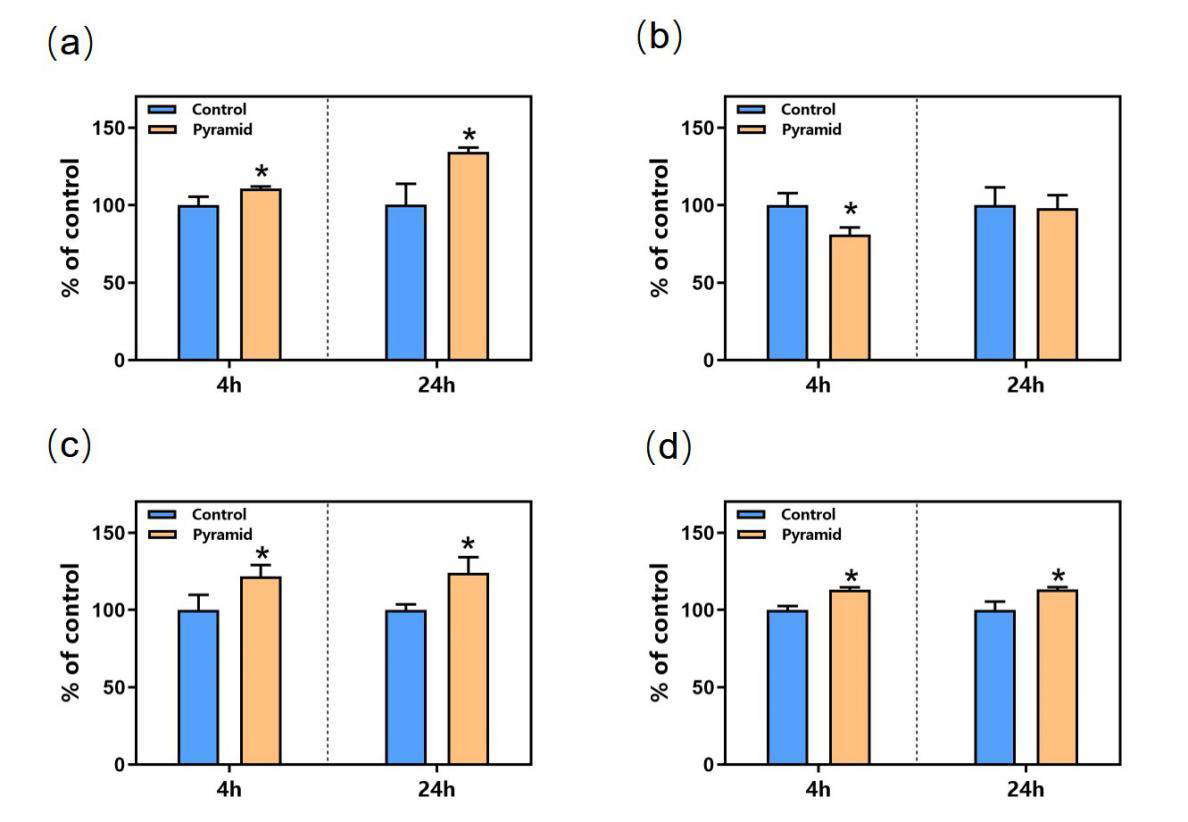
However, the mitochondria were significantly affected by PS as shown in Figure 4. The ATP level of cells in the PS group was upregulated by 20% and 35% at 4h and 24h respectively (Figure 4a) compared to that of the control group. The cellular ROS level was significantly decreased by pyramid at 4h but was not affected at 24h (Figure 4b). MMP level was both upregulated significantly at 4h and 24h (Figure 4c). The activity of GSH, an antioxidant protein, was increased by pyramid at 4h and 24h (Figure 4d), showing a potential role of antioxidant of PS.
Figure 4: Effects of PS on mitochondrion.
(a) ATP level;
(b) ROS level;
(c) MMP level;
(d) GSH level. *,
P<0.05. *Significantly different from the control group.
Effects of Pyramid Structure on mtDNAcn and Telomere Length
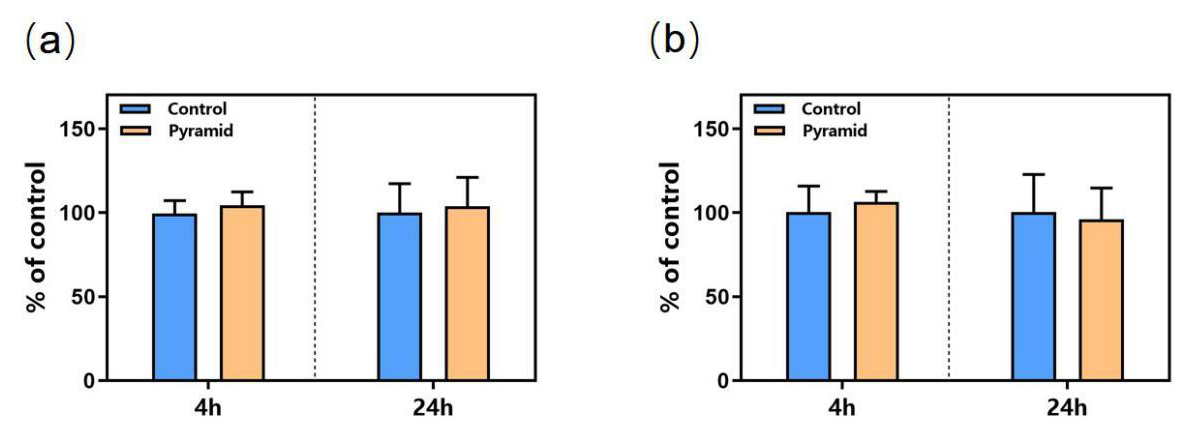
Cellular mtDNAcn and telomere length were detected using the qRT-PCR technology and no obvious changes was observed at 4h and 24h in the cells of pyramid group according to the results (Figure 5). All in all, PS modulated the function of mitochondrion in cells and may protect the cells from oxidative damage by upregulating the level of anti-oxidative protein and downregulating ROS level.
Discussion
In this study, we found that the activity of mitochondria in 293T cells became more active under the influence of PS, and the levels of MMP and ATP were significantly up regulated. At the same time, the overall antioxidant capacity of cells was improved. The PS significantly increased the content of GSH and reduced the level of ROS. No significant changes in cell growth and telomere length were observed in the study, indicating that the PS has a mild effect on cells, which avoids the negative effects caused by strong stimulation. Studies have found that the liver enzyme activity of mice in PS shaped feeding cages is significantly increased [8]. The plasma cortisol content of adult female rats exposed to long-term PS changed significantly, and the antioxidant defense ability of erythrocytes was improved [13,14]. In addition, some researchers found that the PS can accelerate the healing of mouse wounds [4,6], which indicates that the cellular activity of mouse wounds is stimulated, which is very similar to our results. As the “power plant” of cells, mitochondria provide energy for cells, and as a bridge for energy transformation inside and outside cells, they can respond to external stimuli in time. The influence of PS is considered as a torsion field [9], which collects the surrounding fine energy through the spatial layout of the structure itself, and then through the induction of mitochondria in the cell, so as to improve the activity and antioxidant capacity of the cell.
Previous studies mainly focused on the effect of PS on biological system, but there were few studies on the effect at the cell level. In this study, we used biochemical and molecular biological methods to determine that the PS has an obvious promoting effect on cell antioxidant and mitochondrial activity, which is helpful to further study the mechanism of PS effect. The healing effect of this special spatial structure also provides a new way for noninvasive treatment.


No comments:
Post a Comment
Note: Only a member of this blog may post a comment.