Nanomedicine and Nano Formulations for Neurodegenerative Diseases
Introduction
Neurodegenerative diseases are characterized by the progressive degeneration of the structure and function of the central or peripheral nervous system that affects millions of people worldwide. The most common neurodegenerative diseases are Alzheimer’s Disease (AD) and Parkinson’s Disease (PD), but they also include Huntington’s Disease (HD), Multiple Sclerosis (MS), brain tumor, and epilepsy. As of a 2021 report, the Alzheimer’s Disease Association estimates that the number of U.S population with Alzheimer’s disease could be as many as 6.2 million [1]. An estimated 1.2 million people in the United States could be living with Parkinson’s disease by 2030 [2]. Alzheimer’s disease is a neurodegenerative disorder that causes memory loss which interferes with mental function and daily tasks. It is a progressive disease, and the symptoms get worse over time. It is caused by an abnormal increase in some proteins around brain cells, such as amyloid and tau.
Treatments available in the market relieve the symptoms and lessen the disease’s progression but do not cure this condition. Central Nervous System (CNS) diseases are characterized by an imbalance in neurological function, which may cause neuronal death [3]. They can result from mitochondrial dysfunction, the accumulation of misfolded protein, a lack of neurotrophic factor production, endogenous antioxidant enzyme activity depletion, neurotrophin deficiency, and sometimes defects at the genetic and molecular levels. Due to the different mechanisms involved in the CNS conditions, it is challenging to have a single treatment strategy to treat these conditions. Besides, the Blood-Brain Barrier (BBB) is another obstacle that hinders potential drugs from crossing to the brain.
BBB and Challenges to Deliver Medications to the Brain
The BBB is a physiological barrier around the brain comprised of continuous tight junction responsible for the low permeability and prevention of molecules passing to the brain. It helps separate the brain from the peripheral circulation and aids protection from entry of foreign pathogens and maintains the fluid level inside the brain. Many parameters can be responsible for the passage of the drugs across the BBB, such as molecular weight, size, shape, ionization state, lipophilicity, and plasma-protein-binding affinity. The BBB only allows the passage of essential nutrients and small hydrophobic molecules less than 400 Da by passive diffusion. Thus, only a small number of drugs reach the brain. Small hydrophilic molecules are transported by carrier-mediated transport while the larger molecules are transported by adsorption-mediated endocytosis or receptor-mediated transport. The process of targeting the brain utilizing a nanocarrier is not straightforward. One of the challenges in delivering drugs is the premature release from the nanocarrier before reaching the target site.
Another challenge is the interaction with biological system components like serum proteins. These interactions can lead to aggregation, elevate the nanocarrier elimination from the body, and decrease circulation time [4]. Furthermore, targeting specific receptors in the brain may not be satisfactory as some receptors may be expressed in a different part of the body leading to off-target effects, which may cause toxicities and side effects [5]. Because of the BBB structure’s complexity and pathophysiology, it is essential to understand the transport mechanisms through BBB to improve the drug design of the formulations of neurodegenerative diseases as many of them fail to succeed due to lack of efficacy or possibility of toxicity [6].
Transport Mechanisms across BBB
There are many approaches to deliver molecules to the brain, such as passive diffusion, receptor-mediated transport, adsorptivemediated transcytosis, and carrier-mediated transcytosis. Passive diffusion is the pathway of most small-sized lipophilic essential elements to reach the brain in which the transport depends on the concentration gradient. In receptor-mediated transport, the ligand interacts with a BBB receptor, forming a complex that is taken to the cytoplasm by endocytosis. This process is a type of active transport that requires energy. In adsorptive mediated transcytosis, a positively charged ligand interacts with the negatively charged by electrostatic interaction. Thus, adsorptive-mediated brain targeting can be utilized for cationic proteins and peptides. In carrier-mediated transcytosis, essential molecules are delivered to the brain by a specific carrier. This type of transport has been used to provide medication to the brain by a modification to resemble the endogenous molecules to bind to the carrier and be transported to the brain [4].
Nanotechnology in Neurodegenerative Diseases
Nanotechnology is the design, characterization, formulation, and applications of materials by managing their size and shape in the nanoscale range (1 to 100 nm). Nanosystems are fabricated using top-down lithographic and nonlithographic fabrication techniques and content in size from micro- to nanometers. The person who initiated thinking about this field was Richard P. Feynman, who presented a talk about extreme miniaturization and manipulating things on a small scale titled “There’s Plenty of Room at the Bottom.” Afterward, nanotechnology has expanded in different fields, including medicine and engineering. As for nanomedicine, we use nanoparticles as a tool to diagnose, prevent, and treat various diseases. Nanoparticles have a high ratio of surface area to volume, which results in different optical, magnetic, and biologic properties inside the human body. Nanoparticles can be classified into organic (e.g., Liposomes) or inorganic nanoparticles (e.g., gold nanoparticles). Polymeric nanoparticles are made of polymers that are biocompatible and biodegradable and can protect the drug from degradation [7].
Besides, nanoparticles can help to deliver water-insoluble drugs which have low bioavailability. For example, micelles are nanosized colloidal dispersions prepared by amphiphilic polymers. They have a hydrophobic tail and hydrophilic head, allowing the hydrophobic core to carry hydrophobic drugs in the body. Furthermore, nanocarrier can be formulated by polymers that can control a sustained-release effect that may be beneficial in many conditions. The FDA has approved many nanoparticles for use in humans, either as nanocarriers for therapeutic applications or as contrast agents for diagnostic imaging. NPs can be modified with polymers and ligands, which improve binding affinities with the gene and can enhance their targeting capability. They also can be coated to their targeting capability. Nanoparticles have a unique size, and physicochemical properties allow them to penetrate anatomical barriers and offer sustained release action. In neurodegenerative diseases, nanotechnology focuses mainly on targeting the brain and offering the local release of drugs after crossing the BBB [8,9].
Organic Nanoparticles
Liposomes
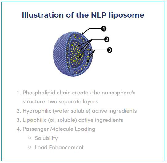
Liposomes are nanocarriers that compose of a phospholipid bilayer with an aqueous core. They can encapsulate both hydrophilic and hydrophobic therapeutic agents. They can be either Small Unilamellar Vesicles (SUVs), Large Unilamellar Vesicles (LUVs), or Multilamellar Vesicles (MLV) [10]. Liposomes are biocompatible, biodegradable, and have low toxicity. They also can protect the encapsulated drug from degradation resulting in higher bioavailability. They can cross the BBB via active transport by binding to a specific receptor or transcytosis [11]. Another approach to bypass the BBB is the intranasal route which can reach the brain via the olfactory and trigeminal nerves. The intranasal delivery of liposomes has many advantages, including fast delivery to the brain, passing the first-pass metabolism, and avoiding systemic side effects. [12]. Arumugam et al. compared the drug delivery of rivastigmine liposomes via oral and intranasal routes to the brain. The results showed a higher level of the drug in the brain for the intranasal route indicates the higher bioavailability using that route of administration.
The rivastigmine liposomes also possess sustained release action, which can reduce the frequency of the administration [13]. The same results were obtained in a later study using Electrostatic Stealth (ESS) liposomes which exhibited higher bioavailability of the drug in the brain compared to the drug solution. The liposomes also did not affect the nasal mucosa and showed a sustained release action and can be a promising intervention for AD [14]. The same intranasal approach was tested for other medications for AD, such as donepezil, due to its lower bioavailability. Al Asmari et al. developed donepezil-loaded liposomes, which had a sustained release effect compared to the drug solution and possessed good stability over three months. The intranasal administration of the liposomes did not cause any histological changes in different organs, including the olfactory nerve. It also had sufficient drug delivery to the brain, suggesting its usefulness in AD conditions [15]. Another approach to improve the therapeutic activity of therapeutic agents is to conjugate liposomes with specific ligands to enhance brain targeting. Mourtas et al. conjugated curcumin-loaded liposomes with transferrin antibody to target amyloid deposits and BBB. The decorated nanoliposomes showed better translocation across BBB and high affinity to the amyloid deposits, which shows that this formulation can be used for AD [16].
Polymeric Nanoparticles
composed of biodegradable polymers like poly(lactide) (PLA), poly(lactide-co-glycolide) (PLGA) copolymers, poly (ɛ-caprolactone) (PCL), or other natural polymers such as alginate, chitosan, gelatin, and albumin. Polymeric nanoparticles are classified to either nanosphere in which is dispersed in the polymeric matric, or nanocapsule, in which the drug is loaded into an oily core and surrounded by a polymeric membrane. Like liposomes, they can be functionalized with various ligands to enhance their targeting or protect them from the opsonization of the immune system. Like other nanoparticles, polymeric nanoparticles have many advantages, including controlled release effect, improved bioavailability, and therapeutic efficacy. They can be prepared by different methods, including solvent evaporation method, emulsification, or nanoprecipitation. Ahlschwede et al. developed PLGA modified curcumin nanoparticles for AD disease. The nanoparticles were prepared using a modified emulsion technique and then conjugated with [Gd]DTPA-chitosan.
The nanoparticles were also conjugated with anti-amyloid antibody (IgG4.1) and K16ApoE. PLGA nanoparticles successfully entrapped the curcumin, which has an anti-amyloidogenic effect and showed an entrapment efficiency of 72%. The IgG4.1 bonded to chitosan as it has amine groups, and the nanoparticles showed high binding affinity to amyloid proteins. The conjugation with K16ApoE allowed higher translocation across hCMEC/D3 endothelial cell monolayers showing that the addition of K16ApoE enhanced the uptake of the nanoparticles. The nanoparticles also offered a specific MRI contrast for amyloid plaques detection [17]. In another study, curcumin was loaded in Se-PLGA nanospheres and were tested in 5XFAD mice and were able to lower the amyloid-β load by Specific binding to Aβ plaques proposing that they can be effective nanoformulation for AD [18]. PLGA-PEG nanoparticles were also used to encapsulate curcumin and were conjugated with B6 peptide, which enhanced the permeability across BBB and enhanced the bioavailability of curcumin.
The nanoparticles were tested in APP/PS1 transgenic mice, and the results showed enhancement in the learning and memory capability. The study suggests the possibility of using these nanoparticles for AD [19]. PLGA nanoparticles were also used to load other possible therapeutic agents like EGCG, which has low bioavailability. Cano et al. developed PEGylated PLGA nanoparticles loaded with EGCG and ascorbic acid. The oral administration of these nanoparticles showed a neuroinflammation reduction and an increase in drug levels in the synapses, which improved memory and learning. Therefore, the developed nanoparticles can be a potential therapy for AD [20]. PLGA nanoparticles were also loaded with quercetin and complexed with Zn. The nanoparticles decreased Aβ fibrillogenesis and reduced related toxicity. Furthermore, the nanoparticles were associated with enhanced memory and learning capabilities, suggesting that they are candidates for AD conditions [21].
Polymeric nanoparticles were also used in PD studies to offer more specific and long-term treatment. Sridhar et al. synthesized chitosan nanoparticles loaded with selegiline in order to increase their bioavailability. The developed nanoparticles showed a 20- fold increased concentration in the brain following intranasal administration. The nanoparticles were also beneficial for increasing dopamine and glutathione concentrations in the brain, which indicates that these nanoparticles can be a promising treatment for PD [22]. PEI nanoparticles were also used for PD by complexing with siRNA. The formulation was delivered via intracerebroventricular infusion into Thy1-aSyn mice, which resulted in a decline in the neuroinflammation in the brain parenchyma and ependymocytes and a reduction of SNCA protein and SNCA mRNA expression by 50 and 65%, respectively. α-synuclein (SNCA) is a presynaptic protein that causes the accumulation of toxic oligomers in the brain leading to PD. The results of this study propose that gene therapy can be a potential therapy [23]. PLGA loaded L-dopa was also tested via intranasal administration to 6-OHDA–Wistar rats. The formulation has a long half-life and shows better bioavailability. Besides, the coordination and motor function in the nanoparticles treatment group was better than the control group showing that this nanoformulation can be a promising candidate for PD [24].
Solid Lipid Nanoparticles
Solid Lipid Nanoparticles (SLNs) have emerged as novel nanocarriers that can deliver the drugs across the BBB to the brain. They can be used through different routes of administration, such as oral, inhalational, and parenteral, to reach the brain and target neurodegenerative diseases. They consist of lipid or modified lipid (triglycerides, fatty acids, or waxes) with a 10–1000 nm diameter. Their solid hydrophobic lipid core allows dispersion of both hydrophilic and lipophilic drugs [25,26]. SLNs have various types depending on the distribution of drugs within them. They are either drug-enriched shell model, drug-enriched core model, or homogeneous matrix model. In the drug-enriched shell model, the drug is distributed around the shell, which offers burst release of the drug in the outer layer; however, the use of a low concentration of surfactants can help control the release of the drug. In the drugenriched core model, the drug is concentrated in the central core surrounded by outer lipid layer offering sustained release action.
In the homogeneous matrix model, the drug is distributed within the lipid matrix by strong molecular interactions. This type is usually used to disperse lipophilic medications without the use of surfactants [6]. SLNs provide controlled drug delivery, high efficacy, and superior targeting [27]. SLNs were developed to overcome the problems associated with polymeric Nanoparticles, such as avoiding the usage of organic solvents, therefore, reduce systemic toxicity [28]. Thus, they are among the safest and cheapest nanocarriers for drugs that enable a safe and nontoxic way to cross BBB. Their efficacy depends on their size, structure, physicochemical properties, and the method they were produced by. In recent years many researchers have been published for SLNs targeting neurodegenerative diseases. Misra et al. developed SLNs loaded with galantamine hydrobromide, an acetylcholinesterase inhibitor used in Alzheimer’s disease. This medication has poor bioavailability in the brain and can cause cholinergic side effects. The SLNs, made of biodegradable and biocompatible components, overcame these limitations by improving bioavailability and enhancing drug delivery to the brain [29].
Dhawan et al. loaded the SLNs with quercetin, a flavonoid with antioxidant activity and a drug candidate for Alzheimer’s disease. Quercetin-loaded SLNs were successfully formulated using the microemulsification technique and exhibited superior memory retention in the in-vivo experiment suggesting that they can be a potential intervention for AD [30]. In another study, Ferulic Acid (FA), which has antioxidant activity, was loaded into SLNs, which decreases ROS generation and cytochrome c release. This formulation is a promising intervention for AD as it can lower free radical generation and inhibit oxidative stress damage [31]. Kakkar et al. incorporated curcumin in SLNs to enhance its oral bioavailability and, therefore, its therapeutic effect. The SLNs could reverse brain alterations induced by AlCl3 exposure making it a potential treatment for AD [32]. Several formulations were also developed for Parkinson’s disease to enhance the drug’s bioavailability. Currently, there are many FDA-approved medications for this condition, such as levodopa, ropinirole, bromocriptine, and apomorphine; however, they have limited therapeutic efficacy to the brain.
Tsai et al. investigated the effect on bioavailability when apomorphine is loaded to SLNs. Results demonstrated that the bioavailability increased by 12-fold with the enhancement of the therapeutic effect. Besides, the formulation was tested in a Parkinson’s disease model, suggesting that SLNs is a promising approach to delivering apomorphine orally [33]. Esposito et al. developed SLNs loaded with bromocriptine which offered prolonged release over 48 hours, stabilizing plasma drug level [34]. Other non-oral routes were also suggested as a route of administration to deliver SLNs to the brain, mainly the intranasal route. Pardeshi et al. investigated ropinirole SLNs to evaluate their therapeutic efficacy. The formulation did not affect the nasal mucosa and offered a sustained action that lowered the administration frequency and was comparable to the product available in the market [35].
Dendrimers
Dendrimers are nanosized drug carriers symmetrical, highly branched polymeric molecules consisting of a central core and repeated units attached to the center and known as generations. Dendrimers are prepared using linear polymers forming an organized structure of repeated units that encapsulate functional molecules. They are synthesized by two methods either a divergent approach in which dendrimers are assembled from the initiator core and extended outward by a series of reactions, or by a Convergent approach in which dendrimers are constructed from the periphery to the core, resulting in a third-generation dendrimer. Drugs are loaded by either covalent conjugation or electrostatic adsorption [36]. Dendrimers can have positive, negative, and neutral surface charges. The positive charge is the most toxic as it may cause low biocompatibility and cell lysis; thus, sometimes dendrimers are coated with PEG to decrease their toxicity. PAMAM dendrimers are the most well-known dendrimers synthesized and made commercially available.
Their core consists of ethylenediamine, and the branches are amide groups. PAMAM dendrimers with positively charged groups may cause cytotoxicity; however, G5 and lower generation PAMAM dendrimers are considered nontoxic [37]. Dendrimers can be used to treat neurodegenerative diseases, either bypassing the BBB or traversing the BBB. Dendrimers can also be conjugated with ligands to improve biocompatibility and to target the BBB. Lgartua et al. encapsulated carbamazepine in PAMAM dendrimers within their ethylenediamine core which offered sustained release action and was safe for administration. The formulation was stable for 90 days and reduced the toxicity of the free drug [38]. The same team also investigated the co-administration of tacrine and PAMAM dendrimer generation 4.0 and 4.5. Tacrine causes hepatotoxicity upon its oral administration; nonetheless, dual therapy did not show any toxicity effects on human red blood cells and reduced the cytotoxicity of tacrine on the Neuro-2a cell line.
This reduction was not associated with a decrease in the activity as this novel therapy had an excellent anti-acetylcholinesterase activity thus can be a potential treatment for Alzheimer’s disease [39]. In another study, low-generation dendrimers conjugated with lactoferrin were employed to deliver memantine to the brain. The nanoformulation was successfully taken in the brain model and showed a sustained release action. The treatment in the AD mice model revealed enhancement of the response, higher acetylcholinesterase (AChE) activity, and better memory which suggests that this formulation has a positive impact on AD [40]. Dendrimers were also synthesized to target Parkinson’s disease. Huang et al. prepared a poly-L-lysine (DGL)- dendrimers-based gene delivery system conjugated with angiopep, targeting brain receptors. This formulation was tested on the rotenone-induced chronic model of Parkinson’s disease and showed improved recovery, indicating its potential benefit as non-invasive gene therapy for PD [41]. In another study, Sheikh et al. employed polylysine-modified polyethylenimine (PEI-PLL) to deliver VEGF expressing plasmid. The synthesized formulation was tested on cell culture and animal PD models and exhibited a positive effect on the dopaminergic system. The results demonstrated the advantageous impacts of PEI-PLL mediated VEGF gene delivery for PD [42].
Nanoemulsion
Nanoemulsions (NEs) are colloidal dispersion consisting of the water phase and oil phase stabilized by surfactants at specific ratios. NEs have many properties, such as good biocompatibility and stability, and they can be administered by various routes. They also can incorporate both hydrophilic and hydrophobic drugs due to their nature. Shah et al. investigated rivastigmine microemulsion administration via the intranasal route to bypass BBB. In-vitro and ex-vivo data demonstrated high drug diffusion, indicating better permeation via the intranasal route [43]. Azeem et al. formulated a transdermal nanoemulsion loaded with ropinirole to improve its bioavailability. In vivo evaluation of the nanoformulation showed its safety and a significant increase in the pharmacokinetic parameters (AUC, Cmax, and Tmax), demonstrating a higher release. Besides, relative bioavailability compared to oral tablets revealed a two-fold increase reflecting enhancement in its antiparkinson activity [44]. Nanoemulsion was also tested for delivering anti-TNFα siRNA to overcome neuroinflammation of neurodegenerative conditions.
The intranasal administration of the nanoformulation had higher uptake in both in vitro and in vivo compared to siRNA alone. In addition, the nanoemulsion reduced the levels of TNFα in the LPS-induced inflammation model [45]. Another study investigated the nanoemulsion of rosmarinic acid coated with chitosan as a potential neuroprotective therapy. Results reveal that the formulation prevented cellular death and alleviated oxidative stress and neuroinflammation in vitro and in vivo [46].
Inorganic Nanoparticles
Gold Nanoparticles
Gold nanoparticles (AuNPs) are one of the widely studied metal nanoparticles due to their wide range of applications. Gold in its nano range diameters has unique properties and can be synthesized by various simple techniques. They have been employed in drug delivery, thermal ablation, radiotherapy, and diagnostic assay [47]. As the main challenge in neurodegenerative disease is crossing BBB to deliver therapeutic agents, some studies evaluated the gold nanoparticles in BBB models. Ruff et al. assessed the potential of AuNPs conjugated with β amyloid in crossing BBB in vitro model. Results demonstrate that small AuNPs improve the BBB integrity while large nanoparticles or negatively charged nanoparticles hampered BBB crossing [48]. These results agree with another study that tested different insulin coated AuNPs sized (20, 50, and 70 nm) in an in vivo model. The highest migration and accumulation in the brain was for the 20 nm AuNPs, which disclose the importance of the nanoparticle size in designing AuNPs for neurodegenerative diseases [49].
Many researchers investigated AuNPs as drug cargo to the brain. Glutathione encapsulated gold nanoparticles were developed to prevent the aggregation of Aβ amyloid in AD. These nanoparticles successfully crossed the BBB and inhibited the Aβ amyloid with no toxicity. Interestingly, this study suggests a different effect of the L and D chiral nanoparticles and highlights the importance of stereochemistry in developing nanoformulations [50]. In other studies, the antioxidant and the anti-inflammatory effect of gold nanoparticles was evaluated in the okadaic acid-induced AD model in rats. The AuNPs reduced oxidative stress and the neuroinflammation caused by okadaic acid, which indicates that they can be a potential treatment for AD [51]. Gold nanoparticles have also been exploited in gene therapy for PD to improve their targeting. Hu et al. synthesized gold nanoparticles conjugated with plasmid DNA via electrostatic adsorption. The in vitro tests showed successful transfection by endocytosis, and the in vivo PD model revealed successful transmission across BBB [52]. Furthermore, gold nanoparticles can be a helpful tool for the detection of Aβ amyloid in AD. Zhou et al. developed a quantitative colorimetric sensor that can detect Aβ aggregation based on the color change of AuNPs. The system is easy to use, has low cost, and has high sensitivity [53].
Silver Nanoparticles
Silver Nanoparticles (AgNPs) are other popular metallic nanoparticles with many biomedical applications due to their distinctive physical and chemical properties. They are helpful and can be used as antibacterial, antifungal, antiviral, anti-inflammatory, and anti-cancer agents [54]. However, many studies have stated that Ag NPs can cause neural death upon their entrance to the brain. Some researchers evaluated the inflammation and the neurological effects of Ag NPs on microglia. Results of the study show that up taken Ag NPs became coated with non-reactive silver sulfide (Ag2S), and Ag NPs have an anti-inflammatory effect by reducing reactive oxygen species (ROS), nitric oxide, and TNFα. The formation of Ag2S was found to render the Ag ion toxicity [55]. On the other hand, several studies suggest that Ag NPs have a neurotoxic effect when targeting CNS. Khan et al. investigated brain endothelial cells and astrocytes after Ag NPs exposure and found an upregulation of many proteins that cause neurodegeneration and multiple inflammatory pathways. In addition, there was a downregulation of protein which maintains hemostasis [56]. These results come in agreement with another study that examined the Ag NPs effect on neural cells. After administration, there was an increase in IL-1β and C-X-C motif chemokine 13, which indicated inflammation. The was also an upregulation in amyloid-β (Aβ) plaques, the pathological characteristic of AD [57].
Silica Nanoparticles
some research asserts that silica nanoparticles cause neurotoxicity, detrimental effects on neural cells, and lead to neurodegeneration. Upon intranasal instillation of SiO2-NPs to mice model, it was found that there was cognitive damage along with an increase of the anxiety level. Furthermore, nanoparticle exposure caused neurodegenerative symptoms, including neuroinflammation, higher tau phosphorylation, and exocytosis deterioration [58]. However, a subclass of silica nanoparticles called Mesoporous Silica Nanoparticles (MSNs) got attention due to its advantages, including large surface area, high drug loading, and functionalization capability [59].
It was found that PEG–PEI functionalized MSNs can cross BBB in vitro model, and they did not have a toxic effect [60]. PEGylated quercetin-loaded silica nanoparticles were developed and tested in a copper-induced oxidative stress model. The nanoparticles showed higher antioxidant activity in neuronal culture. This neuroprotective activity suggests its usefulness in neurodegenerative diseases [61]. Silica nanoparticles were also conjugated with different ligands to enhance the uptake by BBB. Song et al. prepared PEGsilica nanoparticles conjugated with lactoferrin (Lf), a cationic iron-binding glycoprotein. This functionalization allowed higher transport of nanoparticles across the BBB in the in vitro model, suggesting that this type of conjugation may be beneficial for nanocarriers for brain conditions [62]. In another study, Karimzadeh et al. synthesized MSNs loaded with rivastigmine hydrogen tartrate and functionalized with succinic anhydride and 3-aminopropyltriethoxysilane. The formulation was able to protect the drug increasing its stability and bioavailability; nonetheless, further optimization is needed to reduce the reduction of viability associated with these nanoparticles [63].
Cerium Oxide Nanoparticles
Cerium oxide nanoparticles (CeONPs), also known as nanoceria, have an antioxidant and radical-scavenging activity which can be helpful for the treatment of neurodegenerative diseases. Small cerium oxide nanoparticles which have a size lower than 5 nm can cross BBB [64]. Nanoceria can be taken and accumulated in neural cells reducing the level of reactive nitrogen species. They can also mitigate Aβ-induced mitochondrial fragmentation and neuronal cell death, indicating their beneficial effect in AD [65]. It was also reported that nanoceria has a neuroprotective effect human Alzheimer’s disease model, which is helpful for AD [66]. Cimini and his colleagues developed PEG-coated CeONPs conjugated with an antibody targeting Aβ aggregates, and results show that nanoparticles improved neuronal survival via the BDNF signaling pathway [67].
Potential Neurodegenerative Nanomedicine in Clinical Trials
Only a few studies are testing nanoparticles for neurodegenerative diseases were found on clinical trials.gov. One is titled “31-MRS Imaging to Assess the Effects of CNM-Au8 on Impaired Neuronal Redox State in Parkinson’s Disease” which is testing the effects, safety, pharmacokinetics, and pharmacodynamics of gold nanocrystals in patients who have been diagnosed with PD. A second study titled “Study of APH-1105 in Patients with Mild to Moderate Alzheimer’s Disease” is evaluating the safety, tolerability, and efficacy of intranasal delivery of APH-1105 for the treatment of AD [68].
Challenges
Despite its many advantages, many challenges face the application of nanotechnology. Sometimes nanoparticles can cause side effects or even toxicity due to their interaction with the biological system. This interaction can depend on size, surface charge, or concentration Metal nanoparticles tend to be more toxic than organic nanoparticles. For instance, it was reported that iron Nanoparticles could cause significant cytotoxicity in the PC12 cells neuronal model, and they produced neurodegeneration and oxidative stress in mice models [69,70]. Also, zinc oxide nanoparticles induce apoptosis in neural stem cells, and manganese and Cu nanoparticles may produce reactive oxygen species (ROS) [71,72]. Another challenge is the lack of an animal model that resembles the features of neurodegenerative diseases. These models do not precisely replicate all the disease features and thus fail the translation from the preclinical to the clinical trials. The development of a perfect animal model mimicking the pathogenesis of the disease, taking into consideration all crucial characteristics, is a key for testing the new nanoformulations.
In addition, there are many hurdles to translating nanomedicine to clinics due to the lack of translational advisory organizations. Usually, the are three main areas in which the project may fail either the choice of the research area, selection of therapeutic agent, or the peer-review process. This is why translation expertise is an essential key to academic research. These hurdles need translational evaluation criteria to overcome, such as safety, technical, competitive, regulatory, reimbursement, and commercial evaluation. The safety evaluation should be concerned about the safety of the nanoparticles and if they will interact or leave any residues or cause harm for patients. Thus, nanoformulations that have not been used before in humans must have a significant advantage to be considered and translated to the market. Besides, long-term safety and biocompatibility are critical for successful nanomedicine in the market. Another technical challenge is that the nanoformulation produced in the lab will be hard to scale up for manufacturing, and large-scale production can be expensive. batch-to-batch variation can also hamper the production process. Another challenge is the competitive market. The improvement in potency or fewer side effects is not sufficient. The new drug should leave better therapeutic outcomes. The manufacturers should know where the competition is likely to be when their drugs enter the market. If the manufacturer is the first or second in the market, it will not be hard to secure a share in the market. But after that, there must be a clinical advantage to compete in the market [73,74].
Conclusion and Future Prospective
Despite the extensive research that was done for discovering an effective treatment for neurodegenerative diseases, the medication available today in the market only manages the symptoms, and there is no cure till now. Thus, exploring other possible innovative approaches for potential treatment is necessary to improve patients’ quality of life. The nanotechnology and nanomedicine field has enabled the synthesis of various nanoformulations with superior properties, which can be beneficial to neurodegenerative diseases. Around 100 novel nanoformulations were approved by the FDA including liposomes and lipid nanoparticles, PEGylated polymeric and inorganic nanoparticles are available in the market for treating different conditions. Nanoformulations have many advantages, such as protecting the drug from degradation, increasing solubility and bioavailability, improving active agents’ therapeutic effectiveness, and decreasing toxicity. Both organic and inorganic nanoparticles were investigated for neurodegenerative disease conditions.
Organic nanoparticles offer a high degree of safety as it consists of biocompatible and biodegradable polymers. They also have the advantage of the ease of preparation and can be manipulated to improve the release of the drug from its cargo. On the other hand, inorganic nanoparticles have unique physical properties and high surface area, nonetheless, have a lower safety profile as they increase the risk of oxidative stress and neuroinflammation. Additional coating with some polymers such as PEG can improve their safety. Nanoparticles can also be conjugated with nucleic acid and other ligands to enhance the targeting to the brain and enhance the translocation across the BBB. Many studies have reported that drug-loaded nanoparticles had higher concentrations in the brain, therefore, higher therapeutic efficacy against neurodegenerative. Others stated that nanoparticles had potent neuroprotective effects in the case of AD and PD. The application of this advanced technology will likely continue to improve, offering new therapeutics which provide superior clinical outcomes and enhanced patient-focused formulations. However, there are still some challenges that must be taken into consideration.
Validation of the in vitro and in vivo protocols is essential to translate these nanoformulations from the lab to clinical trials. The generation of ideal models mimicking the pathophysiology of the diseases is a crucial factor for the translation of nanoformulations. It is also necessary to set specific guidelines for the quality, safety, and efficacy evaluation of these products and perform appropriate clinical trials to ensure the lowest side effects to facilitate marketing these nanoformulations successfully. These models should emphasize on the long-term effect of nanocarriers on different organs in the human body as well as the environment. Besides, cost-effectiveness in developing nanoformulations must be taken into consideration for commercial success in the market.
For more Articles on: https://biomedres01.blogspot.com/


