Monday, October 17, 2022

Repurposed Nystatin to Inhibit SARS-Cov-2 and Mutants in the GI Tract

Repurposed Nystatin to Inhibit SARS-Cov-2 and Mutants in the GI Tract

Introduction

The severe acute respiratory syndrome coronavirus 2 (SARSCoV- 2) is originated from Wuhan in late 2019 and resulted in pandemic with crisis in public health and in World economy. More than 220 million cases and 4.5 million deaths have been registered by the Center for Systems Science and Engineering at John Hopkins University, in the beginning of September 2021. This virus belongs to the Beta coronavirus genus of Coronaviridae family of enveloped, single-positive-stranded RNA viruses, which may cause infection in lung and the gastrointestinal tract (GIT), as well. They are sensitive to bile-acids, to intestinal proteases, which may alter membrane lipid distribution and function of proteins attached to lipid rafts, making them more ineffective [1-3]. SARS-CoV-2 causes mainly upper respiratory tract symptoms however the virus presence in the GIT is remarkable. GIT symptoms are also well known in 2% to 79% [4-8] in COVID-19 and the GIT symptoms were associated with the severity of the disease [9].

A meta-analysis of 21 studies with 5285 patients draws attention to COVID-19 to be more serious in patients with GIT symptoms exploring the relationship between GIT symptoms and the severity of COVID-19 [10]. Another meta-analysis of publications found that stool samples from 48.1% of patients tested positive for virus RNA and stool samples from 70.3% of these patients tested positive for virus RNA even after respiratory specimens tested negative [11]. Diarrhea has been listed as a symptom of COVID-19 in the guideline of the Centers for Disease Control and Prevention since 22 February 2021 [12]. First the American College of Gastroenterology has drawn attention to “patients with new-onset digestive symptoms after a possible COVID-19 contact should be suspected of the illness, even in the absence of cough, shortness of breath, sore throat…” [13]. This important guideline, now is well strengthened by the tool described here, to cure this illness, in GIT importantly. In spring of 2021 Leal et al. reported on GIT symptoms present in 30% of the European patients, among which diarrhea was the most frequent with almost 18% incidence [14].

Similar study on the American population resulted in 22.4% incidence [9]. Gastrointestinal mucosal damage (degeneration or necrosis) [15], bleeding [7,16,17], edema [18-20] were present in severe COVID-19 cases suggesting a direct effect of the virus on GIT cells. Elevated liver enzymes was observed during hospitalization and progression to severe liver injury have also been noted in COVID-19 patients. The severe decompensated liver disease increased the severity of COVID-19 and vice versa [21]. Despite the fact of above-mentioned clinical symptoms only a few study reports on the investigation of GIT tissue samples [22]. Papoutsis et al. identified and characterized mutational variations of SARSCoV- 2 by enrichment next-generation sequencing (NGS) from stool samples suggesting that the gut is an ideal environment for the virus, where it may also mutate [23]. The role of gut during the SARS-CoV-2 pathogenesis was proved by detecting viable and infectious viruses from stool samples [24-26].

Several studies report on the isolation of virulent virus after the resolution of GIT symptoms even when it was not detectable from respiratory samples. This phenomenon based the ability of virus replication in GIT which may contribute to long-term consequences after the disease [27,28]. Multisystem inflammatory syndrome in children is thought to driven by zonulin-dependent loss of gut mucosal barrier which is the consequence of the prolonged presence of SARS-CoV-2 in the GIT [29]. All these data highlight the importance and the urgent need of the local antiviral treatment of the GIT for which we are looking for a solution. The mechanism of the SARS-CoV-2 viral infection is investigated thoroughly. The key factor for viral entry and life cycle is the cholesterol in the human plasma membrane [30]. Enveloped viruses primarily engage plasma membrane fusion or endocytosis for entering the host cell [31]. Lipid raft domains are involved in this process and serve as a platform for docking the viruses to the host cell. Li et al. found that lipid rafts may contribute to SARS-CoV-2 infection during the replication process in Vero E6 cells [32].

The augmented cholesterol/ fatty acid ratio of lipid rafts enhances the fusion of coronaviruses to the host cells while the decreased ratio blocks this process [33]. Meher et al. reported on the SARS-CoV-2 infection rate and binding affinity was increased by raising the cholesterol level in human plasma membranes [30]. Wang et al. proved that decreasing membrane cholesterol inhibits. SARSCoV- 2 entry [34]. Sanders et al. found that in the case of SARS-CoV-2, cholesterol is essential for the spike-mediated fusion, and for the pathological multinucleated cell (syncytia) formation as well [35]. Wang et al. showed that loading cells with cholesterol from serum, using a cholesterol transport protein (apoE) enhanced the entry and the infectivity of the SARS-CoV-2 spike protein pseudotyped retro virus [36]. Cholesterol rich SARS-CoV-2 entry sites showed almost twice the total endocytic entry sites. Additionally, they found that in virus-producing cells, the cholesterol optimally positions a protease enzyme, furin for priming the virus. Inhibiting cholesterol transport has been found to inhibit SARS-CoV-2 replication in late endosomes/lysosomes [37].

Also, depletion of the available cholesterol of plasma membranes inhibits the virus-membrane fusion [34]. In the mechanism of receptor mediated cell entry, the role of angiotensin converting enzyme II (ACE2) was proved [38,39]. Organs such as of the respiratory tract, GIT, bile duct and liver have a high expression of ACE2 receptors making them a specified target for SARS-CoV-2 [21] A single-cell RNA- Seq analysis showed that the ACE2-positive-cell ratio in digestive tract organs was significantly higher compared to the lung in COVID-19 patients [40] and the bile duct endothelial cells expressed higher quantities of ACE2 receptor than the liver endothelial cells [41]. This data may correlate with that ACE2 is an important regulator of intestinal inflammation [42]. Toelzer et al. report on the cryo–electron microscopy structure of SARS-CoV-2 spike (S) glycoprotein (S-protein) revealing that the receptor-binding domains tightly bind the essential free fatty acid linoleic acid (LA) in three composite binding pockets.

LA binding stabilizes a locked S conformation, resulting in reduced ACE2 interaction in vitro [43]. The possible role of high-density lipoprotein (HDL) particles during SARS-CoV-2 infection was also studied. Hu et al. hypothesized that because of the immunomodulatory effects of HDL-cholesterol [44,45], it may be involved in the immune cell regulation, thus resulting in the decreased HDL levels in COVID-19 patients [46]. SR-B1 is a cell-surface HDL receptor that mediates the selective uptake of receptor-bound HDL particles therefore SR-B1 has a critical role in hepatitis C virus entry [47]. Based on this mechanism a potential role of SR-B1 in SARS-CoV-2 infection was raised. Wei et al. found that the SR-B1 overexpression in Vero E6 cells increased SARSCoV- 2 infection. Additionally, the S-protein is bound to cholesterol with high S1 subunit affinity. However, S-protein did not bind to ApoA1 protein [48], main component of HDL which enhanced the entry of SARS-CoV-2. These data suggested that HDL might form a bridge between SARS-CoV-2 and SR-B1 during viral entry [48].

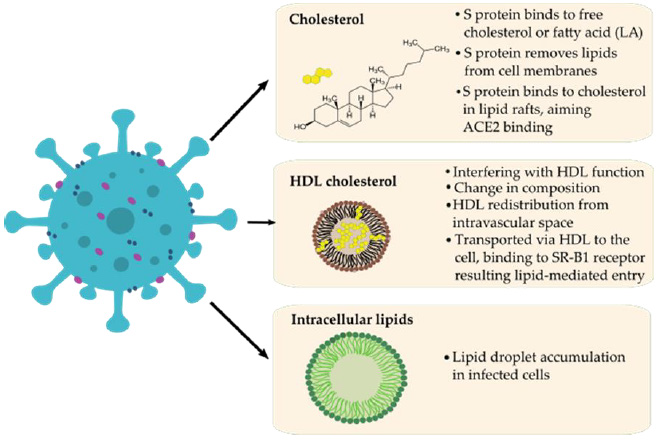
The role of HDL in COVID-19 was manifested in the change of HDL plasma concentration and proteome composition and functionality as severe patients were associated with HDL dysfunction manifesting in aggravated inflammatory endothelial conditions [49,50]. Another evidence for lipid involvement in COVID-19 pathogenesis was suggested by Nardacci et al. They observed lipid droplets accumulation in cells during SARS-CoV-2 infection, both in vitro and in lungs of COVID-19 patients [51]. Correlations between SARS-CoV-2, cholesterol, HDL, lipid rafts and fatty acids are summarized in (Figure 1). Compounds affecting cholesterol were shown to interfere with viral infectivity at different stages of viral replication. For example, cholesterol depletion proved to disrupt the HIV-1 virion membrane [52]. Baglivo et al. report on natural-based substances (cyclodextrins and phytosterols) that were used to reach membrane destabilization through cholesterol reduction and disturbed the process of SARS-CoV-2 entry into the host cells [53].
biomedres-openaccess-journal-bjstr

Figure 1: Possible correlations between SARS-CoV-2 (left) and lipids (LA) / HDL-cholesterol (right) during pathomechanism based on the literature.
biomedres-openaccess-journal-bjstr

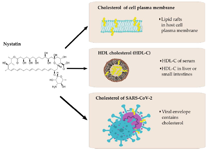
Figure 2: Possible interactions between nystatin (left) and cholesterol of different origin (right).

Another cholesterol-lowering agent, fenofibrate recently has been proved to be effective in the reduction of SARS-CoV-2 infection in vitro. Treatments for reducing the plasma cholesterol levels (i.e., by the use of statins) are also suggested by healthcare professionals for a long while in COVID-19 care [54]. Nystatin is a polyene macrolide antibiotic whose mechanism of action is to bind to sterols and forming ion channels in the fungal membrane leading in this way to leakage and cell death. This compound possesses a high affinity to bind to the ergosterol of the fungal plasma membrane, but the binding to human cholesterol is also significant [55,56]. Possible interactions between nystatin and cholesterol are presented in (Figure 2). Nystatin is not appreciably absorbed from the mucous membranes of GIT, thus has no systemic effect from oral formulations and therefore can be used to treat fungal infections of the GIT. Consequently, it may modify the cell surface through lipid rafts `floating’ on the cell surface, binding to their cholesterol content to prevent the SARS-CoV-2 viral particle from entering the cell [57,58].

This may require a high concentration of active ingredient that could be systemically problematic based on its toxicity, but not in local application. It is also reported in the literature that polyene macrolides, such as nystatin, may also be able to alter the fat metabolism of mammalian cells, thereby interfering with the replication of the virus [32]. In any case, nystatin-based pharmaceuticals are already indicated for use as complementary therapy as preventing candidiasis in patients assigned to antibiotic therapy. Nystatin had been previously examined regarding to its potential antiviral effect in HIV-1 infection model in H9 cells by Selvam et al [59]. Nystatin A inhibited viral replication effectively, in certain concentrations that did not affect cell viability. The results suggest that Nystatin A merited attention as an antiviral drug for the treatment of HIV-1 infection. Nystatin in the antiviral indication alone was used against HIV in a clinical trial also [60]. Nystatin had also been proved to be effective in reducing the cytopathic effect manifesting in cell-cell fusion during infection of SARS-CoV-2 pseudovirus [35].

In addition to the antifungal and antiviral effects of nystatin, its immunomodulatory effect was also proved [61,62]. We recommend that nystatin may be a safe and effective agent in the treatment of infections caused by enveloped viruses, including COVID-19. Since this drug affects the virion structure as well and is not absorbable in the GIT, the development of nystatin resistance has less chance and nystatin may has a direct antiviral effect on viruses during infection, while might have only negligible effect on host tissues. To verify the antiviral activity of nystatin against SARS-CoV-2 in vitro, the virus inhibitory effect of nystatin was investigated performing SARS-CoV-2 antiviral assay on Vero E6 cells according to the method of Manenti, et al. [63,64].

Materials and Methods

Materials

Nystatin was used in pharmaceutical grade. 50.000μg/ml stock solution in DMSO diluted 100-fold in Dulbecco’s Modified Eagle’s Medium (DMEM)‐high glucose supplemented with 2mM L-Glutamine, 100 units/ml penicillin‐streptomycin mixture without FBS.

Cell Cultures

Vero E6 cells were purchased from Veterinary Diagnostic Directorate of the National Food Chain Safety Office, Hungary. The cells were cultured in Dulbecco’s Modified Eagle’s Medium (DMEM)‐high glucose (Sigma) supplemented with 2mM LGlutamine (Sigma), 100 units/ml penicillin‐streptomycin mixture (Sigma), and 10% of FBS, at 37°C, in a 5% CO2 humidified incubator. Adherent sub‐confluent cell monolayers were prepared in growth medium DMEM high glucose containing 2% FBS in T175 flasks or 96‐well plates for propagation or titration and neutralization tests of SARS‐CoV‐2.

Viral Growth in Cell Culture

Vero E6 cells were seeded in T175 flasks at a density of 1 × 10⁶ cells/ml. After 18 to 20 hours, the sub‐confluent cell monolayer was washed twice with sterile Dulbeccos’s phosphate buffered saline (DPBS). After removal of the DPBS, the cells were infected with 3.5ml of DMEM 2% FBS containing the virus ata multiplicity of infection of 0.01. After 1 hour of incubation at 37°C in a humidified atmosphere with 5% CO2, 50 ml of DMEM containing 2% FBS were added to the cells. The flasks were daily observed, and the virus was harvested when 80%‐90% of the cells manifest cytopathic effect (CPE).

Virus and Titration
biomedres-openaccess-journal-bjstr

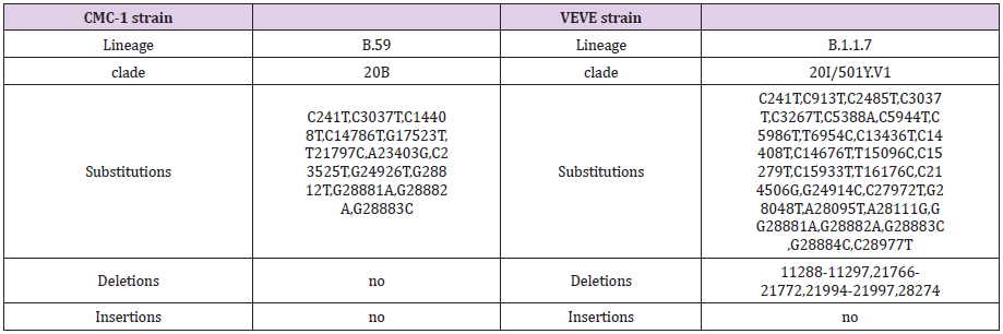
Supplementary Table S1: Informations of used SARS-CoV-2 strains.

SARS CoV‐2 virus strains (Wuhan and British mutants, CMC- 1 and VEVE) were obtained from the Complex Medical Centre, Budapest, Hungary. For detailed informations of strains, see Supplementary Table 1. The virus was titrated in serial dilutions of 1log to 11log (10-1 to 10-11) to obtain 50% tissue culture infective dose (TCID50) on 96‐well culture plates of Vero E6 cells. The plates were observed daily for a total of 4 days for the presence of CPE by means of an inverted optical microscope. The end‐ point titer was calculated according to the Reed & Muench method based on eight replicates for titration [65].

Treatment of Virus
biomedres-openaccess-journal-bjstr

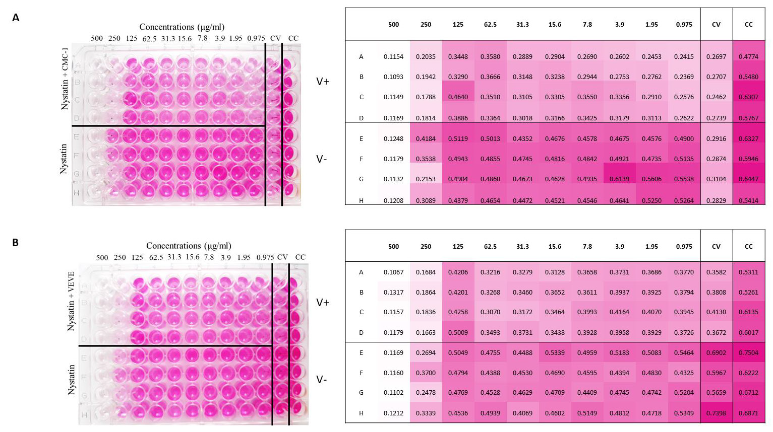
Figure 3: Colorimetric read-out of SARS-CoV-2 antiviral assay of nystatin. The cell plate contained a semi-confluent Vero E6 monolayer with SARS-CoV-2 infections of Wuhan strain, CMC-1 (A) and British strain, VEVE (B).

Two‐fold serial dilutions of nystatin, starting from 500μg/ml were prepared. All dilutions contained 1% DMSO. The dilutions were mixed with an equal volume of viral solution containing 100 TCID50 of SARS‐CoV‐2 in Dulbecco’s Modified Eagle’s Medium (DMEM)‐high glucose supplemented with 2mML-Glutamine, 100 units/ml penicillin‐streptomycin mixture. The nystatinvirus mixture was incubated for 30 min. at 37°C in a humidified atmosphere with 5% CO2. Layout of SARS-CoV-2 antiviral assay 96 well plates (Figure 3) were prepared according to the following. A1- H12 wells contained Vero E6 cells (0.2 x 10⁶ cell number per well). Column 12 contained the control cells (CC): cells without virus and nystatin treatment. Column 11 contained the virus control (CV): 100 TCID50 of SARS-CoV-2 without nystatin treatment. Column A1- D10 contained virus treated with nystatin dilution series. 100μl of the mixture at each dilution of nystatin was added in quadruplicate (A1-D1, A2-D2, etc.). Column E1-H10 contained cells treated with the nystatin dilution series in quadruplicate without virus (E1-H1, E2-H2, etc.). The bisecting dilution series of nystatin concentration was 500μg/ml-0.97μg/ml. The plates were incubated for 4 days at 37°C in a humidified atmosphere with 5% CO2.

Colorimetric Read‐Out of SARS-CoV-2 Antiviral Assay

After 3 days of incubation, the supernatant of each plate was carefully discarded and 100μl of a sterile DPBS solution containing 0.02% neutral red was added to each well. After 1 hour of incubation at room temperature, the neutral red (Renal) solution was discarded, and the cell monolayer was washed twice with sterile DPBS containing 0.05% Tween 20. After the second incubation, the DPBS was carefully removed from each well; then, 100μl of a lysis solution (made up of 50 parts of absolute ethanol, 49parts of MilliQ and 1 part of glacial acetic acid) was added to each well. Plates were incubated for 15minutes at room temperature and then read by the spectrophotometer infinite F50 at 492 nm and by using Tecans iControl Software. The plates were inspected by an inverted optical microscope before olorimetric read-out.

Results

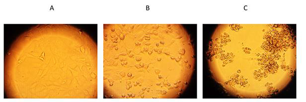
Antiviral effects of nystatin on the replication of SARSCoV- 2 in cell culture to confirm that nystatin inhibit SARS-CoV-2 replication, Vero E6 cell culture was infected with two mutants of SARS-CoV-2 pretreated with nystatin in vitro. SARS-CoV-2- early Wuhan and British mutants were treated with increasing concentrations of nystatin, and protection from cytopathic effects (CPE) observed visually and with colorimetric read-out. Nystatin showed a reduction of the viral CPE dose dependently (Figure 3). After the cells were stained with neutral red, their optical density at 492nm was measured (Figure 3A). At 96h post infection, cells were examined with an inverted optical microscope (magnification, x100). The cell control (CC) refers to cells without compound treatment and virus infection (Figure 4A).
biomedres-openaccess-journal-bjstr

Figure 4: Validation of viable cells by inverted optical microscope.
(A) Cell control, CC;
(B) Cells with SARS-CoV-2 Wuhan strain treated by 62.5 μg/ml nystatin;
(C) virus control, CV.

Vero E6 cells after 96 h of SARS-CoV-2 infection are shown on the Figure 4C. as the virus control (CV). 62.5 μg/ml nystatin treated SARS-CoV-2-infected cells are shown in Figure 4B. The results showed that nystatin inhibited SARS-CoV-2 infection, with an effective concentration. (EC) of 62.5μg/ml against Wuhan CMC-1 and British mutant VEVE strains (Figure 5). To evaluate the cytotoxicity of nystatin to cells, Vero E6 cells were treated with different concentrations of nystatin up to 500 μg/ml. Taking into account the toxic effect, the concentration at which nystatin inhibited the virus growth, the EC was determined. We found that nystatin was only slightly toxic up to 250μg/ml.
biomedres-openaccess-journal-bjstr

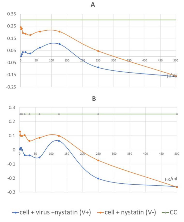
Figure 5: Graphical illustration of the corrected absorbance values. IC50 of nystatin may be detected at 62.5 μg/ml for the Wuhan strain, CMC-1 (A) and 125 μg/ml for the British mutant, VEVE (B).

Calculation of Dose Response to Nystatin
biomedres-openaccess-journal-bjstr

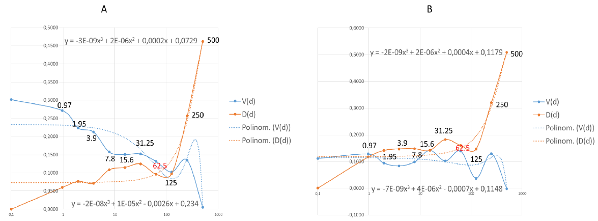
Figure 6: Effective dose (EC) of nystatin was calculated by data analysis with mathematical functions.
Points where the functions are stable are the 62.5 in both Wuhan (A) and British (B) mutant strains. The functions are not stable at the grater points therefore the effective and safe dose is 62.5μg/ml in both case. The values shown in the diagram are the nystatin doses correlated to function.

Values of the measured absorbance are proportional to the number of living cells. To evaluate the plates, the absorbance values of the four replicates were averaged. CV was then extracted from all values to obtain corrected absorbance (Figure 5). In the case of CV, almost complete cell death was observed. In the case of CC, the cell layer confluently overgrown the wells. Where the corrected absorbance was positive greater than cutoff (63) – OD (CC) /2 – there were cells that appeared alive confirmed by microscope (Figure 4B). Based on our investigation, it can be determined that treatment with nystatin up to 125μg/ml was not cytotoxic, but at all concentrations reduced the live cell concentration compared to the cell control (CC) to an extent that was considered constant independent of the concentration used. Nystatin treatment has been shown to be effective against CMC-1 (Figure 5A) and VEVE (Figure 5B) SARS-CoV-2 strains at concentrations of 62.5 and 125 μg / ml, respectively. Since the accurate antiviral IC50 could not be determined from reason of nystatin-host cell effect, the EC value of nystatin was modelled mathematically (Figure 6).

Mathematical Data Analysis of EC Value of Nystatin

EC value of nystatin was modelled by a mathematical data analysis (Figure 6). Functions were generated such as:
i. V(d) a dose-dependent viral function, where (d) is an independent variable. This function determines how much the agent changes the number of cells by the effect of the dose on the number of viruses, which reduces the number of cells by a viral function definition. Thus, it describes the change in cell number by the virus in the dose function.
ii. D(d) is a function that determines which cell number definition is affected by the dose and directly by the agent on the cell number. To get the D(d) function need to know A, that is the average of CC: 0.5808. Describing by a function, there is present both virus and agent: A-V(d) -D(d) this means A-D on the plate rows. To determine D the following definition can be determined:

Equation Definition of D functional.

A polynomial of degree 3 functional can be fitted to both. It can be seen that at high doses the destructive effect of the virus decreases, while with increasing dose the D function reduces the number of cells increasingly. The optimum must be found where the dose level of the agent is still safe. The closest dose value to the intersection (Figure 6A) of the trend line is 62.5. 125 can also be considered as a close point, mathematically, and may be a good solution, but it is the limit of a drastic change in value for both trend lines, so it is less safe to use this dose (Figure 6A). The same phenomenon can be seen in (Figure 6B). The last point where the functionals shown stability is 62.5. This is the concentration of which nystatin shown the highest efficacy with low toxicity. The IC50 calculated according to the standard protocol (Figure 5) was not exact to estimate the antiviral dosage of nystatin in this test system. The values of effective antiviral dose therefore was based on mathematical modelling suggesting 62.5μg/ml in both case. In general use, nystatin dose is 100 mg-600 mg daily. Since nystatin is a non-absorbable drug this means a dilution in 5-6 liter GIT juice daily [66] approximately. In this experiment we determined the EC under the maximum daily dose of nystatin.

Discussion

During the SARS-CoV-2 pandemic period of time, - as the virus’ name Severe Acute Respiratory Syndrome shows accordingly- COVID-19 is considered mainly as a new disease in pulmonology, and viral infection, invasion of GIT are oversight and undervalued, equally. However, the increasing concentration of excreted viral fragments in wastewater, as shown by tests data, might predict the soon increase in community infection rate, days, or weeks earlier than the number of clinical cases with symptoms of COVID-19 are set on, in communities [67]. The web search analytics as infodemiology study for abdominal symptoms in COVID-19 reflects the early sign of infection (diarrhea) presenting the involvement, i.e., the viral infection of gastrointestinal tract of patients, at the same time [68,69]. In addition to the additional GIT manifested clinical cases, notable number of asymptomatic and presymptomatic virus carriers are also to be considered as uncontrolled role-players in pandemic [70].

The pandemic of SARS-CoV-2 requires an intensive search for new antiviral agents. Many compounds are currently being investigated for their efficacy in COVID-19 therapy. Considering the severity of COVID-19 there is a need to find effective but less toxic drugs for a rapid treatment. Therefore, the possibility of nonsystemic therapy came to the fore. In this report the antiviral effect of nystatin antibiotic was investigated against Wuhan and British mutant strains of SARS-CoV-2 tested on Vero E6 cells. The presented results demonstrate that nystatin at a non-cytotoxic dose may protect cells against infection with SARS-CoV-2. This protection is associated with inhibition of virus replication that was manifested by an augmented living cell rate (Figure 3).

Nystatin is a polyene macrolide antibiotic of which the broad-spectrum antifungal activity and low fungal resistance are characteristics [71-75]. Nystatin is not or slightly absorbed [76,77] and non-toxic allowing its application in the GIT. It is used worldwide since 1961, in different pharmaceutical formulations and dosage forms in antifungal indications, including prophylaxis. It might be a safe agent as the annual number of reports on adverse reaction counts less than 100 per year in average [78]. The antiviral effect of polyene macrolide antibiotics against a variety of lipidenveloped RNA and DNA viruses was proved [59,79,80]. Nystatin bind irreversibly with sterols [81,82]. This is a direct structural and non-metabolic effect that consequence is the low resistance rate [83,84]. The binding of nystatin to cholesterol in cell membranes causes changes in plasma membrane fluidity and cell permeability therefore decreases infectivity of lipid-enveloped viruses.

The drug-sterol interaction is a well-known phenomenon that has already been demonstrated in our earlier studies on the macrolide antibiotic primycin [84-89] where the membrane disorganization was due to the ergosterol-primycin bound stabilized by hydrogen bounds [87]. The inactivation of SARS-CoV-2 is dependent on the dose of nystatin: the accurate dose of inhibition is probably due to a complex effect that is strongly depend on the binding action of host-cell membrane and/or viral envelope cholesterol ratio. Similar effect of nystatin was described against HIV-1 [59]. Interestingly, if viral envelope cholesterol was reduced the infectivity of SARS-CoV-2 decreased while depletion of cell membrane cholesterol had no effect on the virus infectivity [35]. Thus, we suppose that the envelope cholesterol/nystatin molecule ratio may influence that drug efficacy. In dose determination we had to consider a slight restrictive effect of nystatin (Figure 3) on test-cells connected to the inhibition effect of virus. Based on this 62.5μg/ml nystatin concentration showed to be the effective dose which improved the infected Vero E6 cells’ viability.

Considering potential therapeutic usage, the properties of nontoxic and LADME - which represents the main pharmacological characteristics of any pharmaceutical product - of nystatin should be interpreted. Liberation, Absorption, Distribution and Metabolism steps are hardly explainable due to the poor water solubility and lack of absorption of nystatin. Liberalization and distribution are limited to the GIT, metabolism is restricted to the interaction with the gastrointestinal juice and its fecal elimination is known. Based on the well-established antimicrobial indication, the daily dose of nystatin is 100-600 mg which is continuously diluted or concentrated depending on secretion or reabsorption of the GIT segments. In this way, the total dilution may be around 5-6 liter in GIT juice daily [66], thus the determined effective antiviral concentration is only about the half of the maximum allowed daily concentration of nystatin. This means there is no reason to change the well-established dosage regimen in a potential SARS-CoV-2 GIT disinfection therapy either [88-90].

In our opinion, GIT disinfection pharmaceutical products may contribute to a step forward in overcoming the pandemic on different levels. Individuals (patients with GI symptoms) shall benefit sooner recovery and less severe or fatal, recurrent, and postcovid cases may be experienced. For communities, one may expect a smaller number of asymptomatic carriers, making lockdown periods more effective and contribute to a better control on pandemic. From regulatory point of view, the issue of emergency use market authorization to efficient, properly documented (i.e. cytopathic assay evidenced SARS-CoV-2 antiviral) nystatin formulations shall be considered. To summarize.

Considering the urgency of the ongoing COVID-19 pandemic, detection of various new mutant strains and the future potential re-emergence of novel coronaviruses, repurposing of authorized drugs such as nystatin could be worthy of attention. Vaccines mainly have systemic effect, they do not neutralize viruses in GIT, one of the general sources of viral infection. Our results suggest that a non-systemic nystatin therapy – based on virus inactivation, in cytopathic assay proven formulated drugs may provide a solution for COVID-19 therapy through GIT disinfection. The use of this safe drug in preventive, prophylactic and therapeutic treatment might offer healthcare and economic advantages and benefits additional to the worldwide vaccination.

For more Articles on: https://biomedres01.blogspot.com/

Saturday, October 15, 2022

Moderna COVID-19 mRNA Vaccine and its Observed Cardiac Side Effects in Adolescent and Children

Moderna COVID-19 mRNA Vaccine and its Observed Cardiac Side Effects in Adolescent and Children

Introduction

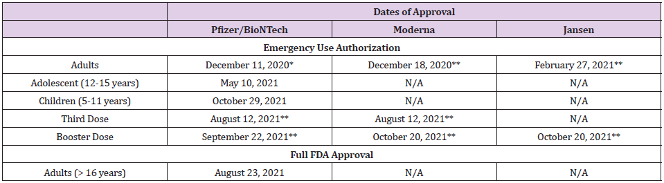
Infection with SARS-CoV-2 has been the deadliest pandemic in the U.S. infecting over 46 million and claiming the lives of over 749,000 people [1]. To date, the U.S. Food and Drug Administration (FDA) has only approved drugs and/or biological agents for the treatment of patients with COVID-19 under Emergency Use Authorization (EUA). Therefore, the three COVID-19 vaccines that have been approved for use in the U.S. are the most effective tool currently available for the prevention of SAR-CoV-2 infection (Table 1). To date, over 428 million doses of COVID-19 vaccines have been administered resulting in full vaccination of over 193 million people and over 222 million have received at least one dose of these multi-dose vaccines (Table 2). Moderna’s COVID-19 vaccine is a lipid nanoparticle–encapsulated mRNA-based vaccine that encodes the prefusion stabilized full-length spike protein of the SARS-CoV-2. Its safety and efficacy was unequivocally established in a Phase 3 clinical trial involving over 30,000 subjects who were randomly assigned in a 1:1 ratio to receive either vaccine or placebo [2]. The vaccine was delivered to established COVID-19 negative volunteers in two-dose protocol 28 days apart. The vaccine was 94.1% effective in preventing COVID-19 disease among these clinical trial participants with 11 cases of COVID-19 in the vaccine group and 185 in the placebo group developed infection with the SARS-CoV-2 virus. At the time of the analysis of these 196 COVID-19 cases, none in the vaccine group and 30 in the placebo group were classified as severe.

biomedres-openaccess-journal-bjstr

Table 1: Dates of Approval of Various COVID-19 Vaccines in the United States.

Note: *> 16 years
** > 18 years

biomedres-openaccess-journal-bjstr

Table 2: COVID-19 Vaccination in the United States1.

Note: 1as of November 08, 2021

Based on the data submitted to the FDA, the Moderna vaccine was approved under EUA for vaccination of adults (>18 years of age) on December 18, 2020 [3]. The primary series of vaccination included two doses 28 days apart. Since its approval, over 70 million people have been fully vaccinated using the Moderna vaccine with no serious adverse effect. Based on the data related to the breakthrough infections in fully vaccinated people, on August 12, 2021, FDA authorized a third dose of Moderna vaccine in highrisk individuals and on October 20, 2021, a single booster dose was authorized to be administered at least 6 months after completion of the primary series to individuals who fall into one of the following three categories; 65 years of age and older; 18 through 64 years of age at high risk of developing severe COVID-19; and 18 through 64 years of age with frequent institutional or occupational exposure to SARS-CoV-2 [4,5]. Since these approvals, over 5.9 million people have received an additional dose of the Moderna vaccine. Rare cases of myocarditis and pericarditis have been reported following vaccination after the second dose of both Pfizer/BioNTech and Moderna mRNA vaccines in adults, adolescents, and children [6- 8]. Despite of these observations, both FDA and the U.S. Centers for Disease Control and Prevention (CDC) approved the use of Pfizer/BioNTech for adolescent and children. In June 2021, Moderna submitted data to the FDA requesting EUA for use of its vaccine in adolescent. According to a press release by Moderna, Inc., the “Phase 2/3 study of its COVID-19 vaccine (mRNA-1273) in adolescents has met its primary immunogenicity endpoint, successfully bridging immune responses to the adult vaccination. In the study, no cases of COVID-19 were observed in participants who had received two doses of the Moderna COVID-19 vaccine using the primary definition. In addition, a vaccine efficacy of 93% in seronegative participants was observed starting 14 days after the first dose using the secondary CDC case definition of COVID-19, which tested for milder disease. This study, known as the TeenCOVE study, enrolled more than 3,700 participants ages 12 to less than 18 years in the U.S” [9].

However, despite these observations, Scandinavian authorities suspended or discouraged the use of Moderna’s COVID-19 vaccine in young people because of an increased risk of heart inflammation, a very rare side effect associated with the shot. Sweden suspended the use of Moderna for recipients under the age of 30 years and in Denmark, people under the age of 18 years will not be offered Moderna vaccine, and Norway recommended that those under the age of 30 years to get the Pfizer/BioNTech vaccine instead. Based on available data, FDA has delayed its approval of Moderna COVID-19 vaccine for adolescent suggesting that additional safety data will be required before any recommendation could be formulated. Similarly, on October 25, 2021, Moderna, Inc., also released interim data from the Phase 2/3 study, called the KidCOVE study, of mRNA- 1273 in children 6 to under 12 years of age. This interim analysis showed a robust neutralizing antibody response after two doses of mRNA-1273 at the 50μg dose level with a favorable safety profile [10]. Moderna plans to submit these data to the FDA and the European Medicines Agency (EMA) and other global regulators in the near term. At the recommendation of the FDA, Moderna, Inc., has however, agreed to delay submission of its request for an EUA for its mRNA vaccine for children 5-12 years of age. The need for further evaluation of clinical data by the FDA and the CDC to fully comprehend the cardiac side effects of Moderna mRNA vaccine in adolescent and children is important not only to ensure that it is safe but also to continue to sustain public confidence. The latter is critical in the face of unprecedented and unsubstantiated public resistance specifically for COVID-19 vaccination and to create a safe environment for children in the classrooms. However, based on the aforementioned information, it is our recommendation that both the FDA and the CDC must make every effort to conclude this evaluation process posthaste.

Our position has been supported by numerous leading scientific organizations who have co-signed a statement which states that “As physicians, nurses, public health, and health care professionals, and, for many of us, parents, we understand the significant interest many Americans have in the safety of the COVID-19 vaccines, especially for younger people. Today, the CDC Advisory Committee on Immunization Practices (ACIP) met to discuss the latest data on reports of mild cases of inflammation of the heart muscle and surrounding tissue called myocarditis and pericarditis following COVID-19 vaccination among younger people. The facts are clear: this is an extremely rare side effect, and only an exceedingly small number of people will experience it after vaccination. Importantly, for the young people who do, most cases are mild, and individuals recover often on their own or with minimal treatment. In addition, we know that myocarditis and pericarditis are much more common if you get COVID-19, and the risks to the heart from COVID-19 infection can be more severe” [11].

For more Articles on: https://biomedres01.blogspot.com/


Friday, October 14, 2022

Physical-Recreational Games to Increase the Participation of Children in the Recreational Activities of the Bengochea District

Physical-Recreational Games to Increase the Participation of Children in the Recreational Activities of the Bengochea District

Introduction

Recreation must serve to sow knowledge, to promote spiritual values such as solidarity and a sense of unity among people. Several researchers at national and international level have addressed the subject making contributions to recreation in which they stand out: (Sosa, 2000; Perez, 2003; Waichman, 2009; Domínguez, 2018) [1-4], among others. In a general sense, everyone considers that Recreation works to teach, educate and raise the quality of life of the community in an organized and harmonious way, thus promoting the achievement of greater cultural, political and social development.
According to the studies carried out by Sosa from the 90s to the present about Physical Recreation, and of which these authors fully agree on the validity it has when it states in the (2000) that: 1. Physical Recreation is the set of physical activities defined by the physical recreational reasons that constitute a subsystem of Recreation and is part of the physical culture of the population with emphasis on active rest, health, the creation of motor habits, fun and creative individual development. This type of Recreation is characterized by a certain level of motor skills, which generates a high level of recreational physical prominence (p. 87).
The present work has as background the research of the authors Vilaú, et al. [5-8]. The aforementioned research allows to reinforce the idea of the work that can be developed in terms of Recreation in the communities, aimed at children, due to the little motivation and the lack of participation on the part of these in the physical-recreational activities in the communities, since in the vast majority of the planned activities the same games are executed, without looking for other alternatives or variants that stimulate the participation of infants, based on their tastes, interests and needs. In the observations made in the community, the interviews with the children, relatives and neighbors of the community, the following probability situation could be appreciated: limited participation in the physical-recreational activities of the children of 9 – 12 years of the circumscription # 9 of the Bengochea Distribution, of the municipality of Santa Clara. The above allows us to formulate the following scientific problem: How to increase the participation in physical-recreational activities of children aged 9 -12 years of the district # 9 of the Bengochea Distribution, of the municipality of Santa Clara?.
For the development of the research, the following system of objectives is proposed:
1. Diagnose the initial state of participation, tastes and preferences to physical-recreational activities of children from 9 – 12 years of age in district # 9 of the Bengochea Distribution, in the municipality of Santa Clara.
2. Select physical-recreational games to increase the participation in the physical-recreational activities of children from 9 – 12 years of age in constituency #9 of the Bengochea Distribution, in the municipality of Santa Clara.
3. To assess the participation in the physical-recreational activities of the children of 9 – 12 years of the circumscription # 9, from the application of the physical-recreational games.
The importance of this research is to promote that the game is an essential means within recreation, provided that it is known at what stage it is most important to play and what are the purposes of the game, such as: teaching, culturizing, transcending, distracting, investigating, developing, generating, creating; everything depends on the form, the type of game and the attitude that is adopted before the essential activity of the playful. According to the diagnosis made and the practical experience of the authors, it is evident that most of the children aged 9 – 12 years of the constituency # 9 of the Bengochea Distribution, are limited to participating in physicalrecreational activities, since those that are carried out are not of their interest or preference, since passive entertainment has become widespread due to the marked fondness for video games, the use of new information and communication technologies, such as computers, Xbox, tablets, smartphones, and other electronic equipment, increasingly inducing new generations to addiction to cyber games, which is counterproductive with the needs of the physical and psychological development of this age group.

Methodology Used

For the development of the research, the authors relied on the concepts issued by Hernández et al. [9], using methods and techniques, which allowed to obtain an accurate information of the problem raised, of the theoretical level, the analytical-synthetic; inductive-deductive that were applied for the theoreticalmethodological foundations of recreation, physical recreation and games, as well as to carry out an objective study of the participation of children in physical and recreational activities, in addition to the preparation of the professional of Physical Culture and other managers. The research involves a first population composed of 12 girls and 17 boys aged 9 – 12 years, a second population composed of 3 professionals of the Physical Culture of the Sports Combined “Martyrs of Barbados” and 15 subjects of the community, 11 relatives and 4 neighbors, all belonging to the constituency # 9 of the Bengochea Distribution, Santa Clara municipality.

Analysis of the Results and Discussion

biomedres-openaccess-journal-bjstr

Figure 1.

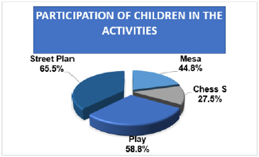
Physical-recreational activities such as board games, simultaneous chess, to play and street plan were observed, carried out in the district # 9, of the Bengochea Distribution for 8 months from which the following results were obtained: The relatives and neighbors of the community felt concerned about how the children used their free time in activities not proper to their age, which was interfering in their bio-psycho-pedagogical and social development, since a systematization was not achieved in the physical-recreational activities that were carried out by the physical recreation teachers. It should be noted that disclosure does not disclose in advance all activities, which can influence the poor participation of children in the activities being carried out. As for the preparation of recreation teachers, although they have mastery of their work, it was found that not everyone prepares for the realization of the activities, nor do they carry out actions to increase the participation of children in recreational activities, since sometimes they showed demotivation, lack of creativity and interest in the search for alternatives or new games for the execution of the activities. In the aspect related to the participation of children in recreational activities in a general sense, Mal was evaluated; in the board games, only 5 girls and 8 boys attended for 44.8%. Regarding the simultaneous chess, 3 girls and 5 boys participated, for 27.5%. The highest participation was observed in the “A jugar”, with 7 girls and 10 boys, which represents 58.6% and in the Street Plan 9 girls and 10 boys were present, which represented 65.5% (Figure 1).
The evaluation of this aspect with respect to participation was influenced by the poor attendance of children aged 9-12 years to recreational physical activities, as well as the organization of the area for the development of activities, since the necessary conditions were not created in terms of the use and creativity of means and implements, the area for the execution of the activities was not correctly delimited, in addition there were no varied offers, since generally the planned activities were carried out and the space was not used to insert other small, recreational, traditional and / or pre-sports games. When applying the interview, it was possible to know that there are 6 children (20.6%) whose reasons are related to going to the cinema or the park with their friends, 5 (17.2%) watch tv, 8 (27.5%) use new technologies to play on the computer or Xbox, as well as connect on social networks with their smartphones, only 10 (34.4%) prefer to participate in physicalrecreational activities. The 4 neighbors of the community that represents 26.6%, raised that currently children from 9 – 12 have a marked interest in new technologies and the use of social networks, since in the area where they live there is a wifi antenna, in terms of support and realization of them to the recreational activities that are planned, participation may be better, in order to avoid other activities, which are not appropriate for their age, nor are they beneficial for the development of children, since sometimes it causes in some children, manifestations of inappropriate behaviors.
The relatives who represent 72.7% stated that they like their children to perform recreational activities, as well as games and sports, but that currently they do not participate for reasons of time, although they prefer to have their children quiet at home watching television or making use of new technologies, since they are motivated by electronic games or being connected in social networks, since in the area there is wifi and on the other hand 3 of the relatives who represent 27.2% do not like to pressure their children, and try to support them in what they like to do, referring to the activities of the age. In the results of the interviews conducted with the children, relatives, neighbors and recreation teachers, it was found that they had poor participation in the physicalrecreational activities that were offered, so that the children participated in other activities, many of which are not appropriate for their age, demonstrating the addiction to electronic games and social networks, this motivated the search and selection of several physical-recreational games that contributed to the increase in participation in the activities of children from 9 – 12 years old, from the constituency # 9 of the Bengochea Distribution of the municipality of Santa Clara.
For the selection of traditional games, the general characteristics according to Watson (2008) [10] were taken into account, which are:
a) They are played by children for the sheer pleasure of playing, who decide when, where and how to play.
b) Respond to children’s basic needs.
c) The rules are easy to understand, memorize and comply with, but they are also negotiable since they can be varied.
d) They do not require many materials and the necessary ones are not very expensive.
e) They are simple and easy to share with other players.
f) They can be practiced at any time and place.
g) They arise for a season, disappear and then reappear.
Some games are practiced more in winter times, since they involve greater physical and body movement, while others arise in times of greater heat (summer).

Below are 9 of the 15 Physical-Recreational Games that Were Put into Practice

The Three Towers

A. Development: At the signal of the teacher the participants formed in teams, but in trios, run to the cone, which is at a distance of 6 meters, then they enter a ring that take it to the waist and move to the little flag that is placed another 6 meters to which they turn, until they reach the cone where they leave the hoop and run in a trio to the finish line to give the exit to the next trio of the team, which will perform the same action.
B. Rules: The race is held in trios; no child can get ahead.

Balloon Up and Down

A. Materials: Balloons, water Organization: rows in teams Development: the participants with their legs apart, will be placed one after the other and at a small distance, at the signal of the teacher the first of each row will hold a balloon full of water and pass it over the head and the second will pass it underneath, until reaching the last child who will come running until the beginning of the row and will perform the same action until everyone has executed it.
B. Rules: Always the first of each row passes the balloon overhead and prevent it from breaking.

Wall, Hunter and Deer

A. Materials:

i. None Organization: Rows in teams Development: they begin by telling them that the wall is when the flexed arms are placed in front and the palms of the hands are open, the hunter is when one of the arms extends to the front reseling a rifle and the deer is when the hands are placed on the head making horns, the wall for the bullet fired by the hunter, the deer jumps the wall and the bullet kills the deer, then the teams are formed, these internally will agree and choose what action they will perform, then they are placed in rows facing each other, but on their backs, and to the sound of the teacher’s whistle the participants turn straight representing the wall, the hunter or deer who will accumulate points.
B. Rules: Only one action can be represented by the participants. If a team participant makes a mistake and does not do the action of the rest of his teammates, the team loses. The team that manages to accumulate the most points win.
C. Variants: It can be done with other animals or objects such as stone, paper and pencil.

Three-Foot Race

A. Materials: Rope. Organization: duos in teams Development: the participants in pairs, will be placed next to each other and will tie their ankles with a rope, and at the signal of the teacher they will run to the indicated distance, they will turn the cone and when they reach the finish line they will release the rope of the ankles and give it to the other couple.
B. Rules: They must run harmoniously without letting go. Teammates can help tie the rope. The team that finishes first and does it best wins.
C. Variant: You can divide the teams in two and place themselves in front of each other and do it in relay.

Transport of Objects in Duos

A. Materials: Cones, bowling, swiss, balls, flags. Organization: teams, duos Development: at the signal of the teacher the first pairs of children will move through the area carrying three different objects with their chests, but without using their hands, when they reach the finish line, they must pass the objects to another couple, and so on until they have all finished.
B. Rules: Couples cannot use their hands but can be helped by another participant. If an object falls it must be picked up and then continue the game. The team that first finishes having transported the objects without touching it wins.
C. Variant: You can transport the objects head-to-head, back to back, hip to hip, etc.

The Basket

A. Materials: Box (basket), cards Organization: rows in teams Development: the participants will be seated behind a starting line, at the signal of the teacher the first of each team will run to the basket and inside this there will be several cards with different syllables where the players upon arrival will have to form a word and say which one was the one that formed, because another player can not recreate it.
B. Rules: They must go out to the teacher’s signal. Players cannot form the same word twice. The team that has formed the most words win.
C. Variants: The teacher can say the word for the participants to form and the one who first creates it wins, it can also be done with mathematical operations.

To Fill the Knob

A. Materials: Buckets, knobs, disposable cups, water, flags Organization: rows in teams Development: the teams formed behind the starting line will have a plastic knob and at a distance a bucket with water is placed with a disposable glass inside, at the signal of the teacher the first participants run out with the knob, when they reach the place where the bucket is, they pour a glass of water and run back to give exit to the next child, so on until they all culminate.
B. Rules: The team with the most water on the knob wins. Participants must exit behind the starting line. Only one glass of water per player can be poured into the knob.
C. Variants: It can be executed by measuring time and players can repeat as many times as possible until the time given by the teacher is consumed.

Rock, Paper and Scissors in Motion

A. Material: Rings, chalk Organization: rows Development: they are placed in teams by rows facing each other, and at a distance of 20 meters hoops are placed or drawn with chalk, one after the other, changing direction every four implements. At the teacher’s signal the first students jump out on each hoop, as quickly as possible and when confronted by the participants say: “Rock, paper or scissors. One, two, three” each take out what they want. The participant who loses goes to the opposing team and the other continues, because from the other team another child must leave very quickly to prevent him from reaching the goal and challenge him with rock, paper or scissors, and so on everyone must perform the game until it ends.
B. Rules: The stone beats the scissors, the scissors the paper and the paper the stone. To leave a participant must wait for their child to lose or reach the finish line. You should jump on the hoops with both legs together.

The Chain with Rings

A. Materials: Rings Organization: rows or circles Development: two teams are formed and the participants hold hands forming a row or circle, between two children a ring is placed and at the signal of the teacher they must pass the hoop between the children to get from one end to the other in case of being in rows, and if it is in a circle that reaches the beginning where it began.
B. Rules: They must pass the hoop through the body without letting go of their hands. The team that first manages to pass the hoop among all the participants wins.
It is necessary to emphasize that in the organization and execution of the physical-recreational games it was not necessary that all the participants were in the teams, since the children repeated the actions of the game, in case their team was incomplete, since the proposed objective was to participate and complete all the proposed games. After applying the physical-recreational games, the evaluations issued by the participants were collected where they expressed the Positive, Negative and Interesting (PNI) of the games performed, in addition to observing the increase in participation, since the attendance ranged between 27 – 28 children for 96.5%, being evaluated very well where they raised how good they felt, the desire to continue playing, since many of the proposed games did not know them and others had done them in physical education classes, but in previous degrees, they also recognized the benefits of performing active games, observing the acceptance and impact of physical-recreational games, before the activities they carried out in their free time, as being most of their time dependent on new technologies, all this influenced the good organization of the areas, the creative use of media, the selection of new physicalrecreational games, according to the tastes and preferences of the children. During the practice of physical-recreational games, the children showed interest in the creation of variants, strengthening the educational work through the opportunities they had to express freely, creatively and voluntarily their criteria to express how they would like to perform the games, in addition there was a great enthusiasm on the part of the relatives, community managers, as well as teachers, in the dissemination and execution of physicalrecreational activities.
1. The motivated, active and conscious participation of the children was achieved under the principle of voluntariness, where they were managers, actors and protagonists of the physical-recreational games they executed.
2. The girls suggested holding traditional costume parties, modeling and games which were supported by the parents.
Most of the children did not have absences, indicating the satisfactory participation in the planned physical-recreational games, which managed to motivate its participants.
3. It is necessary to emphasize that in the execution of some physical-recreational games the children played with their relatives, where they demonstrated their experiences by stating that they “revived their childhood”.
4. The motivation for the realization of the physical-recreational games selected from the interest shown by the children, were supported by the relatives and neighbors of the community.
5. The execution of the physical-recreational games allowed to be more united, both with the children of the same sex, as well as the interaction with those of the opposite sex, which helped to develop traits such as tolerance, consideration and mutual respect attending to the individual possibilities of each one and in a general sense group work was favored.
6. The participation of the children in the different physicalrecreational games contributed to the formation of knowledge, habits, sports skills and the development of values.

Conclusion

In the diagnosis carried out, it was found that the participation of children aged 9 – 12 years of the constituency # 9, of the Bengochea Distribution, in the physical-recreational activities, was limited since they preferred to participate in other activities. The physical-recreational games were selected from a collective construction, taking into account the interests, tastes and preferences of the children of 9 -12 years of the circumscription # 9, of the Bengochea Distribution, which increased the participation to the physical-recreational activities. The participation in the activities, from the application of the physical-recreational games caused an atmosphere of socialization among the children, relatives, and other members of the community, where all the participants intervened, opined and supported, being the main managers, actors and protagonists.

For more Articles on: https://biomedres01.blogspot.com/

Thursday, October 13, 2022

Can Probiotics Play an Important Role Against COVID-19?

Can Probiotics Play an Important Role Against COVID-19?

Introduction

Probiotics are live microorganisms, which if administered at adequate amounts, confer beneficial physiological effects [1]. Previous study underscored the positive impact (whether directly or indirectly) of probiotics on the ACE enzymes [2]. During the process of food fermentation, probiotics make bioactive peptides which interfere with the ACE enzymes through blocking the active sites [3,4]. The debris of the dead probiotic cells can also work as inhibitors to ACE [1], suggesting that probiotics are possibly potential blockers to the ACE receptors, which act as gateway for SARS-CoV-2 to attack gastrointestinal cells. Imai and colleagues reported that ACE blockers could be used to decrease respiratory distress syndrome [5]. The prebiotics are defined as ‘substrates that are selectively utilized by host microorganisms conferring a health benefit’ [6]. Similar to probiotics, prebiotics can be orally administered into microbially colonized body sites to reach the intestine, or by a direct way to the skin or vaginal tract [6]. Prebiotics include lactosucrose, oligosaccharides, isomaltooligosaccharides, fructans, xylooligosaccharides, resistant starch, lactobionic acid, galactomannan, arabinooligosaccharides, psyllium, polyphenols and polyunsaturated fatty acids [6-8]. The health benefits of prebiotics to the gastrointestinal tract such as stimulation of immune system and inhibition of pathogens are because of their ability to modulate the activity and composition of human microbiota [1]. Prebiotics, which enhance probiotics survivability and growth, may have an excellent potential effect against COVID-19 [1]. Prebiotics could block the ACE enzymes, which may have a direct effect on gastrointestinal symptoms caused by COVID-19 [1]. There are many ongoing registered trials aiming to investigate the efficiency of probiotics in treating COVID-19 patients [9].
Some COVID-19 patients showed intestinal microbial dysbiosis characterized by decreased probiotics such as Lactobacillus and Bifidobacterium. Prebiotic or probiotic supplementation, and nutritional support has been recommended to re-normalize the balance of intestinal microbiota and decrease the risk of secondary infection due to bacterial translocation [10]. Probiotic supplementation could be a promising strategy given previous studies of the potential application of probiotics in treatment and prevention of various viral infections [1,11,12]. The elderly and disordered microbiota patients are the most susceptible groups to COVID-19. Thus, it is suggested that probiotics supplementation in those groups could increase the ability of the gastrointestinal microbiota in modulation of immunity and help in prevention of viral infections including COVID-19 [1]. Competition with pathogens for nutrients, production of anti-microbial substances, enhancement of the intestinal epithelial barrier, and adhesion to the intestinal epithelium, and modulation of the host immune system might explain clinical success of probiotics [13,14]. Saavedra and colleagues conducted randomized control trial of 55 infants and found that enteral supplementation with a combination of Streptococcus thermophiles and Bifidobacterium bifidum decreased the incidence of diarrhea and rotavirus shedding [15], which may indicate interference with entry of the virus into cells and/or inhibition of viral replication in the intestine. Although probiotics were not administered to the respiratory tract, this mechanism may play a role in lowering dissemination of SARS-CoV-2 through the gut. Therefore, direct inhibition may be impossible at the respiratory tract. Having said that, lungs have their own microbiota and a gut-lung connection has been previously reported whereby microbe-microbe host-microbe, and immune interactions could affect the course of respiratory diseases [14,16].
Growing evidence showed that the gut-lung axis plays a pivotal role in the pathogenicity of viral and bacterial and infections, as the intestinal microbiota could enhance the activity of alveolar macrophage, thus having a prophylactic role in host defense against pneumonia [17]. Respiratory tract infections such as influenza are linked with a dysbiosis in the microbial communities of the both gastrointestinal and respiratory tracts [18,19], which could alter immune function and facilitate secondary bacterial infection [14]. Previous studies reported that COVID-19 could be associated with intestinal dysbiosis leading to inflammatory reactions and poorer response to pathogens [20,21], the case exists for probiotics that could restore gut homeostasis [22]. Arroyo and colleagues evaluated the efficacy of oral administration of Lactobacillus fermentum CECT5716 or Lactobacillus salivarius CECT5713, two lactobacilli strains isolated from breast milk, compared with the efficacy of antibiotic therapy in treatment of lactational mastitis [23]. They found that females took the probiotics improved more and had reduced recurrence of mastitis than those who took the antibiotic therapy.
The gut microbiome plays a pivotal role in systemic immune responses, including those at distant mucosal sites such as the lungs [24,25]. Administration of certain lactobacilli or bifidobacteria helps in clearance of influenza virus from the respiratory tract [24,26]. Probiotic strains increase type I interferon levels, the activity and number of T cells, NK cells, antigen presenting cells, as well as the levels of systemic and mucosal specific antibodies in the lungs [24,27,28]. Growing evidence showed that probiotic strains could regulate the dynamic balance between proinflammatory and immunoregulatory cytokines that facilitate viral clearance with minimum immune response-mediated lung damage [14]. This seems be particularly important as a way to inhibit acute respiratory distress syndrome, which is the most feared complication of COVID-19.
Chong and colleagues reported that Lactobacillus plantarum DR7 suppressed plasma pro-inflammatory cytokines (TNF-α, IFN-γ,) in middle-aged adults, and enhanced anti-inflammatory cytokines (IL-10, IL-4,) in young adults, along with decreased levels of oxidative stress and plasma peroxidation [29]. This type of modulation is considered to be very important, especially for many COVID-19 patients, who have from cytokine storm. Orally administered probiotic strains appear to involve the immune response originating from the intestine, a main site of the body’s defenses. Thus, probiotic strains, which could improve the integrity of tight junctions, for example through butyrate augmentation, a fuel for colonocytes, may in theory decrease SARS-CoV-2 invasion [14]. Zuo and colleagues found that faecal samples with signature of low-to-none SARS-CoV-2 infectivity had higher abundances of short-chain fatty acid producing bacteria, Bacteroides stercoris, Parabacteroides merdae, Lachnospiraceae bacterium 1_1_57FAA, and Alistipes onderdonkii [30]. A recent study tested the impact of short-chain fatty acids (acetate, propionate and butyrate) in the infection by SARS-CoV-2 [31]. They found that short-chain fatty acids did not change SARS-CoV-2 entry or replication in intestinal cells. These metabolites had no effect on permeability of intestinal cells and had only little effect on the synthesis of anti-viral and inflammatory mediators. Although this may seem discouraging, we propose that testing real short-chain fatty acid-producing bacteria (not short-chain fatty acids only) may give good results. Testing bacteria is different from testing metabolite, especially that there are many current pieces of research that speak about virus-bacteria interactions [32,33].
Ren and colleagues reported that faecal and oral microbial diversity was remarkably decreased in confirmed COVID-19 patients versus healthy controls [34]. They found that there was a reduction in butyric acid-producing bacteria and an increase in lipopolysaccharide- producing bacteria in COVID-19 patients in oral cavity. Researchers reported that confirmed recovery COVID-19 patients showed depletion in 47 lipid molecules, including sphingomyelin (SM)(d40:4), SM(d38:5) and monoglyceride(33:5), and enrichment of phosphatidylcholine(36:4p), phosphatidylethanolamine (PE)(16:0p/20:5) and diglyceride(20:1/18:2) versus confirmed COVID-19 patients. This is the first study that explores the alterations in the human oral and gut microbiomes and lipidomics in COVID-19 patients, which may be involved in the development and progression of COVID-19 and could be also useful as an auxiliary diagnostic tool. Previous clinical and experimental studies reported that some probiotic strains have antiviral effects against common respiratory viruses, including respiratory syncytial virus, rhinovirus, influenza [12,28,35,36]. Although these mechanisms or effects have yet to be tested on the SARS-CoV-2, this should not refute considering this new line of investigation, especially when effects of probiotics against other coronavirus strains such as transmissible gastroenteritis virus have been reported [37-40]. Research is urgently needed to assess the effect of probiotics and prebiotics against SARS-CoV-2, which may lead to a better understanding of the bacterial dynamics in the gastrointestinal tract.

For more Articles on: https://biomedres01.blogspot.com/

Antimalarial Aloe Compounds

  Antimalarial Aloe Compounds Introduction Among the most prevalent diseases caused by protozoan parasites, malaria is caused by parasites o...