Thursday, June 29, 2023

Re-Irradiation Spine SBRT in Oligoprogression Breast Cancer: A Case Report


Re-Irradiation Spine SBRT in Oligoprogression Breast Cancer: A Case Report

Introduction

The clinical state of oligometastases was first described by Hellman and Weichselbaum in the 1990s [1]. They suggested that there is an intermediate tumor stage between localized lesions and the widespread metastatic disease and proposed the concept of “oligometastatic disease” (OMD). Some studies have described OMD as a maximum of five treatable lesions. Recently, Guckenberger M, et al. redefined a new OMD classification system [2]. SBRT has experienced exponential development in recent years, as its ablative capacity has demonstrated a benefit in certain patients including OM and OP patients [3]. Oligoprogression is a limited tumor progression in some tumor sites with continued response or stable disease in other sites. SBRT allows the administration of high antitumor biologically effective doses. There are different dose fractionation schemes used depending on the anatomical location, size and tumor histology, among other factors. In general, the most common fractionation used in SBRT is over 6 Gy per fraction delivered 1-5 fractions. The radical treatment of metastatic lesions includes surgery, radiation therapy and combination therapies.

Case Presentation

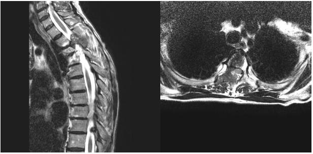
We present a case of a 59-year-old woman with a history of infiltrating metastatic ductal carcinoma of the right breast. The subtypes of ER negative, PR 50%, Her2+ and ki-25% were identified at the time of diagnosis in March 2013. CT revealed multiple pulmonary and hepatic metastases. Prior to the diagnoses, the patient started systemic treatment with Navelbine given orally + Herceptin. Following two years with stable disease, on 15 January, CT revealed the progression of a unique bone metastasis of the vertebral column on vertebra T3. MRI confirmed the bone metastasis on the T3 right lateral vertebral body with bulging of the medullary canal. Given the oligoprogression of the disease, a radical treatment with SBRT was performed until reaching a dosage of 18 Gy in a single fraction of the T3 vertebral body at Hospital Vithas del Consuelo. The spinal cord received a maximum dose (Dmax, 0.01 cc) of 5.7Gy. Following treatment with SBRT, systemic treatment with Navelbine given orally and Herceptin resumed. In February 2019, follow up with CT indicated local progression of the previously treated lesion on vertebra T3. An MRI was performed showing an osteolytic lesion in T3 affecting the body, pedicle, and right lamina, with an extradural tumor mass occupying the spinal canal in 50% of T3 and medullary compression/deviation from right to left (Figure 1). Based on these findings, the tumor in the spinal canal was resected, with subsequent post-operative SBRT on the surgical site.

biomedres-openaccess-journal-bjstr

Figure 1: These images represent vertebral metastases before SBRT.

a) Saggital plane MRI before treatment.

b) Axial plane MRI before treatment.

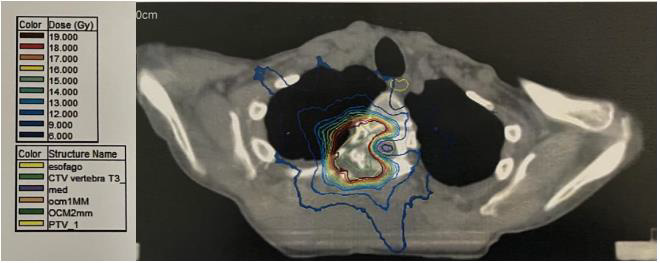
On 1 March 2019, the extradural intra-spinal tumor mass was resected, decompressing the spinal cord and separating it from the surgical site using two Teflon sheets (the sheets surrounded the thecal sac). The postoperative period was without complications. After the resection, on 25 March 2019, SBRT was performed on the surgical site of the D3 vertebra. Fixation and immobilization were carried out using a body stereotaxic frame (BodyFix®, Elekta). The simulation was performed by CT and MRI. IRMT was used with stereotaxic coordinates on the tumor bed until reaching a dosage of 18 Gy in a single fraction (Figure 2). The spinal cord received a maximum dose (Dmax, 0.01 cc) of 7.3Gy. Following treatment with SBRT, the patient resumed systemic treatment with Navelbine given orally and Herceptin resumed. As of the last follow-up on 25 October 2021, the patient remained stable, with no evidence of local or distant progression.

biomedres-openaccess-journal-bjstr

Figure 2: Patient’s planning image with dose distribution.

Discussion and Results

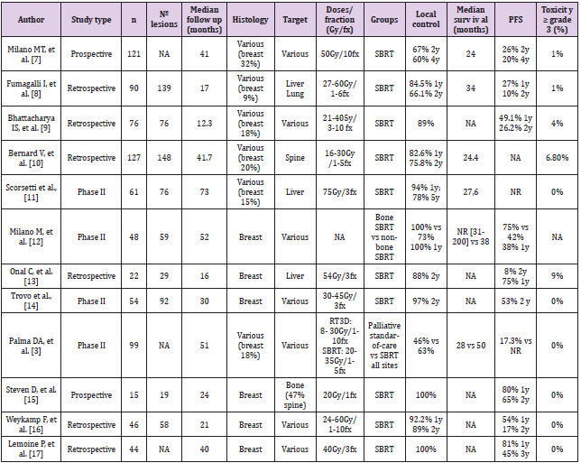
Breast cancer (BC) is the most common cancer in women, with 2,26 million estimated new BC cases worldwide in 2020 [4]. Bone is the most common site of metastasis for BC [5]. Nevertheless, metastasis confined to the bone have a more favorable prognosis than other types of distant metastasis [6]. As we previously mentioned, SBRT and its ablative ability offers a greater tumor control compared to conventional palliative radiotherapy (CPR) [3]. A non-systematic review has been carried out on the topic of SBRT targeting oligometastases in BC. Table 1 summarizes the publications reviewed from PubMed database within the last 10 years. The potential benefit of SBRT in oligometastatic BC has increased the evidence supporting local control (LC), overall survival (OS) and progression free survival (PFS) in these patients. After this review, 12 articles were included, four of them were randomized clinical trials (RCT) [3,7-9], 2 were prospective studies [10,11] and 5 were retrospective studies [12-17]. The sample size is remarkably different among studies ranging from 22 to 227 patients. The follow-up is also distinct ranging from 17 to 73 months. Furthermore, 6 out of 12 articles include multiple histologies [3,7,10,12-14]. LC after SBRT is achieved in 60 to 100% of the cases [10,11], and OS varies from 24 to 50 months [3,18]. When focusing on studies including only BC patients [8,15,9,11,16,17] LC varies from 73% to 100% [8,11].

biomedres-openaccess-journal-bjstr

Table 1: A review of the different retrospective and prospective trials of SBRT in oligometastases breast cancer.

OS median ranges from 28 to 50 months and it was not reached in one of the studies [8]. PFS varies from 38 to 81% [15,9,16,17] and 2-year PFS from 17,3 to 65% [3,11]. A single study included exclusively bone lesions (47% spine metastases) in BC patients [11]. This prospective study reports up to 100% LC rates and a 2-year PFS of 65% of bone metastases treated with radiosurgery (20Gy in 1 fraction) [11]. Milano M et al. demonstrated a better LC (100% vs. 73%), OS (not reached vs 38 months; p=0.002) and PFS (75% vs 42%) after the treatment of extracranial bone lesions compared to those not involving the skeleton (adrenal gland, liver, lung or lymph nodes) [8]. Other authors also report differences in terms of LC depending on the target [3]. Palma et al. reported an improvement in LC (46% vs 63%; p<0.05) and OS (28% vs 50%; p<0.05) after SBRT to all metastatic sites compared to CPR standard-of-care in oligometastatic patients. No increase in toxicity was observed after SBRT [3]. In this context, Sprave and collegues showed no detriment in the quality of life (assessed through QOLBM22, QLQ FA13 and QSC-R10) following SBRT compared to CPR in vertebral metastatic lesions [19]. Grade 3 or higher toxicity reported in the articles reviewed ranges from 0% to 9%. In patients with spinal instability, cord compression, or neurologic deficits, the standard of care is surgery followed by radiation therapy.

Some authors report excellent results with the use of SBRT in patients who have undergone surgical intervention for spine metastases [19,20]. Separation surgery, as our case, refers to providing sufficient surgical circumferential decompression of the spinal cord to create at least 1–2 mm of space between the spinal cord and disease to optimize the SBRT dose distribution. SBRT in oligometastatic patients shows favorable results. In our patient, after radical surgical treatment and SBRT, a complete response was achieved. After more than two years of follow-up the patient remains with neither local nor distant recurrence. Finally, we have to consider that the studies reviewed show heterogeneity both in the target location and treatment site (lung, liver, bone…). Owing to the limitations mentioned above, few robust conclusions can be drawn to the date. There are currently several ongoing clinical trials, such as NRG BR002 (NCT02364557), AVATAR (ACTRN12620001212943) [21] or STEREO-OS (NCT03143322) [19], that will provide more data in relation to the SBRT in oligometastatic BC patients [22,23].

Conclusion

Patients with oligometastatic or oligoprogressive breast cancer are candidates for radical treatment modalities. SBRT has demonstrated promising LC, PFS and OS in these patients with an acceptable toxicity. In this context, there are currently ongoing phase III studies in order to provide stronger evidence.


For more Articles on: https://biomedres01.blogspot.com/

Wednesday, June 28, 2023

Cannabis Abuse and Suicide in Non-Affective Psychosis: A Recent Literature Mini-Review


Cannabis Abuse and Suicide in Non-Affective Psychosis: A Recent Literature Mini-Review

Introduction

Schizophrenia is a severe chronic psychiatric disorder characterized by delusions, hallucinations, disorganized thought and/or behavior and negative symptoms [1]. Compared to the general population, schizophrenic patients have and increased risk of suicide [2-4]. According to the WHO, suicide is the most common cause of death among patients affected by a psychotic disorder [5]. There is a close relationship between schizophrenia and suicide [6]. Bleuler defined suicidal impulse as “the most severe of schizophrenic symptoms” [5]. It has been reported that schizophrenia reduces overall life span by approximately 10 years. Suicide is the largest single contributor to this reduced life expectancy. Suicide is the most common cause of death among people with schizophrenia [5]. There is also a strong correlation between schizophrenia and substance abuse disorder (SUD), including cannabis. In fact, cannabis is the most common substance of abuse in the world, being easily available at low cost [7]. Substance use and psychosis appear to be linked by reciprocal interactions, in terms of the development, evolution, and severity of the disorders [8-10]. According to some studies, patients with schizophrenia have an increased risk of developing a substance use disorder [11]. On the other hand, substance use can represent a trigger for a psychotic episode, or a precipitating factor for an already florid psychosis [12-15].

According to the current evidence in the literature regarding cannabis use disorder (CUD) and synthetic cannabinoids (SCs), not only they seem to be a possible risk factor for the development of a psychotic disorder, but also are likely to be involved in the progression of the disease as well as determining the severity of symptoms [16-21]. Evidence also seems to suggest that the abuse of cannabis or synthetic cannabinoids may increase violent behaviors both directed towards others and themselves [22-24]. For this reason, we decided to conduct a literature search with the aim of better clarifying the existence of a possible relationship between suicide and cannabis use or abuse, as well as explaining and evaluating nature and characteristics of this correlation.

Methods

Data source

We searched the PubMed database from 2010 to November 2021 using the following search string: “(“Suicide”[Mesh] OR suicide[tiab] OR “suicide attempt*”[tiab] AND “suicidal behaviour*”[tiab] OR suicidality[tiab] OR “suicidal ideation*”[tiab] OR “self injurious behaviour*”[tiab] OR “self-injurious behaviour*”[tiab] OR “suicide idea” [tiab] OR “self harm*”[tiab] OR “suicidal thought*”[tiab] OR “suicidal thinking*”[tiab] OR “self injur*”[tiab] OR suicidality[tiab]) AND (“Schizophrenia Spectrum and Other Psychotic Disorders”[Mesh] OR schizophrenia[tiab] OR “nonaffective psychos*”[tiab] OR “non-affective psychosis” [tiab] OR “non affective psychosis”[tiab]) AND (Cannabis[tiab] OR “Cannabis use disorder*”[tiab] OR Marijuana OR cannabinoid*[tiab] OR endocannabinoid*[tiab] OR CUD[tiab] OR “Cannabis”[Mesh] OR “Marijuana Abuse”[Mesh] OR “Endocannabinoids”[Mesh] OR “Cannabinoids”[Mesh] OR “Synthetic cannabinoid*”[tiab])”.

Eligibility Criteria

For our mini review we focused on studies regarding patients diagnosed with schizophrenia or schizophrenic spectrum disorders or diagnosed with non-affective psychotic disorder induced by the use of SCs, who used cannabinoids and attempted suicide or had suicidal ideation. We included all studies that reported the effects of cannabis on suicide risk, including ideation or attempts. We excluded those regarding self-harm that did not have suicide as a goal. We included only reports written in English. Specifically, we included all articles that reported on the effects of cannabis in general, THC, CBD, and synthetic cannabinoids. We applied the following exclusion criteria: articles in which self-injurious acts were mentioned but without suicidal intent; articles in which patients were not diagnosed with schizophrenia or schizophrenic spectrum disorders or diagnosed with non-affective psychotic disorder induced after using SCs; articles that did not include specifically cannabinoids use but included drug abuse as a general category. Titles and abstracts were screened for inclusion by three researchers (A.C., E.C. and A.P.). A fourth investigator (V. R.) was assigned for those cases in which there was no agreement about whether the manuscript met the criteria for inclusion.

Results

The research yielded a total of 49 articles which have been screened according to the inclusion criteria. Among these, 8 studies published between 2010 and 2021 were selected. Study designs distributed as follows: reviews (2), cohort studies (1), meta-analysis (1), cross-sectional analysis (2), systematic review (1); longitudinal studies (2); case reports (1). An Australian cross-sectional analysis (“Is cannabis a risk factor for suicide attempts in men and women with psychotic illness?”), performed on a sample of 1790 people diagnosed with schizophrenia or schizophrenia spectrum disorder, shows a positive correlation between cannabis use in adult males and suicide attempts. This study indicates a different gender susceptibility in response to cannabinoids regarding suicide risk, thus laying the foundations for further investigation to understand whether regular cannabis use has an influence on specific biological mechanisms that could explain the differences observed between men and women. However, the article concludes that there may be confounding factors and (depression, loneliness, homelessness and hallucinations) that need to be considered [25]. The Danish court study “Associations between substance use disorders and suicide or suicide attempts in people with mental illness”, performed on 35 patients, reports that cannabis is associated with an increased risk of suicide attempt in people with schizophrenia (HR: 1.11, 95% CI: 1.03-1.19).

As previously suggested, the associations between SUD and suicide attempts can be explained either by causal associations or by shared genetic and environmental vulnerabilities that predispose to both outcomes [26]. The longitudinal study “Suicidal behavior in first-episode nonaffective psychosis: Specific risk periods and stage-related factors” considers cannabis use, along with depressive symptoms, as the predominant risk factor for suicidal behavior over time. In particular, the article emphasizes the risk of cannabis use during first-episode psychosis (FEP), being this a phase characterized by a particular vulnerability to cannabis abuse, which appears to be an important risk for suicide attempts. Therefore, it would be important to assess the intervention on substance use during FEP as a valuable strategy to achieve the goal of reducing if not preventing suicidal risk [27]. The longitudinal study “Persistent cannabis use among young adults with early psychosis receiving coordinated specialty care in the United States” confirms the correlation between chronic cannabis use and increased suicidal ideation and risk. This article also underlines the importance of an intervention on the use of cannabis in the field of suicide risk prevention and quality of life [28]. In the Mendelian randomized study “Studying individual risk factors for self-harm in the UK Biobank: A polygenic scoring and Mendelian randomization study” polygenic scores (PSs) were generated to index 24 possible individual risk factors for self-harm, including suicide risk.

The results identify PSs, which appear to be predictors of selfinjurious acts. Concerning lifetime cannabis use, the study shows that it actually is a predictor of risk for suicidal self-injurious acts [29]. The only selected study involving synthetic cannabinoids follows the same trend as the studies before mentioned. The crosssectional analysis “Clinical characteristics of synthetic cannabinoidinduced psychosis in relation to schizophrenia: a single-center cross-sectional analysis of concurrently hospitalized patients” [22] specifically studying the effects of synthetic cannabinoids, states that a psychosis induced by this specific kind of drug exhibits a very high rate of suicidal ideation and acts. The two articles dealing exclusively with the CBD molecule show conflicting results. In the first study “The effects of cannabidiol (CBD) on cognition and the other symptoms in outpatients with chronic schizophrenia: a randomized placebo-controlled trial” [30], conducted by administering 600 mg/day of CBD for six weeks to schizophrenic patients stabilized with antipsychotics, no increase in suicidal risk was observed compared to the placebo-treated control group. Thus, this article found no association between CBD and increased suicidal risk. On the other hand, the study “A Review of Human Studies Assessing Cannabidiol’s (CBD) Therapeutic Actions and Potential”14, aiming to test the efficacy and safety of CBD, proves that the impact of this substance requires further investigation. In particular, the authors emphasize the need to understand in a more detailed way the effects of CBD on suicidal ideation, which seems to be a rare but dangerous adverse event when used in combination with anticonvulsant drugs.

Conclusion

From our review only few data have emerged regarding the correlation between cannabis use and suicide risk in patients with schizophrenia or other schizophrenia spectrum diseases, therefore we can’t draw definitive conclusions. Nonetheless these studies seem to point toward a positive correlation of cannabis use with increased suicide risk. It is not clear whether a single active component or rather a set of active metabolites is the explaining cause of the increased suicide risk, and unfortunately the data and the limited knowledge of the substance consumed by the patients included in the selected studies do not allow us to have a better understanding of the underlying phenomena. We think that greater clarification on these issues could be critical in reducing suicidal risk in patients with schizophrenia or schizophrenia spectrum disorders.


For more Articles on: https://biomedres01.blogspot.com/

Friday, June 23, 2023

Juggling Between the Cost and Value of New Therapies: Does Science Still Serve Patient Needs


Juggling Between the Cost and Value of New Therapies: Does Science Still Serve Patient Needs

Opinion

Decades of hope for a cure vanished into thin air when cost outweighed the value of the first gene therapy for thalassaemia, obliging the manufacturing company to withdraw it from Europe. This may create a precedent for other curative therapies that are currently in the pipeline after many years of research, raising questions over their future acceptance by payers and the fulfilment of their purpose: to cure as many patients as possible.

Frequent transfusions, chronic pain, absence from school and work, discrimination, mental health issues are just a few of the daily challenges that patients with thalassaemia face. Standard care with lifelong regular blood transfusions, iron chelation therapy and multidisciplinary care has achieved an increase in life expectancy [1,2]. However, a curative therapy would further allow patients to lead a new life with equal opportunities and challenges, as every other person not suffering from a severe chronic disease. The Thalassaemia International Federation (TIF), representing the united voice of people with thalassaemia and their families globally, has been striving for more than three decades to empower research on a curative approach for thalassaemia.

Haematopoietic stem cell transplantation (HSCT) offers the possibility of cure, but bears specific limitations, i.e. HLA-identical sibling matching and young age [3]. Gene therapy may overpass these challenges, covering more patients and a larger age span. Research on genome-based therapies persisted for decades and this journey has been difficult and immensely challenging until a few years ago [4,5]. The small US-based biotech company bluebird bio that undertook the improvement of a vector produced by Leboulch in 1994, finally succeeded in what predecessors failed [6], partly because it paid attention to the patients’ perspective and their everyday journey with this debilitating disease. Stakeholders always knew that an innovative and complex therapy for thalassaemia would be expensive but always supported its development. Governments and academia provided grants, the industry invested in product’s improvement, healthcare professionals and patients monitored the pipeline and hoped for access. But when the European Medicines Agency granted the gene therapy product of bluebird bio, called ZyntegloTM, a conditional market authorisation in May 2019, everyone focused on numbers and cost-effectiveness studies [7] However, no health economist would ever capture accurately the real cost of thalassaemia in terms of pain, uncertainty and fear.

The withdrawal of an authorised gene therapy from Europe will most probably slow down or even halt the access of people with thalassaemia to curative approaches, rendering the future of thalassaemia treatment gloomy at the very least. And if the developed countries of Europe cannot afford an innovative therapy, what will happen to low- and middle-income countries, where the 80% of the global thalassaemia population lives? Depriving patients from a chance to be cured is at the minimum unethical and constitutes a violation of human and patients’ rights endorsed for decades now by relevant European Union and international bodies. It is also a discriminatory behaviour against people with thalassaemia, given that patients in other disease areas do receive innovative therapies bearing a hefty price tag. It is of utmost important for all stakeholders engaged in the development of medicines, and especially the industry and academia, to seek early and transparent dialogue during the long course of drug development to identify timely safety and cost hurdles.

The developers of medicines, after having invested considerable funds in developing and commercialising a product, should not be left exposed to failure but be given the necessary space, time and motivation to mitigate problems in market access. Additionally, a central, special fund on innovative therapies should be created and managed by the European Commission to compliment national funds of Member States for novel therapies. Governments need to develop synergies and discuss pricing early on, considering the lessons of the past and the challenges to come. As countries do not have unlimited financial resources, governments may opt for joint procurements in the context of regional alliances, such as the Valletta Declaration or the BeneluxA initiative, that increase their negotiating power for the purchase of expensive therapies. Finally, treating physicians and patients should become actively and meaningfully involved in the development process from the very early stages to provide concrete information on medical and other priorities, the potential number of patients that could benefit from innovative therapies, and short and long-term plans for the access of eligible patients to such therapies. Everyone has a role to play and we all share the same responsibility for the sad decision of bluebird bio to withdraw its services (even temporarily) from Europe. Life cannot be measured using mathematical models. Science needs to be available, accessible and most importantly, at the service of patient needs.


For more Articles on: https://biomedres01.blogspot.com/

In Vitro and in Vivo Antimicrobial Effects of Aloe Vera Fermented Hericium Erinaceum KU-1 for Food Borne Pathogens and Helicobacter Pylori


In Vitro and in Vivo Antimicrobial Effects of Aloe Vera Fermented Hericium Erinaceum KU-1 for Food Borne Pathogens and Helicobacter Pylori

Introduction

Aloe vera is most commonly known representative of aloe which belongs to the Liliaceae family and has been used as a remedy in many cultures. It is short-stemmed perennial and has leaves with sharp thorns. Aloe contains ingredients such as aloe ulcin, alomicin, aloetin, and aloesin, so it is widely known to have effects for anti-ulcer, anticancer, antiviral, antibacterial, and antifungal [1]. Hericium erinaceus is an edible mushroom and is used for gastrointestinal diseases by regulating gut microbiota and immune system [2]. It has the rich diterpenoid compounds, steroids, and polysaccharides and is known to have the antimicrobial activities against Helicobacter pylori, a human gastrointestinal pathogen [3,4]. As such, the mixture of the mycelium of Aloe vera and Hericium erinaceus was expected to have a strong antimicrobial effect, but it has not been studied yet. Therefore, we aimed to study the antimicrobial effects of Aloe vera fermented Hericium erinaceum KU-1 for food borne pathogens and Helicobacter pylori in vitro and in vivo.

Food borne pathogens, such as E. coliSalmonella and Listeria, are major causes of gastrointestinal disease worldwide. Enterohemorrhagic E. coli O157:H7 causes hemorrhagic colitis, which is occasionally complicated by hemolytic uremic syndrome [5-7]. It is estimated that E. coli O157:H7 infection causes over 73,500 cases of illness each year in the United States [8]. Salmonella spp. are facultative intracellular organisms that grow primarily inside the macrophages of the liver and spleen. S. enterica serovar Typhimurium is the most commonly isolated Salmonella serotype, accounting for 23% of laboratory confirmed Salmonella cases among the more than 1.4 million infections each year in the United States [9]. This infection is characterized by clinical symptoms such as fever, abdominal pain, and diarrhea in both animals and humans. Listeria monocytogenes is a gram-positive intracellular bacterium that is normally nonpathogenic in healthy individuals. However, in pregnant women, newborn infants, the debilitated elderly or immunocompromised people, it may cause severe clinical disease including meningoencephalitis, septicemia, or abortion [10-15].

It is estimated that Lmonocytogenes infection causes about 2,000 to 2,500 cases of illness and as many as 500 deaths per year in the United States [16]. Helicobacter pylori is characterized by its typical corkscrew-like appearance and is at least twice as long as H. heilmannii [17], with a low infection rate of 0.08-1.0 % in humans [18,19]. Its infection has been described mostly in relation to chronic gastritis [20,21], gastric ulcers [22] adenocarcinoma [23] and MALT lymphoma [24] in men. Antimicrobial chemotherapeutic agents have been widely used to control these gastrointestinal infections. However, widespread use of antibiotics is now being discouraged due to problems including the emergence of drug-resistant strains and chronic toxicity [25]. In addition, antibiotics are often responsible for acute diarrhea due to the loss of normal intestinal microbes and the protection they provide against pathogenic organisms [26]. As alternatives, Hericium erinaceum (HE) such as KU-1 and their mycelium have been administered.

It is well known as lion’s mane or monkey head mushroom have health-promoting attributes including antimicrobial properties [27-29], immunomodulation [30-33], anti-tumor characteristics [34-36], and hypocholesterolemic effects [37,38]. These findings have caught the attention of nutrition, food, and microbiology scientists and have heightened interests to produce functional foods.  We isolated HE KU-1 and their mycelium from Pterocarpus indicus, and Cyclobalanopsis spp. This HE KU-1 was fermented in an appropriate Aloe vera broth, condensed by vacuum evaporation, and mixed with equal doses of each pathogenic strain. In this study, the antimicrobial effects of this HE KU-1 fermented condensate mixture (ALOHEM) were evaluated using in vitro and in vivo models of food borne pathogens and Helicobacter pylori.

Materials and Methods

Microorganisms

coli O157H:7 ATCC 43894 (American Type Culture Collection, Rockville, MD, USA), Salmonella enteritidis CCARM 11066 (Culture Collection of Antibiotic Resistance Microbes, Seoul, Korea), Staphylococcus aureus (KFRI 240, Korea Food Research Institute, Wanju-gun, Jeollabuk-do, Korea), and Listeria monocytogenes ATCC 19115 and ATCC 51774, were used as pathogenic strains.

Mycelial Culture

biomedres-openaccess-journal-bjstr

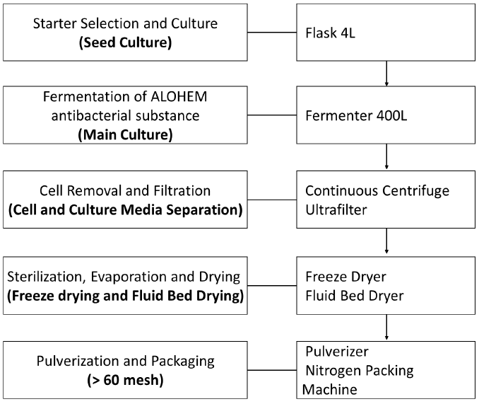
Figure 1: Manufacturing process of ALOHEM.

The preparation process of ALOHEM is as shown in Figure 1. HE KU-1 mycelium was maintained on solid Ko medium [39] which is composed of glucose 18.02 g, arginine 2.613 g, ammonium acetate 2.613 g, CaCl2 0.33 g, KH2PO4 8.5 g, MgSO4·7H2O 2.0 g, FeSO4·7H2O 0.02 g, ZnSO4·7H2O 0.02 g, MnSO4·7H2O 0.02 g, agar 18.0 g and distilled water 1 L at pH 4.5~5.0. A single colony of each HE KU-1 mycelium was inoculated into 10 mL of the Ko broth in a 15 mL conical tube and cultured in a shaking incubator (NB 205, N-Biotec, INC., Bucheon, Kyoungki-Do, Korea) at 200 rpm and 28℃ for 2~3 days [39] HE KU-1 mycelia were fermented in Aloe vera broth on a jar-fermenter (CelliGen 115, Eppendorf, INC., NY, USA) at 150 rpm, 28℃ for 3~4 weeks. 99% of Aloe vera extract (Aloe vera gel W®, KimJeongMoon Aloe Ltd., Jeju-Do, Korea) was tested.

Preparation of Aloe vera Fermented Hericium erinaceum KU-1 Condensate Mixture (ALOHEM)

Following fermentation period of 3-4 weeks at 25~28°C, the HE KU-1 mycelia were isolated from a single colony and tested for their inhibitory effects on various food-borne pathogens. The HE KU-1 mycelia were removed by centrifugation at 2,700 ´ g for 30 min and the fermented media was concentrated 20 fold into a solid content by vacuum evaporation. Each concentrated medium was mixed with an equal weight of Aloe vera extract and lyophilized. The final ALOHEMs were prepared by mixing each condensate of equal weight and stored at 4℃ until use.

Animals

Five-week-old specific pathogen-free (SPF) female BALB/c mice (Seoul National University Laboratory Animal Center, Seoul, Korea) for S. enteritidis infection and ICR mice (Daehan-Biolink, Chungbuk, Korea) were used for H. pylori infections. They were housed in polycarbonate cages in isolators and fed a commercial pellet diet with water ad libitum. Food and water were removed from the cages 5 hours prior to inoculation of the bacteria and/or ALOHEM. All animal experimentation was performed in accordance with the laboratory animal guidelines of Korea University (Seoul, Korea).

In vitro Assessment of Antimicrobial Effects of the ALOHEM

Each pathogenic strain was grown to an optical density (OD) of 0.8-1.0 at 600 nm. Briefly, 200 mL of each bacterium was added to conical tubes containing 10 mL of appropriate concentrations (0, 1.25, 2.5, 5, 10 %) of the AFHCM in TS broth and incubated with shaking at 200 rpm and 37℃. And 1 mL of each culture was taken at 0, 1, 2, 3, 4, 6, and 8 h post incubation and centrifuged at 5,000 rpm for 10 min. The supernatants were discarded and the bacterial cells were floated in 1 mL of fresh TS broth. Finally, the OD was measured at 600 nm. This process was repeated once. To determine whether the antimicrobial effects of the ALOHEM are due to pH, tubes containing various concentrations of the ALOHEM were adjusted to pH 7.2 by the addition of NaOH. Subsequently, 200 mL of cultured S. enteritidis was added to each tube and incubated at 37℃. OD was measured at 0, 2, 4, 6, and 8 h post incubation using the same method as the sample preparation described above. This process was not performed against the other pathogens.

In vivo Assessment of Antimicrobial Effects of the ALOHEM

For S. enteritidis infection models, 1 mL containing 5 ´ 107 CFU of S. enteritidis was added to each tube containing 9 mL of the ALOHEM at various concentrations (2.5, 5, and 10%) or of the TS broth (positive control), vortex-mixed, and used as inocula. The mice were immediately administered with 0.5 mL of each inoculum (2.5 ´ 106 CFU of S. enteritidis per mouse) via the intragastric route while the negative control mice were administered with the same volume of TS broth. Subsequently, the mice of each treated group were administered with 0.5 mL of ALOHEM of their respective concentration once a day for 4 days and the mice of positive and negative groups received 0.5 mL of TS broth. Bacteria from fecal samples were counted by plating on XLD (Difco) agar at 12 h, 1, 2, 3, and 4 days post-inoculation (pi) days and mortality was checked at 21 days pi. 

 pylori originating from pig’s stomach had been kept in the gastric passage of mice [40]. Three H. heilmannii-infected mice were necropsied by cervical dislocation and the stomachs were collected. They were carefully opened and the gastric content was removed by washing with sterile phosphate buffered saline (PBS). The stomachs were then added to a conical tube containing 10 mL of sterile PBS, electrically homogenized (Ultra-turrax T25, Ika, Staufen, Germany), and the resulting mixture was used as an infective inoculum. Then 1 mL of the infective inoculum was added to each tube containing 9 mL of 2.5, 5, and 10% ALOHEM or TS broth (positive control), respectively, and vortex-mixed. The mice were immediately administered 0.5 mL of each inoculum intragastrically while the negative control mice were administered the same volume of TS broth. Subsequently, the mice of each group were administered 0.5 mL of each concentration of the ALOHEM once a day for 2 months pi, at which time all mice were euthanized by cervical dislocation and necropsied. Their stomachs were collected, opened, and the gastric contents were removed as described above. The gastric samples were then fixed in 10% buffered formalin for 24 h, processed, and embedded in paraffin wax as standard protocol. Sections measuring 2 micrometers each were prepared and stained with hematoxylin and eosin (H&E) and modified Steiner’s silver stain (Sigma, St. Louis, MI, USA) for histopathological examination and for the detection of H. heilmannii, respectively.

Results

Antimicrobial Effects in vitro of Various Concentration of ALOHEM

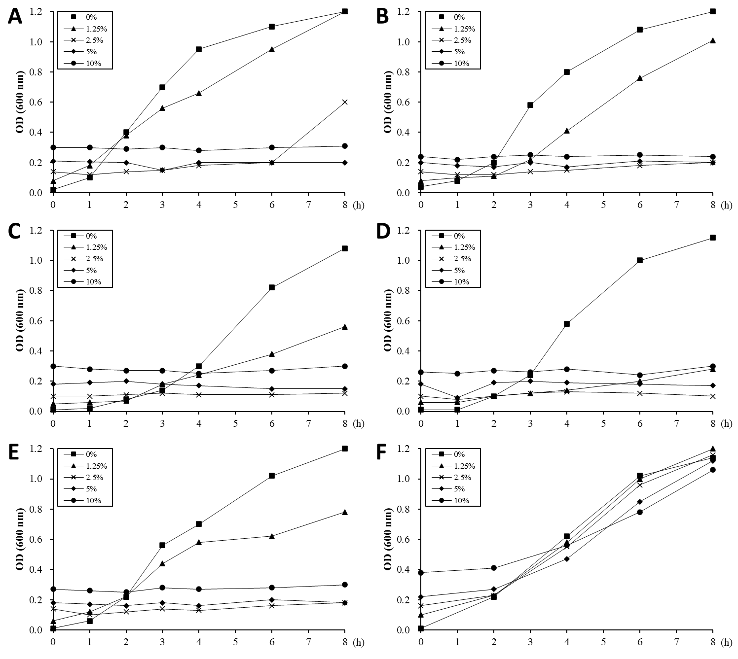
The ability of the ALOHEM to inhibit the in vitro growth of food-borne pathogens was evaluated and the results are shown in Figure 2. Just before the pathogens were added to each tube, the pH levels of the solutions were measured at 4.03 (10% ALOHEM), 4.32 (5% ALOHEM), 4.96 (2.5% ALOHEM), 6.02 (1.25% ALOHEM), and 7.24 (0% ALOHEM). The growth of all strains except E. coli O157:H7 was completely inhibited by the presence of the ALOHEM at concentrations above 2.5%. All strains except L. monocytogenes ATCC 51774 began to grow in the broth containing the concentration of 2.5% ALOHEM 2 to 4 h post incubation, although the growth rate was lower than that of the control broth (0% ALOHEM) (Figure 2A-2E). However, when the pH of the broth was adjusted to 7.2, the presence of the ALOHEM at any concentration did not inhibit the growth of S. enteritidis at all (Figure 2F).

biomedres-openaccess-journal-bjstr

Figure 2: In vitro growth of E. coli O157:H7 (A), S. aureus (B), L. monocytogenes ATCC 19115 (C), L. monocytogenes ATCC 51774 (D), S. enteritidis (E) was inhibited by the presence of ALOHEM. Data show the mean value of two experiments performed. At the initial time of experiment, the pH of each broth containing 0% (-■-), 1.25 % (-▲-), 2.5 % (-ⅹ-), 5 % (-◆-), and 10 % (-●-) ALOHEM was 7.24, 6.02, 4.96, 4.32, and 4.03, respectively. Another experiment was performed to clarify the effects of pH on the antimicrobial ability of the ALOHEM (F). Each broth containing the different concentrations of the ALOHEM was adjusted to pH 7.2 with NaOH. Because the ALOHEM brings about a brown hue, optical density depended on the concentration of the ALOHEM at the start of the experiment (0 h).

Bacteria Count Evaluation by ALOHEM

Antimicrobial effect of the ALOHEM against S. enteritidis and H. heilmannii infection was assessed in BALB/c and ICR mice, respectively. Table 1 shows the viable bacterial count from the fecal samples. The number of isolated bacteria was dose-dependently less in the mice inoculated with an admixture of the ALOHEM and the bacteria than in the mice that received only the bacteria (positive control). However, bacteria were steadily isolated in the mice that received 2.5% and 5% ALOHEM over the first 4 days. Bacteria were isolated from only one mice of the group administered with 10% ALOHEM 4 days following injection. The limitation of the bacterial count was 4.00 log10 CFU/g feces.

biomedres-openaccess-journal-bjstr

Table 1: Bacterial counts from fecal samples.

aValues in parentheses represent the number of mice from which bacteria were isolated. All groups originally consisted of five mice.

Assessment of Mortality by ALOHEM

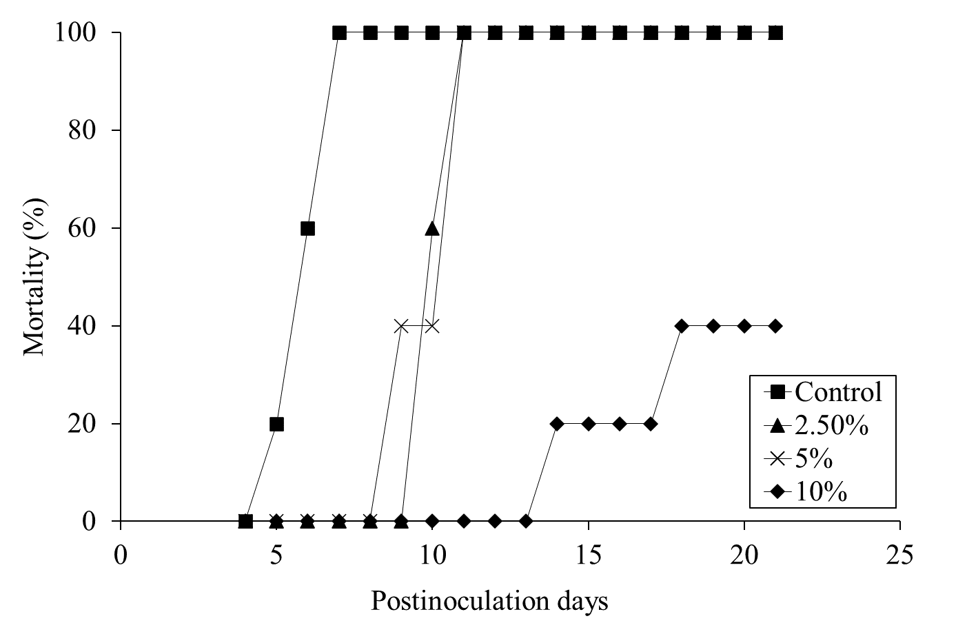
Mortality was assessed for 28 days pi. Daily administration of the ALOHEM or TS broth in each group was terminated when the first mortality occurred in the positive control group on day 5 pi. All mice of the positive control group died within 7 days of injection (Figure 3). Although all mice in the 2.5 and 5% ALOHEM treated group eventually died, mortality occurred between 9- and 11-days pi, later than that of the positive control group. Within the 10 % ALOHEM treated group, one mouse died on day 14 pi and another on day 18 pi, while the rest survived until 21 days pi (Figure 3).

biomedres-openaccess-journal-bjstr

Figure 3: Mortality of the mice dosed with S. enteritidis throughout the experimental periods; the positive control group (-□-), 2.5 % (-△-), 5 % (-ⅹ-), and 10 % (-○-) ALOHEM treated groups.

The Histopathological and Microbiological Findings

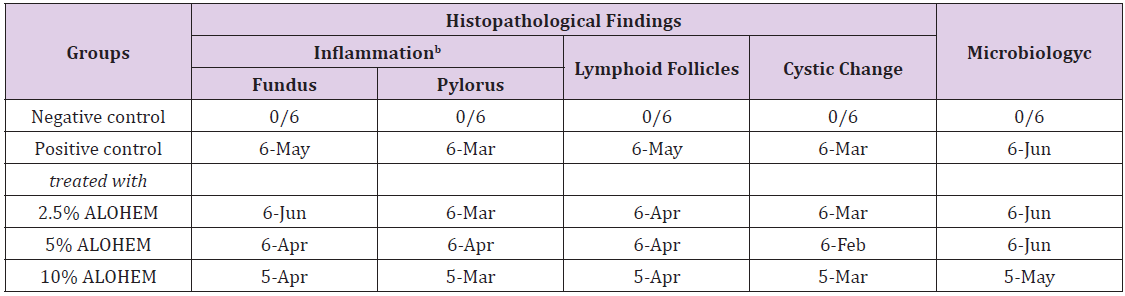
Table 2 summarizes the histopathological and microbiological findings. The results show that the use of ALOHEM did not suppress the bacterial colonization and histological lesions in the stomach at all.

biomedres-openaccess-journal-bjstr

Table 2: Histopathological and microbiological findings of the mice stomachsa.

aexpressed as number of detected mice/ number of tested mice.
bconsisted of infiltration of mononuclear cells in lamina propria and submucosa.
cassessed by Steiner’s silver stain.

Histological Lesions

biomedres-openaccess-journal-bjstr

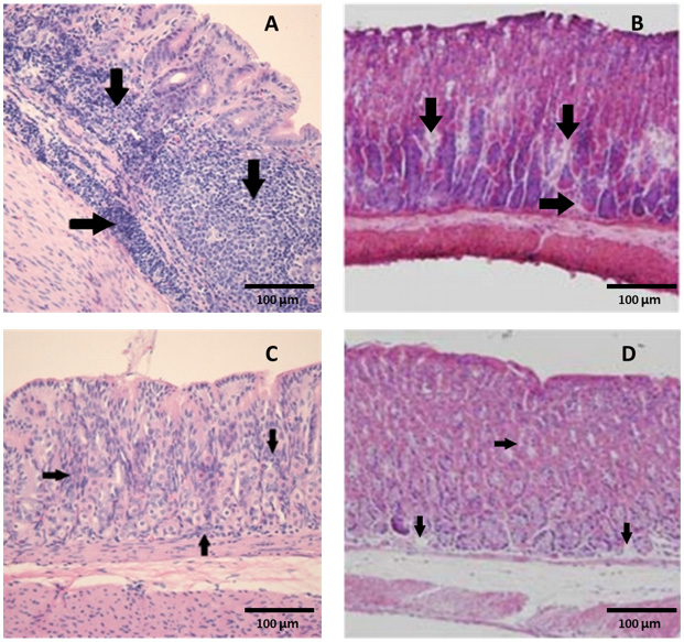
Figure 4:  Marked colonization (arrows) of H. heilmannii in the lumen of the gastric glands of (A) a positive control mouse and (B) a mouse inoculated with an admixture of 10% ALOHEM and H. heilmannii. (C) Infiltration of mononuclear cells and (D) presence of lymphoid follicle in the submucosa of the gastric body of mice inoculated with the admixture of 10% ALOHEM and H. heilmannii. A, B, C; Bar = 250 mm, D; Bar = 62.5 mm.

Regardless of the concentration of the ALOHEM treatment, marked colonization of H. heilmannii was observed in the lumen of the gastric glands, gastric pits, mucosal surfaces, and intercellular spaces of all infected mice (Figure 4A&4B). Histological lesions such as the formation of lymphoid follicles, cystic changes, and mild inflammation in the mucosa or submucosa were randomly observed (Figure 4C&4D). No bacteria and histological lesions were observed in the negative control group.

Discussion

Many studies have recently shown that Hericium erinaceum have antimicrobial effects. The potential mechanisms by which mushrooms might exert their antimicrobial activity includes the production of hericenone, other metabolites such as hydrogen peroxide and short chain fatty acids, and specific antimicrobial compounds such as antibiotics [41,29,38]. In the present study, we have shown that the ALOHEM effectively inhibits the in vitro growth of enteropathogens such as E. coli O157 H:7, S. enteritidisS. aureus, and L. monocytogenesIn vivo experimentation also revealed that the presence of 10% ALOHEM increased the survival rate of mice infected with S. enteritidis. In addition, the survival time of the 2.5 and 5% ALOHEM groups was extended beyond that of the positive control group, even though all mice died within 11 days pi.

This might be because the presence of 2.5 and 5% ALOHEM did not completely inhibit the growth of S. enteritidis and the bacterial growth rapidly progressed after the ALOHEM administration was terminated (4 days pi). Also, the presence of the ALOHEM did not inhibit the colonization of H. heilmannii in the stomach. These divergent results made it difficult to identify the antimicrobial mechanism of the ALOHEM. We first considered the low pH due to the hericenone as the factor, because the pH of each broth was in inverse proportion to the concentration of the ALOHEM. Based on this consideration, the pH of each broth was adjusted to 7.2 by the addition of NaOH and 200 µL of an overnight culture of S. enteritidis was subsequently added to each broth. As a result, bacterial growth was not inhibited in any of the broths. This finding indicates that the antimicrobial effects of the ALOHEM might be due to low pH, which is also the reason that the presence of the ALOHEM did not inhibit the colonization of H. heilmannii in the stomach. H. heilmannii is able to colonize the stomach by increasing the gastric pH through its urease production.

Therefore, it is likely that H. heilmannii mixed with 10% ALOHEM could survive at a low pH level by producing urease and colonizing the stomach. Previous findings [41] have shown that some hericenone produce by Hericium erinaceum spp, although able to inhibit a variety of pathogenic bacteria, do not inhibit the growth of both Salmonella sp. and Vibrio cholerae, when the effect of acids was excluded. Our preliminary study also shows that long periods of ALOHEM administration did not affect clinical signs, or the gross and histopathological lesions of the mice (data not shown), indicating that its ingestion might not pose health risks to humans and in fact be effective in preventing infections due to food borne pathogens.

Conclusion

This study was investigated the antimicrobial effect of an Aloe vera fermented Hericium erinaceum (HE) KU-1 condensate mixture (ALOHEM) using in vitro and in vivo models of food borne pathogens and Helicobacter pylori. The presence of the ALOHEM effectively inhibited in vitro growth of food borne pathogens such as E. coli O157:H7, S. aureusL. monocytogenes, and S. enteritidis. However, when the pH of the broth was adjusted to 7.2, the ALOHEM did not inhibit the growth of S. enteritidis at all. The in vivo antimicrobial effects of the ALOHEM against S. enteritidis and H. pylori were also assessed. In S. enteritidis-infected mice, the ALOHEM decreased the viable bacteria found in the feces and the mortality rate. However, it did not affect the gastric colonization of bacteria and histopathological lesions in mice infected with H. pylori, which is able to colonize the stomach by increasing the gastric pH through its urease production. These findings showed that the ALOHEM might have antimicrobial ability by decreasing pH.
For more Articles on: https://biomedres01.blogspot.com/

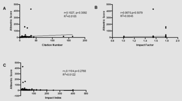
The Correlation between Altmetric Score and Citations in Pediatric Orthopaedic Journal Articles

  The Correlation between Altmetric Score and Citations in Pediatric Orthopaedic Journal Articles Introduction In scientific research, it is...