Friday, August 4, 2023

Blue Cumin Tea: Prophetic Herbal Tea for the Therapy of Hypercholesterolemia in the COVID-19 Season


Blue Cumin Tea: Prophetic Herbal Tea for the Therapy of Hypercholesterolemia in the COVID-19 Season

Introduction

Herbal tea is a term for a mixture of flowers, leaves, seeds, roots, or dried fruit to make a drink which is also called herbal tea. Although called “tea”, this herb or drink does not contain leaves from the tea plant (Camellia sinensis). Herbal teas are usually brewed with hot water to get a fragrant drink. However, herbal teas made from plant seeds or roots often need to be boiled before they are filtered and ready to serve. Even though they contain flowers or dried fruit, teas made from tea leaves such as jasmine tea or Earl Gray tea are not called herbal teas. The mixture of citrus bergamot in Earl Gray tea or jasmine flowers into jasmine tea is intended as a fragrance to vary the aroma of the tea. Herbal teas are available in cans, tea bags, or ready-to-drink herbal teas in boxes. Herbal teas are also often advertised as health drinks to cure various diseases. In addition, ingredients collected from the garden, such as hibiscus flowers, chrysanthemums, or chamomile, and fragrant leaves such as peppermint and rosemary, after drying can be mixed into herbal teas. However, in this study, the authors took a mixture of black cumin seeds and telang flowers which were dried and put in a tea bag with rock sugar. Black cumin seeds have been known for thousands of years as a healer of various diseases as stated in the following hadith narrated by Bukhari, “The black seed can heal every disease, except death”. The author uses the method in herbal teabags because the use does not use alcohol for extraction where Rasulullah SAW explicitly said “Khamr is not a medicine, but it is a disease” (HR. Muslim: 1984) (Figure 1).

biomedres-openaccess-journal-bjstr

Figure 1: Blue Cumin Tea Tjap Maryam and Isa Clinic.

Methods

Blue Cumin Herbal Tea Making

1) Dry 100 grams of black cumin seeds
2) Dry 100 grams of butterfly pea flower
3) Enter 1/2 teaspoon of the mixture of herbal ingredients in a tea bag with 1 piece of white rock sugar
4) Then serve by dipping it in boiling water
*Gifting of telang flowers is intended to give a modern tea color that is more beautiful and also gives a different sensation of freshness in drinking tea.

How to Use

Drink once a day in the morning. This study uses an experimental post test only control group design using humans as research subjects. Inclusion criteria were clinical patients with:
1) History of hypercholesterolemia without comorbidities
2) Over 30 years old
3) Willing to participate in this research
There were 2 treatment groups, namely the group that drank herbal tea and the second group who routinely used simvastatin 20 mg at night. We got each group of 5 volunteers. Then the study was conducted for 1 month.

Results and Data Analysis
biomedres-openaccess-journal-bjstr

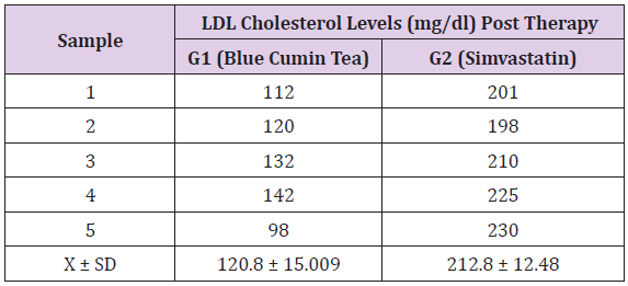
Table 1: LDL Cholesterol Levels (mg/dl) Post Therapy Between Groups.

The data obtained from the examination of serum cholesterol levels were numerical data, then the data was tested for normality using the Shapiro Wilk test and the results obtained that all groups were normally distributed with p > 0.05 (Table 1). After the data is normally distributed, Levene’s test is carried out to find out whether the data is homogeneous or not. And the results obtained p = 0.672 (p>0.05) so it can be said that the variance is homogeneous. Because the data was said to be homogeneous and normally distributed, a one-way ANOVA test was carried out to analyze the differences between groups. Based on table 2, the p value of the one way ANOVA test is 0.0001 (p < 0.05) so it can be concluded that there are significant differences in cholesterol levels between groups.

biomedres-openaccess-journal-bjstr

Table 2: One Way Anova Test Results.

Discussion

Lipoproteins are a combination of nonpolar lipids (triacylglycerol and cholesteryl esters) with amphipathic lipids (phospholipids and cholesterol) and proteins that function to transport lipids in blood plasma. One of the four main groups of plasma lipoproteins is low density lipoprotein (LDL) [1]. LDL functions as a cholesterol carrier for tissues and cells, where cholesterol is needed for membrane synthesis, modulation of membrane fluidity, and regulation of cellto- cell communication [2]. LDL cholesterol has a major role in the development of cardiovascular disease, especially atherosclerosis. The role of LDL cholesterol begins early in atherogenesis, where LDL cholesterol accumulates in the tunica intima of arteries. High LDL cholesterol levels can increase the risk of atherosclerosis [3]. Atherosclerosis can cause various cardiovascular diseases, one of which is coronary heart disease (CHD). Nigella sativa is an herbal plant from the Ranunculaceae family [4]. Many researchers have succeeded in revealing the broad pharmacological potential of this herbal plant. N. sativa is commonly known as black cumin [5,6]. Black cumin seeds and oil are usually used as a tonic and traditional medicine for various ailments [7]. Black cumin seeds contain tymoquinone [8] which can reduce LDL cholesterol serum levels in 2 ways, namely by inhibiting the activity of the HMG-CoA reductase enzyme and increasing the expression of mRNA in the LDL receptor. Simvastatin can only reduce serum LDL cholesterol levels through inhibition of HMG-CoA reductase enzyme activity, so that serum LDL cholesterol levels of patients given black cumin extract are lower than those given simvastatin. This is an interesting finding from this study, wherein the serum LDL cholesterol levels of hypercholesterolemic patients given simvastatin were initially estimated to be lower than those given black cumin extract but on the contrary. Symptoms of hypercholesterolemia such as a heavy neck, difficulty sleeping and tingling can be reduced from the first day of using blue cumin tea, while for simvastatin, patients still often complain of feeling dizzy and heavy.

Simvasratin sold in Indonesia at this time also does not have an official halal certificate from the BPJPH institution. Simvastatin is a drug that is commonly consumed by dyslipidemia patients and works mainly in lowering serum LDL cholesterol levels, but like other drugs, simvastatin also has various side effects for the body. Some of the side effects of simvastatin are liver injury, diabetes risk, and muscle injury. Muscle injuries that occur due to the side effects of simvastatin are myopathy and rhabdomyolysis, and these muscle injuries are more common in patients taking 80mg of simvastatin daily, so the use of a dose of simvastatin 80mg/day has been prohibited. Through the data obtained from this study, blue cumin tea can be used as a substitute therapy that has guaranteed its halalness for those with high serum LDL cholesterol levels [9]. This study shows faster results with the use of blue cumin tea within 1 month and does not have to be consumed continuously like simvastatin and does not cause boredom because of its attractive taste and color. Patients are sometimes quite saturated when consuming continuously in the usual way and with unpleasant side effects.

Conclusion

Blue cumin tea therapy had a significant effect on reducing LDL cholesterol levels in patients with hypercholesterolemia. So that it can be a fairly good halal treatment solution for hypercholesterolemic patients.

For more Articles on: https://biomedres01.blogspot.com/

Thursday, August 3, 2023

Ionization of X-Chromosomes by Galactic Cosmic Ray and the Male Bias at Birth


Ionization of X-Chromosomes by Galactic Cosmic Ray and the Male Bias at Birth

Introduction

The phenomenon of the predominance of boys among born children has been known for more than 300 years. Back in 1778 P.-S. Laplace showed that statistically this difference cannot be considered an accident. Among the proposed explanations, the most popular hypothesis is that we are dealing with a kind of preventive life mechanism designed to ensure the gender balance of populations. According to this logic, the excess number of males should reach a maximum at the beginning of fetal development and then decrease with age. The latest data from large-scale studies do not confirm the idea of genetic prerequisites for gender imbalance [1,2]. Obviously, it is not by chance that no structures have been found in the genetic apparatus of a human cell that could be responsible for the offspring sexual differentiation. Reliable establishment of gender discrepancy at birth not only in humans, but also in different groups of animals allows us to talk about a global pattern. It has not yet been possible to reveal its origin. It also remains unclear what environmental factors can lead to the occurrence of local anomalies of opposite signs. The solution to the problem is further complicated by the fact that significant variations in the sex ratio in humans have been found in recent decades, which are clearly not in any way associated with conscious actions such as selective abortion. The substantiation of the hypothesis of the ionization of the X-chromosomes by galactic cosmic ray (GCR) as the cause of changes in the gender ratio in space and time is given below. Particles of galactic cosmic origin carry an energy close in magnitude to dangerous X-ray quanta, but their effect lasts around the clock, throughout human life, in the open air and at home. First of all, the female body suffers from it.

Materials and Methods

The analysis was based on the demographic statistics of Iceland, Norway, Sweden, Finland and Denmark with Greenland, which was determined by the quality and quantity of the necessary information, as well as the location of the listed countries in high latitudes, where the intensity of GCR increases due to the lower thickness of the atmosphere. For comparison, information on the southern regions was used. The values of the GCR intensity were taken from the observations of the neutron monitor in Oulu (Finland, 65.05 ° C), operating since 1964. Radiation conditions in previous years were characterized using reconstructed data and Wolf numbers, an indicator of solar activity, from which the intensity of GCR is in close inversely proportional dependence (correlation coefficient -0.87), since the Sun modulates the GCR. To test the hypothesis put forward, the method of mental critical experiment was used. If the assumption about the effects of GCR on the organism correctly reflects reality, the following events that cannot be explained from any other point of view should be associated with an increase in their intensity over time:
1) A decrease in the girl birth rate,
2) An increase in female mortality and
3) An increase in the frequency of death of the embryo and fetus of the female sex, and with a decrease - phenomena of the opposite direction.
In addition, it should be expected that the level of risk of negative consequences of exposure to cosmic radiation will be maximum in the northern latitudes. The analysis of demographic events was carried out using the methods of superimposed epochs and minimax (comparison of anomalies of different signs). The ratio of the number of female and male groups was adopted as the main indicator of demographic processes. The periods were considered, the middle of which fell on the years of minimum or maximum solar activity (and, accordingly, the highest or lowest GCR intensity).

Results

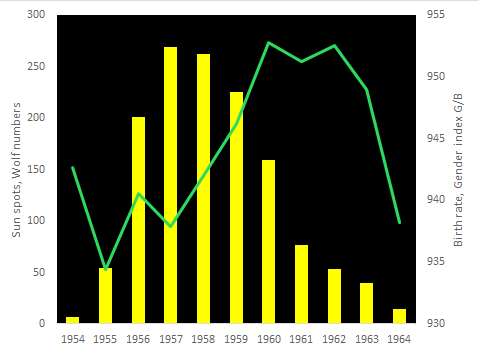
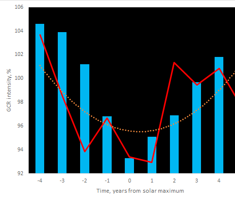
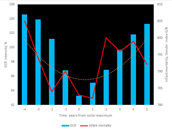
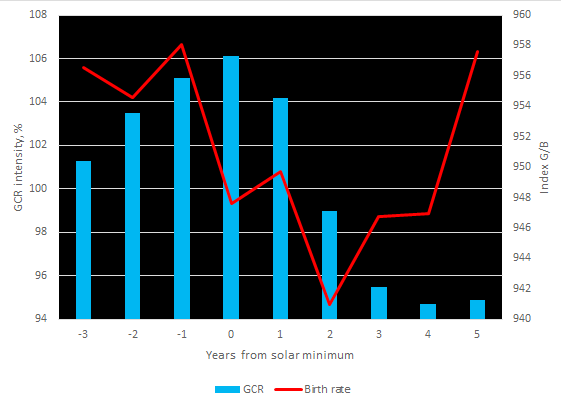
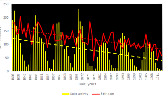
Expected results of the first mental critical experiment is as follows: at a high intensity of GCR, the birth rate of girls should be abnormally low, and at a reduced intensity - above average. Powerful galactic radiation causes a significant drop in the birth rate of girls, as can be seen from the statistics of Iceland, and there is a characteristic delay in the effects of 1-2 years (Figure 1). A longterm increase in the intensity of GCR, accompanying the weakening of solar activity, entails a reduction in the girl birth rate (Figure 2). The gender index, as expected, increases after a period of very low GCL intensity, when the Sun is abnormally active and the solar wind blocks the flows of high-energy particles (Figure 3). Interestingly, in Greenland, after the minimum GCR in 2000, the ratio of newborn girls and boys with a weight of 3000 g or more reached an absolute maximum of 1028 (with an average of 900). The essence of the second thought critical experiment: it can be predicted that galactic rays of high energy increase female mortality. Using the example of Sweden, it is clearly seen that with a positive anomaly in the intensity of GCR, the maximum of female infant mortality naturally occurs, and in years of low intensity of GCR, when the Sun is very active, the situation with female infant mortality improves dramatically (Figure 4). The influence of galactic radiation is well traced by trends in infant mortality. A long-term increase in the intensity of GCR as a result of the weakening of solar activity is associated with an increase in the level of female infant mortality (Figure 5).

biomedres-openaccess-journal-bjstr

Figure 1: The drop in the birth rate of girls in Iceland after a period of high intensity of GCR (reconstruction) during the time of calm Sun of the 11-year cycle. Averaging over the period 1838-2013, 16 cycles, based on data on 554,000 events.

Note: Calculation based on data of Statistics Iceland, WDC-SILSO, Royal Observatory of Belgium, Brussels and Cosmic Ray Station of the University of Oulu.

biomedres-openaccess-journal-bjstr

Figure 2: The tendency for a decrease in the birth rate of girls in Finland as a result of an increase in the intensity of GCR with a weakening of solar activity in 1836-1911. Averaging according to 5293000 events.

Note: Calculation based on data of Statistics Finland, WDC-SILSO and Royal Observatory of Belgium, Brussels.

biomedres-openaccess-journal-bjstr

Figure 3: An increase in the birth rate of girls in Denmark due to low energy level of GCR with abnormally high solar activity in the mid-50s of the twentieth century.

Note: Calculation based on data of Statistics Denmark, WDC-SILSO and Royal Observatory of Belgium, Brussels.

biomedres-openaccess-journal-bjstr

Figure 4: The dependence of female infant mortality on the intensity of UCS by the years of the solar activity cycle. Averaging over the period 1757-2019, for 24 cycles. The polynomial trend of mortality is shown

Note: Calculation based on data of Statistics Sweden, WDC-SILSO, Royal Observatory of Belgium, Brussels and Cosmic Ray Station of the University of Oulu.

biomedres-openaccess-journal-bjstr

Figure 5: The female infant mortality in Finland and the intensity of GCR in last decades. The Linear trends are shown.

Note: Calculation based on data of Statistics Finland and Cosmic Ray Station of the University of Oulu.

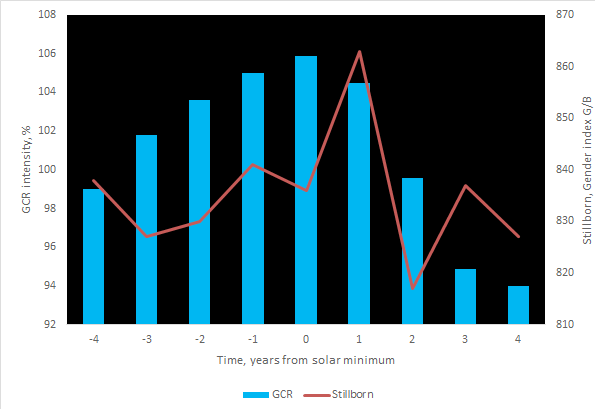
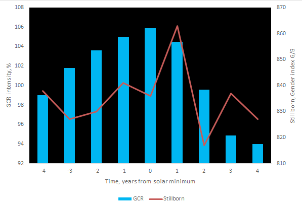
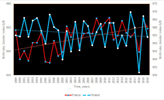
Nowadays, there is an increase in galactic particles energy associated with a weakening of solar activity, which determines the tendency to aggravate the risk of death of newborn girls on the first day of life. Attention is drawn to the coincidence in time of anomalies of different signs: in Norway in 1959, after two years of abnormally high solar activity, the gender indicator of infant mortality decreased to a minimum of 706, and in Greenland, with a minimum of GCR intensity in 1999-2000, the index reached the lowest values, respectively, 444 and 571. It should be noted that the negative impact of high-power galactic rays in the north covers not only newborns, but also the entire female part of the population. Thus, the era of the solar minimum of Dalton at the beginning of the 19th century was marked in Finland by an increase in total female mortality 3-6% higher than average. The third critical experiment should show that high-energy particles penetrating the body, especially in the early stages of its development, are capable of destroying the genetic apparatus and causing mutations, which leads to antenatal mortality and an increase in the frequency of stillbirths. And indeed, an abnormally high frequency of stillbirths among girls is recorded precisely during the years of high intensity of GCR in the calm Sun (Figure 6). In addition, judging by the information from Denmark, the risk of female stillbirths increases in the conditions of contemporarty growth of GCR energy due to the weakening of the solar wind (Figure 7). Finally, it should be assumed that the phenomena discussed above are most pronounced in the northern regions, where the amplitude of fluctuations in the intensity of GCR is significantly greater than in the southern ones. Demographical statistics confirms this (Figure 8).

biomedres-openaccess-journal-bjstr

Figure 6: The relationship of female stillbirths with the intensity of galactic rays over the years of the solar cycle. Averaging over the period 1861-2012 for 14 cycles.

Note: Calculation based on data of Statistics Sweden, WDC-SILSO, Royal Observatory of Belgium, Brussels and Cosmic Ray Station of the University of Oulu.

biomedres-openaccess-journal-bjstr

Figure 7: Modern female stillbirths trend in Denmark.

Note: Calculation based on data of Statistics Denmark.

biomedres-openaccess-journal-bjstr

Figure 8: Ð¡haracteristic differences in the birth rate of girls between the north (Finland) and south (France) of Europe.

Note: Calculation based on data of Statistics Finland and Institut national de la statistique et des études économiques.

Conclusion

These facts - their number could be multiplied - indicate the destructive role of high-energy galactic particles in relation to the female body in the early stages of individual development, which is caused by the ionization of the X chromosomes. The discovered phenomenon not only demonstrates the cause of the global gender imbalance, but also allows us to explain the origin of patterns that remained mysterious, for example, an increase in the number of boys born with the latitude and altitude of the terrain. However, the most important conclusion of the study is the need to classify GCR as the most serious health risk factors in the environment.


For more Articles on: https://biomedres01.blogspot.com/

Wednesday, August 2, 2023

Tumors Originating from Fat Tissue in the Head and Neck Area: A Review of 40 Cases


Tumors Originating from Fat Tissue in the Head and Neck Area: A Review of 40 Cases

Introduction

Lipomas, including spindle cell lipomas, pleomorphic lipomas, angiolipomas, lipoblastomas, and so on are benign tumors originating from fat tissue. Lipomas are the most frequently occurring benign tumors, and most lipomas arise subcutaneously. Liposarcoma is a malignant tumor consisting of a lipoblast. But, the degree of differentiation among all of these tumors are various, and the growth speed, metastasis, and prognosis of these tumors differ substantially by histological type. I therefore undertook this study to clarify the features of lipomas that had been operated on in Kitasato University Hospital (KUH) and diagnosed by reviewing a series of cases.

Materials and Methods

A. Ethical Consideration: patients included in this study were not subject to any harm. I ensured all patients had their privacy protected by ensuring anonymity of the data collected.

B. Study Design: A retrospective study of all patients who underwent tumor removal.

C. Setting: All patients having undergone tumor removal at Department of Otolaryngology-Head and Neck Surgery, KUH between January 2000 and December 2021. Patients were identified through pathology department records.

D. Participants: In each case, I recorded age, sex, pathological results, chief complaints, growth style, size, time until the first visit, primary lesion, treatment, complications and prognosis.

Results

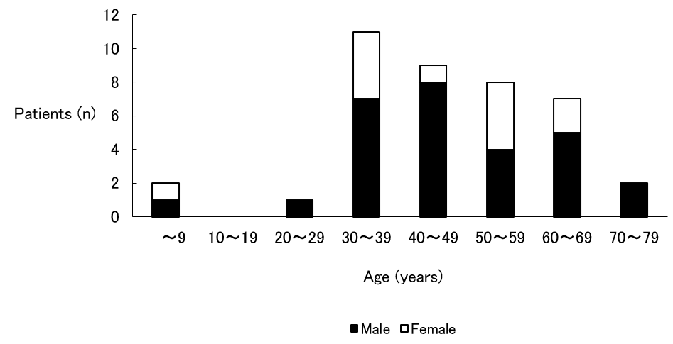
Age and Sex (Figure 1)

biomedres-openaccess-journal-bjstr

Figure 1: Age and Sex.

Note: Ages ranged from 2 to 80 years, and it was most common in the third decade of life. Sex difference was that male predominated.

Ages ranged from 2 to 80 years, with an average age of 47.1 years. Tumor removal was most common in the third decade of life. Sex difference: male (28 cases) and female (12 cases), so male predominated.

Pathological Results

Lipomas were benign in 38 cases (95 %) and malignant in 2 cases (5 %). One liposarcoma was a recurrent case; the other was a fresh case.

Chief Complaints

Swelling of the primary lesion without pain was observed in 37 cases (92.5 %). Swelling of the primary lesion with pain was observed in 1 case (2.5 %); this was a fresh case of liposarcoma. The remaining 2 cases (5 %) were asymptomatic and were accidentally found by image examination or by examination in another department.

Growth Style

Growth style was either unchanged (16 cases; 40 %) or slow (24 cases; 60 %). Two cases of liposarcoma were slow.

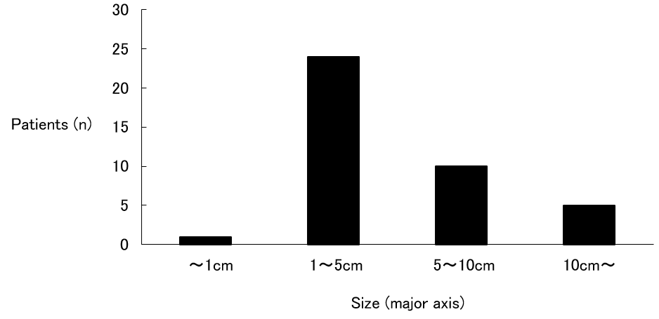
Size (Figure 2)

Lipoma size classes (along its major axis) were counted, and the following results were obtained: smaller than 1 cm (1 case; 2.5 %); 1 to 5 cm (24 cases; 60.0 %), the most common and accounting for more than half of all cases; 5 to 10 cm (10 cases; 25.0 %); and larger than 10 cm (5 cases; 12.5 %). The size of the recurrent case of liposarcoma was 6 cm, and the size of the fresh case was 11 cm.

Time Until the First Visit (Figure 3)

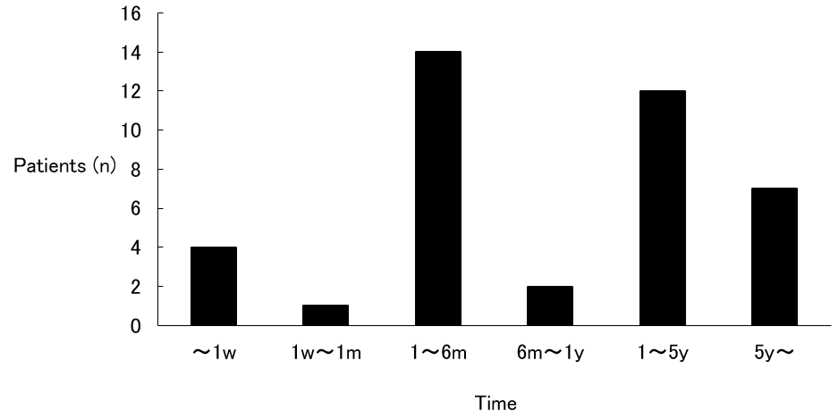
A count of the times until the first visit to KUH was made, and the following results were obtained: within 1 week (4 cases; 10.0 %); 1 week to 1 month (1 case; 2.5 %); 1 month to 6 months (14 cases; 35.0 %), the most common; 6 months to 1 year (2 cases; 5.0 %); 1 year to 5 years (12 cases; 30.0 %); and over 5 years (7 cases; 17.5 %). There were 19 cases (47.5 %) that had been left for more than 1 year. In the recurrent case of liposarcoma, 6 days elapsed, and in the fresh case, 2 months elapsed.

Primary lesion (Figure 4)

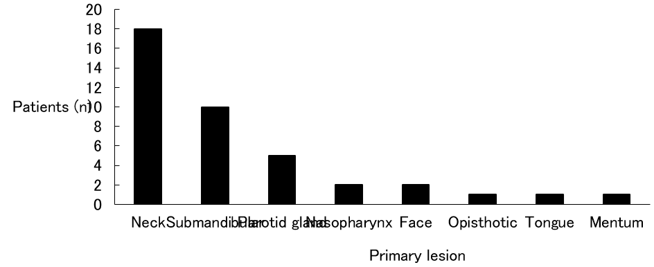
The neck was the most common with 18 cases (45 %) and was followed by the submandibular (10 cases; 25 %), the parotid gland (5 cases; 12.5 %), the nasopharynx (2 cases; 5 %), the face (2 cases; 5 %), the opisthotic (1 case; 2.5 %), the tongue (1 case; 2.5 %), and the mentum (1 case; 2.5 %). Laterality was right side in 27 cases (67.5 %) and left side in 10 cases (25 %), so the right side predominated. The remaining 3 cases (7.5 %) occurred at the nasopharynx and the mentum and were middle. Both cases of liposarcoma occurred at the right neck.

biomedres-openaccess-journal-bjstr

Figure 2: Size.

Note: 1 to 5 cm was the most common and accounting for more than half of all cases.

biomedres-openaccess-journal-bjstr

Figure 3: Time until the first visit

Note: Although 1 month to 6 months was the most common, there were 19 cases that had been left for more than 1 year. w: week, m: month(s), y: year(s)

biomedres-openaccess-journal-bjstr

Figure 4: Primary lesion.

Note: The neck was the most common with 18 cases.

Treatment

All 38 lipomas required only surgery. One of 2 liposarcomas was a recurrent case after surgery at another hospital. Dyspnea had occurred 1 week before the scheduled surgery date at KUH, and an emergency tracheostomy was performed. Surgery was still performed according to schedule 1 week later. In the fresh case of liposarcoma, because internal carotid artery was coarctated by the tumor, it was determined that surgery would be impossible. After a radiotherapy dose of 60 gray, insertion of superficial temporal artery catheter, and administration of 252.5 mg CDDP from the catheter, the tumor reduced. Therefore, surgery would be possible and was performed.

Complications

In cases of lipoma, 3 cases developed complications after surgery. One hemorrhaged after surgery and required a tracheostomy; another resulted in transient palsy of the marginal branch of the facial nerve; and the last resulted in transient palsy of the marginal branch of the facial nerve, fistulation, and an abscess at the wound.

In fresh case of liposarcoma, the recurrent laryngeal nerve (RLN) was involved in the tumor. Therefore, the RLN had to be cut and an RLN palsy occurred.

Prognosis

In the cases of lipoma, although postoperative results were varied and changing, there were no recurrences. In the cases of liposarcoma, the recurrent case died by another disease, and the fresh case is unknown because a change of address occurred. But, there were no recurrences or metastases during the follow-up period.

Discussion

Lipoma is a benign tumor that is composed of mature fat cells that grow relatively slowly. Pathologically, it is composed of crescentshaped and unevenly distributed nuclei, and round or oval cells that have monosegmental vacuolar cell bodies. It forms various sized lobules and is separated by narrow connective tissue. Lipoma is a soft globular or lobulated tumor by palpation, and there is almost no pain. Therefore, if there are no cosmetic problems or difficulties in daily life, patients tend to live for a long time. In this study, there were 19 cases (48.7 %) that lived for more than 1 year. Although treatment requires total removal, in some cases that lipoma grows not only subcutaneously but also inside requires partial removal [1]. The growth styles of all cases were unchanged and slow, and there were no complaints of pain in this study. Further, all patients could be totally removed, and there were no recurrences.

Although liposarcoma is one of the frequently encountered soft tissue tumors, liposarcoma in the head and neck area is rare (5.67 %) [2]. Pathologically, liposarcoma is composed of lipoblasts, and there are various differentiated types ranging from well to poor. Growth speed, metastasis, and the survival rate after 5 years varies markedly by differentiated type. Round cell types and pleomorphic types cause metastasis in the early stages, and prognosis is much worse. Differentiated type and mucous type have a low rate of metastasis, and prognosis is much better. Because the latter is greater than the former, liposarcoma is considered to be a tumor with a relatively better prognosis [3]. There were atypical nuclei abounding in chromatin and both large and small vacuolar and lipoblast cells here and there in the recurrent case. As to treatment, there is general removal and preoperative or postoperative chemotherapy and radiotherapy. Postoperative radiotherapy was not added in the recurrent case because the patient was in advanced age. Preoperative radiotherapy and chemotherapy were both performed in the fresh case. The recurrent case was pleomorphic type, and the fresh case was mucous type. Both cases did not cause recurrence and metastasis during the postoperative follow-up period.

Although differential diagnosis between benign and malignant tumors is based on biopsy, it is said that 10 to 15 % of malignant tumors have local pain or heat, clinically. Although the details of recurrent case were unknown, the fresh case did complain of pain. Further, although 2 liposarcoma cases did not perform computed tomography (CT) or magnetic resonance imaging (MRI) because they occurred in the early 1970’s, Ito, et al. [4] reported that both CT and MRI show lipoma as an almost isolated signal in subcutaneous fat. The margins are clear, and except for well-differentiated types, liposarcomas differ from fat and contain the part of CT is high density, T1 of MRI is low intensity and T2 of MRI is high intensity. Therefore, differential diagnosis between liposarcoma and lipoma is possible, but differential diagnosis between well-differentiated types of liposarcoma and lipoma is difficult.

Conclusion

Clinical files were reviewed for 40 patients who had been operated on and diagnosed as lipoma in the head and neck area. Ages ranged from 2 to 80 years, and the majority of patients were male. Lipomas were benign in 38 cases and malignant in 2 cases. Complications after surgery were seen in 3 cases. Although treatment requires total removal, in some cases require preoperative or postoperative chemotherapy and radiotherapy.


For more Articles on: https://biomedres01.blogspot.com/

Tuesday, August 1, 2023

Using MRI Radiomics and Machine Learning Algorithms in Noninvasive Biopsy of Brain Tumor


Using MRI Radiomics and Machine Learning Algorithms in Noninvasive Biopsy of Brain Tumor

Introduction

Brain cancer is diagnosed via samples acquired from a tumor biopsy. Since the 1970s, anatomical imaging methods (CT, MRI, PET/ CT, and PET/MRI ) have been employed for brain tumor diagnosis, treatment planning, and follow-up studies. Recent technology such as PET/CT and PET/MRI scanners created a significant quantity of detailed neuroimaging data in patients with brain tumors [1-3]. Using AI-based technologies may also speed up picture processing and analysis, enhancing productivity. Radiomics is a particular application within the vast subject of AI that involves computing, identifying, and extracting picture attributes. An overview of feature-based radiomics and machine learning algorithms in noninvasive biopsy of brain tumor using MRI image is presented [2,3].

Radiomics

Robert Gillies and colleagues provide a clear and concise description of radiomics. Radiomics is a high-throughput method that converts medical pictures into radiomics characteristics. This quantitative link may help improve early diagnosis and prognosis prediction in some illnesses, therefore enhancing treatment options. Radiomic analysis includes data gathering, segmentation, feature extraction, exploratory analysis, and modeling [4-6].

Data Selection and Image Preprocessing

Radiomics starts with digital imaging, and MRI is the most often utilized imaging modality; radiomics analysis can be performed on MRI, CT, PET, and ultrasound. Having enough imaging data is helpful for statistical inference, but variation in imaging procedures might impact the quality of retrieved features and radiomics models. Recent research shows that radiomics in MRI are substantially affected by image collection and reconstruction conditions, making them less reproducible. Many studies have demonstrated that many variations exist in radiomics, and preprocessing (normalization, voxel, and pixel resampling ) could improve and solve this problem [7,8].

Segmentation

Segmentation approaches are a crucial part of the radiomics process that separates a lesion from normal tissue. There are three types of segmentation (Manual, semi-automatic, and fully automated). Manual segmentation needs a skilled physician to reduce variability. It is attractive to use semi- or completely automatic segmentation to observe robust characteristics from a particular ROI [9,10]. Nowadays, machine learning softwares (3D-Slicer,Lifx, IBEX) can semi-automatically segment the lesion on MR images [6,11,12].

Feature Extraction

Numerous quantitative radiomics features, such as size and shape-based, histogram, matrix feature (run-length matrix (RLM), size zone matrix (SZM), gray-level co-occurrence matrix (GLCM), and neighborhood gray-tone difference matrix (NGTDM), wavelet feature base could be extracted from the medical images [8,10,12].

Feature Selection

All quantitative features collected from imaging data are not essential for predictive or prognostic models. Also, overfitting occurs when a created model closely fits the test data set; it makes the dependent model more susceptible to noise. Pre-selection of features reduces the probability of overfitting [5]. Radiomics uses both unsupervised and supervised reduction dimensions. The first methods try to remove unnecessary features; the two unsupervised feature selection approaches in radiomics are principal component analysis (PCA) and clustering [5]. There are three popular ways to reduce supervised feature sets:

Filter Techniques

Filter techniques often used include Wilcoxon rank-sum, Fisher score, Chi-squared score, Student’s t-test, and minimal redundancy maximum relevance [13-15]. These approaches investigate the full feature space, considering feature relations to other features in the dataset, and the prediction model is used to score a subset of characteristics [13,14].

Wrapper

Wrapper techniques are hence greedy algorithms. These are forward and backward feature selection, comprehensive feature selection, and search [14,15].

Embedded Approaches

Embedded techniques, mixed filter, and wrapper methods are more accurate than filter methods, quicker than wrapper methods, and less prone to overfitting data. (Regression, treebased techniques like random forest classifier, or the least absolute shrinkage and selection operator (LASSO)) [15-17].

Modeling and Evaluating

Depending on the study’s goal, after feature selection, a model may be created to predict tumor types, machine learning algorithms may develop predictive models. The decision trees and random forest are the most used radiomics algorithms for predicting tumor type. Creating and testing models on the same dataset is a methodological error that leads to overfitting; the train and test datasets and K-fold are in machine learning models can solve this problem [2,16,17].

Conclusion

Radionics is a new imaging biomarker that combines information from radiology, computer vision, and machine learning. With the growth of imaging examination datasets, emerging computational models, there are more demand to use radiomics biopsy in brain tumors. We think that novel diagnostic hypothesis in conjunction with machine-learning algorithms may improve predictive cancer diagnosis and speed up the practical use of quantitative cancer imaging data.


For more Articles on: https://biomedres01.blogspot.com/

Antimalarial Aloe Compounds

  Antimalarial Aloe Compounds Introduction Among the most prevalent diseases caused by protozoan parasites, malaria is caused by parasites o...