Wednesday, August 6, 2025

A Reproducible Method for the Identification of Methandrostenolone Metabolites Using a Humanized Liver Rodent Model


A Reproducible Method for the Identification of Methandrostenolone Metabolites Using a Humanized Liver Rodent Model

Introduction

Anabolic Androgenic Steroids (AAS) are synthetic testosterone derivatives that increase and reduce muscle and fat mass respectively, making them the most prevalent sport performance enhancing substances used by both elite and recreational athletes [1]. Despite being prescription-only drugs, with even their recreational use being illegal in many countries, AAS derivatives are freely available and may be purchased in their pure form or as additives to supplements through numerous online outlets. Their effects on sport performance, and even more worryingly the adverse consequences of their chronic inappropriate use to general health, is very poorly understood. Therefore, there is pressure on antidoping laboratories world-wide to develop reliable, reproducible and sensitive tests for the detection of both the parent AAS, as well as its stable metabolites [2]. One way of producing such tests is to screen for the metabolites of these steroids and identify the ones for use as markers. However, as these drugs are banned substances, ethical issues often make studies in human subjects difficult. The challenge to identifying metabolites for the various AAS and other related sport performance enhancing substances is the availability of an appropriate experimental model.

Several methods to produce these metabolites, both in vitro and in vivo, have been described [3]. While in vitro methods using human hepatocytes have been partially successful, they lack the whole-body environment to generate a full complement of metabolites. Reports on humanized-liver chimeras, where human hepatocytes are transplanted into the spleen of various mice, such as the uPA+/+-SCID, Alb-TRECK/SCID and Por-deficinet PIRK mice models, are available [4-7]. These mice have been used as in vivo models to predict drug metabolism, pharmacokinetics and efficacy in humans. uPA+/+-SCID chimeric mouse with highly humanized liver used in this study has been widely tested and validated in various applications more than the other aforementioned models [7]. Methandrostenolone, (metandienone/methandienone/17α- methyl-δ1-testosterone/17α-methylandrost-1,4-dien-17ß-ol-3- one), a 17α-methylated derivative testosterone, whose metabolism has been most characterized, is an orally active, synthetic AAS. It is also known by its Ciba trade name, Dianabol. This is a popular AAS used for ‘bulking up’, by producing a rapid increase in muscle mass. Daily doses have been reported as being between 20-25 mg, taken for 5 days per week, over a 5-week cycle. It has a half-life of 3.5 hours, with systemic peak levels being reached between 1.5 to 3 hours after ingestion [8]. The primary urinary metabolite of methandrostenolone was reported to be detectable for up to 3 days, with the hydroxymethyl and sulphate metabolites for up to 19-26 days following a single oral (5mg or 25mg respectively) dose [9,10]. Many of these metabolites are unique to methandrostenolone. The current study aimed to confirm previous findings of urinary metabolites of orally administered methandrostenolone generated by the uPA+/+-SCID mice transplanted with human hepatocytes, using pharmacological concentrations of the drug, both as single and multiple doses [5]. Changes in the hepatic mRNA expression of drug-metabolizing enzymes, and the aldehyde dehydrogenase activity in the serum, prior to and after administration of the drug, were also determined.

Material and Methods

Animal Studies

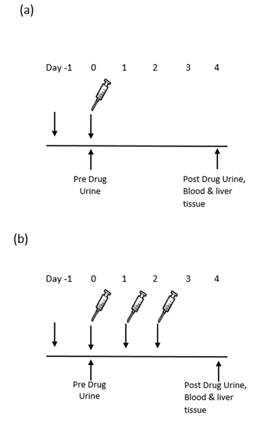
All in vivo experiments and procedures were carried out at KMT Hepatech lnc. (Edmonton, Canada) and approved by the Laboratory Animal Ethics Committee. uPA+/+-SCID chimeric mice with highly humanized liver were generated [11] where primary human hepatocytes were transplanted into the uPA+/+-SCID mice and human albumin concentration was determined prior to drug administration to confirm the engraftment of human hepatocytes. uPA+/+-SCID mice without transplanted human hepatocytes (nonchimeric mice) served as control animals. Chimeric mice were treated with a single or multiple dose of methandrostenolone (n=6) (Toronto Research Chemical, Canada) or 5% ethanol/PBS vehicle (n=6). Similarly, non-chimeric mice were also treated with methandrostenolone (n=4) or 5% ethanol/PBS vehicle (n=4). Methandrostenolone was administered by oral gavage (100μl of 400mg/L, 40μg/gavage) on day 0 (single dose: Figure 1a), and on days 0, 1 and 2 of the experiment (multiple doses: Figure1b). Metabolic cages were used to collect urine samples noninvasively from all animals prior to and on Days 1 and 4 after drug administration. Samples were stored at -20°C. All animals were sacrificed by Schedule 1 protocol at the end of the experiment. Blood was obtained by cardiac puncture without anticoagulant. Livers were harvested and flash frozen. Samples (urine, serum and livers) were collected.

RNA Extraction and CDNA Synthesis

Frozen liver tissue was ground to a fine powder using a prechilled mortar and pestle, with occasional addition of liquid nitrogen. RNA was extracted using TRIzol reagent (Invitrogen, USA), precipitated with isopropanol (Sigma Aldrich, Germany) and washed with 70% ethanol (Sigma Aldrich, Germany). RNA quality was determined by 260:280nm ratio (NanoDrop, Thermo Scientific) and gel electrophoresis (Bioanalyzer, Agilent). RNA Integrity Number (RIN) of 5.5 was considered acceptable. cDNA was synthesized using first strand cDNA synthesis kit (Qiagen, UK).

Microarray Analysis

Pathway specific mRNA PCR arrays (RT2 profiler array Human/ Mouse Drug Metabolism: Phase I Enzymes PCR Array, Qiagen) were used to assess the expression of 84 key Phase 1 drug metabolism genes by real time PCR. Endogenous reference RNA controls were used to normalize the amount of target gene for relative quantification. Human and mouse arrays were used according to manufacturer’s recommendations.

RT-PCR

Transcripts encoding genes identified in the arrays were validated by real-time PCR using ABI SYBR Green master mix (ViiA7 Applied Biosystems, USA). Specific primers were obtained from Qiagen (Qiagen, UK). The level of β-actin in each sample was used to normalize for the variability in RNA quantity. Data was presented as fold change (2-ΔΔCt) after normalization.

Aldehyde Dehydrogenase Activity in Serum

Serum was used to assess NAD-dependent Aldehyde Dehydrogenase (ALDH) activity. ALDH superfamily (19 genes) are key phase 1 cellular detoxification enzymes that oxidize various aldehydes and generate the corresponding carboxylic acid. It comprises 3 major classes: Class 1 and Class 3 are cytosolic and include both constitutive and induced forms; Class 2 is a constitutive, mitochondrial form. The ALDH activity was assayed using a fluorometric method (BioVision’s PicoProbe, USA). ALDH is oxidized by acetaldehyde forming NADH that incorporates to the PicoProbe generating fluorescence (Ex/Em = 535/587) with sensitive detection of < 0.05 mU ALDH activity.

Urine Metabolite Analysis

All urine samples collected from the uPA+/+ -SCID mice were analyzed by UHPLC/HRMS and GC/MSMS for the detection of the parent drug and its major metabolites. Additionally, a blank human urine sample spiked with reference standards of the above metabolites was analyzed to confirm the presence of the detected metabolites.

Sample Preparation

To 0.5 mL of urine, 1 mL of phosphate buffer, pH7, 50 μL of E-Coli β-glucuronidase (Sigma Aldrich) and 50 μL of a methanolic solution of internal standards mixture were added and the mixture was hydrolyzed at 50°C for 1.5 hours. The pH was then adjusted to 9 to 10 with sodium hydrogen carbonate and potassium carbonate (2:1) (w/w followed by liquid–liquid extraction against diethyl ether. The organic layer was separated from the aqueous phase after centrifugation by freezing and evaporation under nitrogen at 50°C and residue was reconstituted in solvent and injected to the UHPLC/ HRMS system. Prior to GC/MSMS analysis, a derivatization step was performed to the dry residue by adding MSTFA/Ammonium iodide/2-Propanthiol (500:2:4) and incubation at 100°C for1 hour.

UHPLC/HRMS Conditions

A Dionex UHLC system (Thermo Scientific) was used for the chromatographic separation and the separation was achieved at 40°C using a Zorbax Eclipse Plus C18 column (Agilent Technologies). A gradient elution program was employed at a constant flow rate of 0.2 mL min-1 with injection volume of 5μL. The high resolution mass spectrometry was performed using QExactive Orbitrap mass spectrometer (Thermo Scientific). An additional targeted MSMS acquisition (product ion scan mode) at 17,500 resolving power was applied for the identification of the parent compound of methandienone(17α-methylandrost-1,4-dien-17β-ol-3-one) (precursor ion m/z 301.22 at normalized collision energy (NCE) 25%) and its metabolites 17-Epimethandienone (precursor ion m/z 301.22 at NCE 25%) and 18-nor-17β-hydroxymethyl,17α- methylandrost-1,4,13-trien-3-one (precursor ion m/z 299.20 at NCE 25%).

GC/MSMS Conditions

The GC/MSMS analysis was performed using Agilent GC 7890 system coupled with Agilent 7000C triple quadrupole mass spectrometer, equipped with a 7693 autosampler, 10μL syringe and split/splitless injector. Chromatographic separation was achieved using a SGE BPX5 column. The oven temperature program was set as ; 160°C for 0 min, ramped at 10°C /min up to 200°C, then ramped at 2 °C /min to 220 °C, ramped at 6 °C /min to 292 °C, 50°C /min up to 310°C and held for 3min. Injection volume was 2 μL in a split ratio 1:10 and the flow of the helium as carrier gas was 1.1 ml/min. The MS system was a QQQ with Electron Ionization at 70eV and multiple reaction monitoring (MRM) acquisition for the detection of Methandienone and its metabolites.

Statistical Analysis

Data were analyzed using Excel or by SA Biosciences in-house software. Significance was defined as p≤0.05.

Results

Microarray Analysis

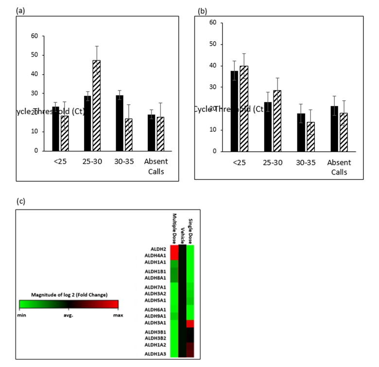
In the animals that had been successfully transplanted with human hepatocytes (significantly elevated systemic human albumin) there was a global up-regulation of the Phase 1 metabolism enzymes in response to methandrostenolone. Sixty five percent of the genes in the Drug Metabolism: Phase I Enzymes array were induced after drug treatment as opposed to 52% following vehicle, this was particularly apparent when looking at genes that showed Ct between 25-30 (Figure 2a). The non-chimeric mice did not mount a xenobiotic response to the drug, probably because their residual liver function was compromised (Figure 2b). single and multiple dosing produced different patterns of gene expression (Figure 2c). More detailed analysis, using ß-actin, B2M and HPRT1 as the house-keeping genes for normalization and excluding samples that did not pass the QC checks, showed the mRNA expression of following genes to be significantly induced by the drug, compared to vehicle: Aldehyde dehydrogenase 7 family, member A1 (ALDH7A1) (1.33 fold difference, 95 % CI 1.27, 1.39: p=0.0003), Aldehyde dehydrogenase 9 family, member A1 (ALDH9A1) (2.26 fold difference, 95 % CI 0.79, 3.73: p=0.04) and Aldehyde dehydrogenase 6 family, member A1 (ALDH6A1) (1.88 fold difference, 95 % CI 0.70, 3.06: p=0.05).
biomedres-openaccess-journal-bjstr

Figure 1: Experimental design and sample collection. Male uPA+/+-SCID chimeric (n=12) and non-chimeric (n=8) mice were administered with 100μl of 400mg/L (40μg/gavage) methandrostenolone by oral gavage, either as a single dose (a) or as multiple dose (b). 24h urine collections were made prior to and after drug administration on Days 0, 1& 4 of the experiment.
biomedres-openaccess-journal-bjstr

Figure 2: Up-regulation of hepatic mRNA expression of Phase 1 enzymes.

a) The mRNA expression of a significant number of the phase I enzymes were up regulated following injection with the drug, compared to vehicle, in the liver of chimeric mice using a human array (Solid bars – Vehicle, Hatched bars - Drug).

b) No such drug effect was seen in the non-chimeric mouse liver using a mouse array (Solid bars – Vehicle, Hatched bars - Drug), suggesting the lack of functionality of the organ in the host.

c) Heat map depicting magnitude of change in mRNA expression. Red=up-regulation; Green=down-regulation. Min=-2; Max=+2. n=6.

Aldehyde Dehydrogenase Activity in Blood

ALDH activity in serum was investigated in response to methandrostenolone using a high sensitivity fluorescent kinetic assay (NADH range 1-5 pmol/μl). Levels of ALDH activity in the samples was extremely low, but detectable. While there was an increase in the serum ALDH activity after the multiple, compared with the single, dose, this did not reach significance {Single 1.32 (0.6) versus multiple 1.56 (0.5) pmol/μl; p=0.45}.

Urine Metabolite Analysis

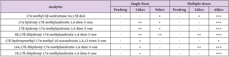
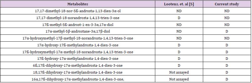
GC/MSMS detected17α-hydroxy-17ß-methylandrosta- 1,4-dien-3-one , 17ß-hydroxymethyl-17α-methyl-18-norandrosta- 1,4,13-trien-3-one and 17ß-hydroxy-17α-methylandrosta-1,4- dien-3-one with the rest by UHPLC/HRMS . The metabolites in the urine samples of the drug-treated animals are summarized in (Table 1). A comparison between single and multiple dosing showed that the latter mainly increased the sensitivity of the detection of all the studied metabolites. None of the mentioned metabolites was detected in the vehicle treated chimeric mice, after single or multiple doses. Following UHPLC/HRMS and GC/MSMS analysis of the urine, data showed the presence of all the previously reported metabolites [4,6], including the stable metabolite, in the drug-treated cohort but not in those treated with vehicle (Table 2).
biomedres-openaccess-journal-bjstr

Table 1: Detected metabolites using GC/MSMS and UHPLC/HRMS. As the analysis was qualitative, data are shown as (-) or (+), with (-) as not detected and variable detection accuracy of 33.3% (+), 66.6% (++) and 99.9% (+++).
biomedres-openaccess-journal-bjstr

Table 2: Comparison of metabolites detected in the current versus previous study [5].

Note: D: Detected; ND: Not detected.

Discussion

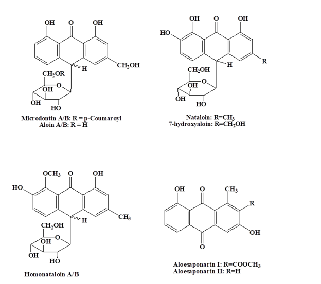
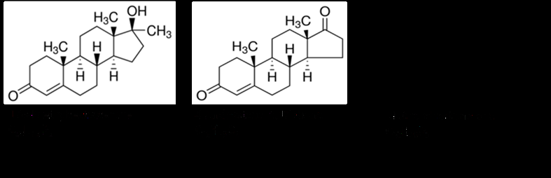
Determination of AAS use in the human sporting arena has been, and continues to be, a significant challenge to scientists. Using a well characterized AAS, methandrosternolone, this study confirmed that the uPA+/+-SCID model with the humanized liver is capable of generating all previously reported urinary metabolites. Furthermore, oral administration of the drug over 3 days (Figure 1) provides a valid experimental set up. Also, both multiple doses, as well as a single dose, leads to detectability of the reported urinary metabolites, as well as elevated mRNA expression of hepatic phase I enzymes. The multiple doses, however, increased the sensitivity of the detection, especially of specific aldehyde dehydrogenases and cytochrome P450. Whilst ALDH activity was detectable in the serum it did not reach significance in the presence of drug and perhaps best investigated in hepatic tissue. Previous studies have also used the same rodent model [5,12], however, with different derivatives of testosterone (4-androstene- 3,17-dione and 17α-methyltestosterone respectively; (Figure 3), concentrations and the numbers of doses. The study by Pozo et al. used 17α-methyltestosterone following a single dose of the drug (40 μg per oral gavage) [12]. On the other hand, Lootens et al. used multiple, suprapharmacological doses (350μg per gavage) of 4-androstene-3,17-dione [5]. The current study used both single and multiple doses, at pharmacological concentrations (40 μg) of methandrosternolone. The multiple doses increased the sensitivity of detection, despite all the reported metabolites being detectable upto 96h after a single dose.
biomedres-openaccess-journal-bjstr

Figure 3: Testosterone derivatives used to investigate human drug metabolism in uPA+/+-SCID mice. Left panel: Derivative used by Pozo, et. al [12]; Middle panel derivative used by Lootens, et. al [5]; Right panel derivative used in current study.

This report clearly provides further support for a reliable in vivo murine model for the identification of transient and stable metabolites of orally administered AAS in humans, especially following multiple dosing. Humanized liver chimeric mice were validated against an array of xenobiotics and were able to be metabolically distinct from recipient mice, while showing metabolic pathways analogous to those expected from human liver. Recent metabolic studies in the chimeric uPA+/+-SCID mouse model, including steroid metabolism, were reviewed in Foster et al [7]. The model closely reflects the human metabolism of some drugs, as well as predict human metabolites, including those generated through more than one metabolism reaction [13]. Using both UHPLC/HRMS and GC/MSMS the current study confirmed detection of previously reported metabolites of related testosterone derivatives. While this study used a targeted approach, it may prove very interesting when combined with non-targeted metabolomics for the identification of novel metabolites, especially for emerging anabolic agents.

For more Articles on: https://biomedres01.blogspot.com/

Tuesday, August 5, 2025

Monoaminergic Neurotransmitters Promote the Percentage of Recovery of Swimming and Ambulation Behaviors following Central Nervous System Lesion in the Leech


Monoaminergic Neurotransmitters Promote the Percentage of Recovery of Swimming and Ambulation Behaviors following Central Nervous System Lesion in the Leech

Introduction

One of the great problems in neuroscience is the fact that central nervous system (CNS) axons are unable to regenerate and successfully reconnect with their targets in superior vertebrates. This failure contrasts with the regeneration abilities in the peripheral nervous system and among inferior vertebrates and invertebrates [1]. However, using peripheral nerve bridges, David and Aguayo [2] demonstrated the regenerative potential of CNS neurons, which is dependent on their environment. This problem of regenerative failure is also of clinical interest due to them any individuals harboring CNS injuries that in many cases, lead to various motor and/or cognitive disabilities. In the field of motor and cognitive rehabilitation, several therapies have been developed to strengthen intact connections and promote the reconnection of injured axons. However, conduct recovery still depends on neuronal regeneration and the successful reconnection of neurons with their targets. Despite the biological and clinical interest of neuronal regeneration and the functional recovery of compromised abilities due to CNS injury, the problem remains to be solved. This issue is complex, multifactorial, and neither its determining elements nor their degree of involvement have been determined. This study focused on the role of monoaminergic neurotransmitters since these molecules are highly involved in the physiological processes of animal mobility and have several effects on neuritogenesis.

Monoaminergic neurotransmitters play an important role in neuritic regeneration. Serotonin acts on neuritic axonal growth, it guides the growth cones, and participates in synaptogenesis [3,4] and many receptors may mediate its mechanism of action [5]. Studies on the somatic secretion of serotonin have posited that this neurotransmitter acts on processes that occur away from the synapsis [6,7] and may participate in neuritogenesis. Further, dopamine is one of the factors regulating neuritogenesis [8-10] and neurogenesis in the mammalian adult brain, including humans [11,12]. Likewise, octopamine (OA) is present in invertebrates such as annelids, mollusks, and arthropods, acting on neuronal and behavioral plasticity. OA has been reported to participate in neuritogenesis [13]. OA is structurally and functionally similar to adrenergic transmitters which is present in vertebrates but not in invertebrates [14,15]. All have been recognized as playing a welldescribed role in animal movement regulation [16]. Their depletion with non-specific drugs such as reserpine, or specific to each neurotransmitter, modifies different features of motor behaviors in animals [17]. An increase in blood serum serotonin leads to behavioral changes in animals in terms of mobility [18] such as aggressiveness in vertebrates and invertebrates. Dopamine is key to movement [19,20] as reflected in Parkinson´s disease, a disease compromising movement due to the degeneration of dopaminergic neurons, and that leads to a dopamine deficit in several areas of the brain [21]. Likewise, octopamine plays a role in the movement and behavior of animals [14,15,22].

All of the above underscore the importance of monoaminergic neurotransmitters in animal motor functions and in neuritic regeneration, suggesting that monoamines overall or independently, may play a relevant role in motor recovery after CNS injury. In the study of neuronal regeneration and functional recovery, invertebrate animals have been used due to their spontaneous neuronal regeneration ability and their recovery of motor function following CNS injury. After CNS injury in the leech, this animal spontaneously recovers motor function [23]. Many behaviors such as swimming, flexion, shortening, drag, feeding, and heartbeat have been described as well as the neuronal circuits associated with these neurotransmitters [17,24]. The addition of serotonin to their environmental water, triggers swimming in animals depleted of this neurotransmitter after 5, 7-dihydroxytryptamine administration [25]. Likewise, in culture, adding serotonin to specific leech neuron cultures induces neuritic regeneration, as in AL1 cells that increase their number of primary neurites, their length, and their ramification; however, in other cells such as Retzius cells, serotonin has an inhibitory effect [26].

The study´s aim was to determine the role of monoaminergic neurotransmitters on the percentage of animals that recovered their swimming and ambulatory functions after CNS injury. The CNS cord was injured, and monoamine depletion was tested with intra-corporeal reserpine injection in the Mexican leech, Haementeria officinalis; this leech, as Hirudo medicinalis, the European medicinal leech, spontaneously recovers motor function after induced injury. Behavioral recovery was evaluated, and the recovery percentage was quantified. Serotonin was also replaced to establish the degree of its effects. In this work we find that these neurotransmitters promote the recovery percentage in swimming and ambulation abilities after CNS lesion. Acutely administered serotonin did not contribute to the recovery percentage.

Material and Methods

Maintenance of the Organisms

Invertebrate organisms of the Phylum Annelida were used: Haementeria officinalis is a Mexican leech from our Neuronal Regeneration Laboratory (NRL) animal colony. The procurement and maintenance of Haementeria officinalis samples has been previously described [26]. These organisms were originally collected in the lakes and dams of the central Mexican plateau by a researcher authorized by the local “Direction of Agricultural Development”. The organisms weighed between 0.5 and 0.8g and were placed in individual 1L containers with water (E-pura, Mexico), labeled, and maintained at laboratory temperatures and conditions. All procedures were approved by the Institutional Committee for the Care and Use of Laboratory Animals (INRCICUAL No. 15/11).

Study Groups and Experiment Series

Seven experimental or repetition series were performed. At each repetition, 5 study groups were created (control, injury, reserpine, injury-reserpine, injury-reserpine-serotonin) with 6 organisms each, to determine the recovery percentage in each group. The “control” animals were neither treated nor injured, the CNS was injured in the “injury” group, in the “reserpine” group, the drug was injected intracorporeally, the drug was injected and the CNS injured in the “reserpine-injury” group, and in the “reserpineinjury- serotonin” group, the animals were injected, their CNS was injured, and serotonin was replaced.

Swimming and Ambulation Motor Behaviors

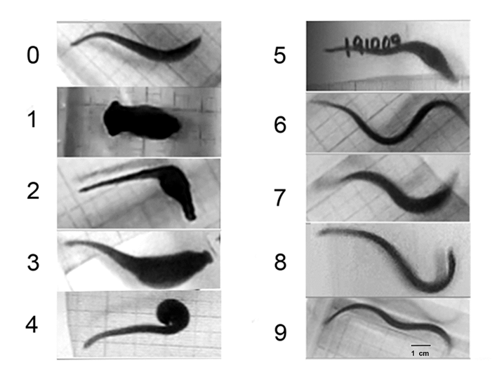
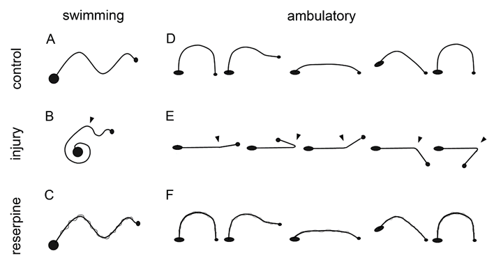
Swimming in control, uninjured, non-injected animals, follows a continuous, sine-wave pattern originating at the head and ending at the tail (Figure 1A), and the animal flattens its body in a dorsoventral axis. After injury, the animal becomes immobile for 1 to 2 days, and subsequently begins to move and swim creating incomplete sine-waves that begin at the head and end at the injury level. The area posterior to the injury does not form a sinus-wave (Figure 1B) and does not flatten. This type of swimming was not considered normal. After several days, the animal recovered and swam forming complete waves from head to tail. Reserpine injection did not affect the animal´s sine-wave swim pattern, but the body´s lateral edges became undulated, like the edges of an oak leaf (Figure 1C). The usual ambulation pattern of the healthy animal on a solid substrate includes release of the anterior sucker, body extension, adhesion of the frontal sucker to the substrate, release of the posterior sucker, body contraction, and adhesion of the posterior sucker to the substrate (Figure 1D). After injury, the animal remains immobile for 1 to 2 days, and subsequently begins to move; injured animals can release the frontal sucker and extend the anterior part of their body, but they cannot adhere the frontal sucker, moving the anterior part of their body above the injury, asynchronically from the rest of the body; over a few days, they can do so, but they are unable to release the posterior sucker (Figure 1E). These movements were not considered normal ambulation. Recovery of normal ambulation was established once the animal could adhere and release both suckers in a synchronous movement that allowed displacement on the substrate. Reserpine injection has no effect on the normal movement pattern (Figure 1F).

biomedres-openaccess-journal-bjstr

Figure 1: Diagram of the healthy, injured, and reserpine-injected leeches´ swimming and ambulation behaviors. A, when swimming, they form a continuous sinewave from the head (small circle) to the tail (large circle). B, in injured leeches, the swimming sinewave originates from the head and reaches the injured site, while the remaining body remains contracted. D, when ambulating, the release the anterior sucker, extend the body, adhere the sucker to the substrate, release the posterior sucker, contract the body, and adhere the posterior sucker. E, during ambulation, the anterior portion of the animal moves from the injury site but independently from the rest of the body. C, F, Reserpine injection has no effect on these behaviors.

CNS Injury

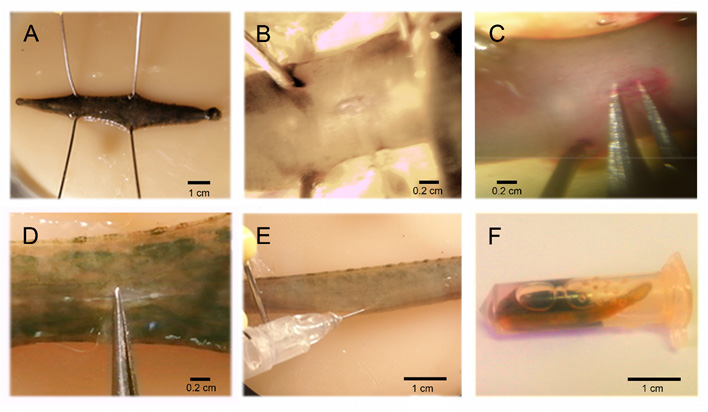
The animals were anesthetized with 9% ethanol, for 20 minutes. They were fixated with 4 pins on a wax dish, and a ventral incision was made at the level of the 7th and 8th ganglia; with microdissection forceps, the connecting nerve between the ganglia were exposed (Figures 2A & 2C), and with another forceps, its central part was obturated causing an injury 300 micrometers in length (Figure 2D). Obturation was performed with the same forceps in all cases, there were no irregularities on contact surfaces, the edges were blunt, and the blade measured 300 micrometers in width. Injury was caused by pressing on the forceps 10 consecutive times, the position of the forceps´ blades was inverted, and pressure was applied 10 more times, the forceps was again inverted, and again, pressure was applied 10 more times. This same procedure was followed in all of the injured groups.

biomedres-openaccess-journal-bjstr

Figure 2: CNS injury procedure, reserpine injection, and serotonin replacement. A, the anesthetized animals were fixated with 4 pins on a wax dish, and a ventral incision was made on the connecting nerve (B). The connecting nerve was exposed with forceps (C), and obturated between the 7th and 8th ganglia (D) causing an injury that compromised axonal inter-ganglionic continuity. E, Reserpine injection (Sigma, 100ug/g weight). The animals were anesthetized with 9% ethanol and placed in a wax dish, the injection was applied to the side of the connecting nerve between the roots of the 9th and 10th ganglia (F). Acute reserpine replacement. This was conducted by placing the animals in a 2 ml container with 500 μl of serotonin solution (2mM. 1hr/day. RT).

Reserpine Injection

The animals´ CNS were depleted of monoamines with reserpine [10]. Reserpine (100 μg/g of the animal´s humid weight, Sigma) was injected with an insulin syringe in organisms anesthetized with 9% ethanol and extended in a wax dish. The 9th and 10th ganglia were located, and the injection was applied between the peripheral nerves of these ganglia, to the side of the connecting nerve (Figure 2E). This procedure was used in the organisms in the groups treated with reserpine. The injury-reserpine group included animals injected with reserpine and injured 24 hours after injection.

Serotonin Replacement

The injured animal group that was injected with reserpine and in whom serotonin was replaced, included organisms injected with reserpine, their CNS was injured 24 hr. later, and during their recovery, they were incubated daily with serotonin (serotonin chlorhydrate, Sigma), for one hour (2mM). This is the concentration at which the resting animals become active and begin to swim vigorously in the containers. The animals were individually placed in 2 mL containers, previously perforated in their upper area to allow air flow, and 500 μL of the serotonin solution were added (2mM. 1h/day. RT. Figure 2F). After incubation, the animals were washed and directly placed in their original containers.

Behavioral Registry and Statistical Analysis

For 30 days after injury, the motor skill development of each organism was registered on video. The presence or absence of swimming and ambulation behaviors in each animal were registered. For data analysis, we recorded for each animal, whether or not they had the normal behavior of swimming or ambulatory, we obtained the percentage of recovery of motor skills in each group of 6 organisms. With the recovery percentages, we obtained statistical parameters of the 7 repetitions, and reported the percentage of recovered animals as the mean ± standard error. The level of significance was established at 95% according to the unpaired Student t test. Calculations were performed with the Sigma Plot 2001 program (Systat Sofware Inc., USA). The data tables show the recovery percentages on days 2, 7 and 21 since they are representative days of the effect of the injury and the recovery of swimming and ambulatory behaviors (Figure 3).

biomedres-openaccess-journal-bjstr

Figure 3: Series of images showing motor recovery after CNS injury. On day zero, before CNS injury, the animals formed a continuous sinewave from the head to the tail. After injury, the animals remained immobile for up to 24 hrs. (Day 1). The animals move asynchronically from the site of injury (day 2). When swimming, sinewaves are generated from the head to the injury site, and the rest of the body is dragged without forming sine-waves. On day 7, many of the injured animals had restored their ability to swim forming continuous waves from the head to the tail. On day 21, animals retain the ability to swim (image not shown). The behaviors of days 2, 7 and 21 were used for data analysis. Scale bar=1cm.

Results

Recovery of Swimming and Ambulation Behaviors After Injury

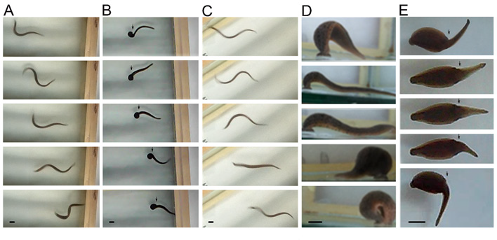
When swimming, the animals flatten their bodies dorsoventrally, and generate continuous sinus-wave shaped undulations from the head to the tail (Figure 4A). After injury, the organisms remain immobile for 24 to 48 hours at the bottom of the container, and subsequently begin their recovery; the animals can swim but using only the body portion anterior to the injury (Figure 4B). Seven days after injury, many animals recovered their swimming abilities forming sinus-wave from head to tail (Figure 4C). When ambulating at the bottom of the container, they move by adhering their suckers and contracting and extending their bodies in alternate movements (Figure 4D). During the first few days after injury, the animals are able to adhere the posterior sucker, extend their body and attempt to adhere the anterior sucker. However, they are unable to contract the body. They generate movement attempts in different directions with the anterior sucker but are unable to release the posterior sucker nor coordinate body contraction or extension (Figure 4E). They can adhere both suckers but independently from the extension and contraction of the body, as seen by the separation of movement from the area of injury.

Monoamine Depletion with Reserpine Prolonged the Effect of CNS Injury

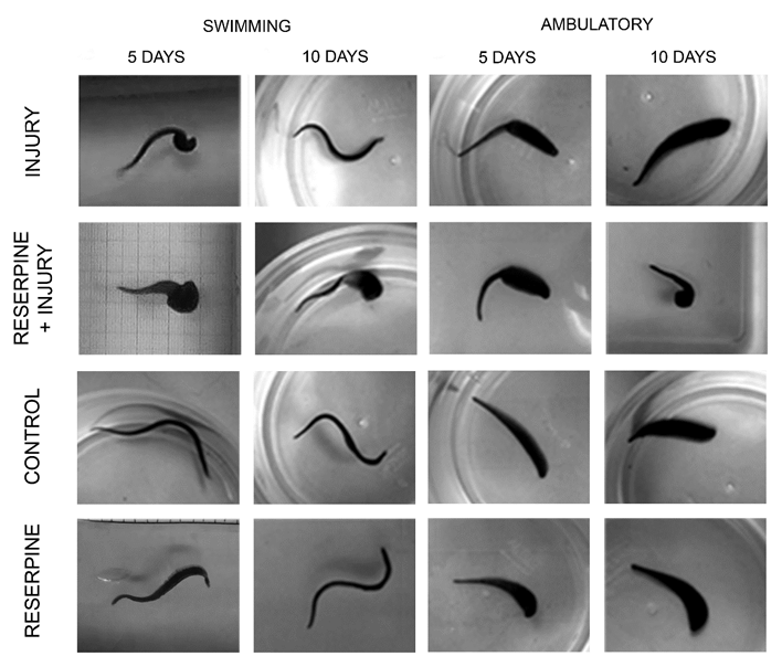
Five days after injury, these animals swim by contracting the posterior part of their body, and only generate sine-waves from the head to the injury site, but 10 days later, they can swim forming complete sine-waves (Figure 5). Likewise, on day 5, the animals still lack ambulatory synchronicity, that is recovered by day 10. Monoaminergic neurotransmitter depletion with reserpine injection, leads to persistence of swimming and ambulatory disability 10 days after injury (Figure 5). Reserpine injection did not modify the sine-wave swimming pattern nor ambulation synchronicity.

biomedres-openaccess-journal-bjstr

Figure 4: Series of images showing swimming and ambulation behaviors.

A) While swimming, the animals form sine-waves. The sine-wave begins at the head (right in images) and ends at the tail (left side).

B) Central nervous system (CNS) injury led to loss of the sine-wave´s continuity, originating in the head to the area of injury (arrowhead).

C) One week later, swimming abilities are recovered, forming full sine-waves from head to tail.

D) In ambulation, the posterior sucker is adhered (right in images), the animal´s body extends, and the anterior sucker adheres to the substrate; the animal´s body contracts, and the posterior sucker is released and adheres again close to the anterior sucker. This is a continuous movement that allows the animal´s displacement on the substrate.

E) Nervous system injury causes movement asynchrony in the anterior body and caudal to the injury. While the posterior sucker is adhered to the substrate, the anterior portion moves independently from the area of the injury (arrow).

biomedres-openaccess-journal-bjstr

Figure 5: Behavior comparisons 5 and 10 days after CNS injury. Five days after injury, swimming and ambulation remain compromised in injured animals and in those injected with reserpine and injured. After 10 days, they recover swimming fully and ambulate with synchronic movements. However, reserpine injection induced persistence of the injury´s effects. Reserpine injection did not affect the sine-wave swimming pattern nor ambulation synchrony.

Reserpine Injection Decreased the Recovery Percentage After Injury

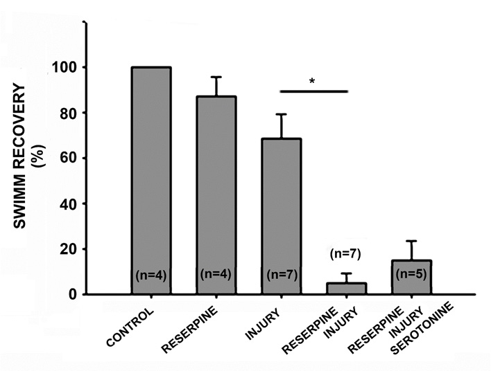
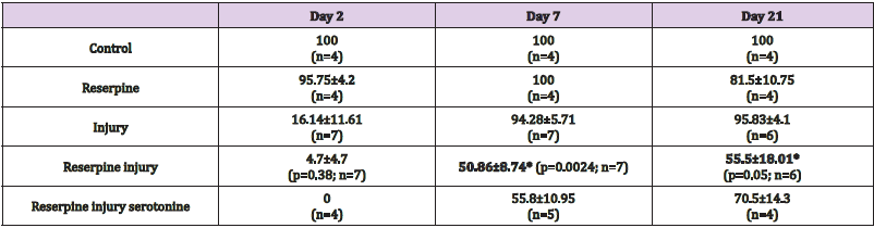
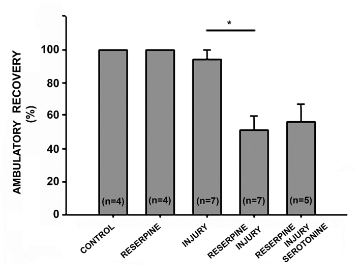
Two days after CNS injury, almost all animals were unable to swim or deambulate (Tables 1 & 2). Seven days after injury, 68.42±11.11% of injured animals can swim forming a continuous sinus-wave from the head to the tail and also recover ambulatory synchronicity (94.28±5.71%) at the bottom of the container, with alternate body extension and contraction, and adhering and releasing the suckers. The animals injected with reserpine and injured, the recovery percentage in swimming abilities decreased (4.71±4.71% p=0.0002) compared with the animals that were only injured (Table 1, Figure 6). Seven days after reserpine injection, the percentage of ambulation recovery decreased in the injured animals (50.86±8.74%, p=0.0024) in comparison with the recovery of animals that were only injured (94.28±5.71%. Table 2. Figure 7).

biomedres-openaccess-journal-bjstr

Figure 6: Graph of the percentage of swimming recovery 7 days after injury. The injection of reserpine did not affect swimming behavior, and all animals swam like those in the control group. Among the injured animals, 68.42% recovered the ability to swim forming continuous waves from the head to the tail. Monoaminergic neurotransmitter depletion associated with reserpine injection decreased the recovery percentage to 4.71% (p=0.0002, n=7), and the acute replacement of serotonin did not modify this recovery percentage (14.6±9%. p=0.31, n=5).

biomedres-openaccess-journal-bjstr

Table 1: Recovery percentage of swimming after intra-corporal injection of reserpine (100μg/g) and CNS injury. Data is presented as average values and standard errors. Bold numbers refer to the significant between-group differences: injury and reserpine-injury.

biomedres-openaccess-journal-bjstr

Table 2: Recovery percentage of ambulation after intra-corporal injection of reserpine (100μg/g) and CNS injury. Data is presented as average values and standard errors. Bold numbers refer to the significant between-group differences: injury and reserpine-injury.

biomedres-openaccess-journal-bjstr

Figure 7: Graph of the percentage of ambulation recovery 7 days after injury. Reserpine injection did not affect the animals´ ambulation behavior, and 100% of animals could move on the substrate just as the control group. Among the injured animals, 94.28% recovered the ability to ambulate continuously by adhering the suckers in an alternate manner and extending and contracting the body. Monoaminergic neurotransmitter depletion with reserpine injection decreased the recovery percentage to 50.86% (p=0.0024, n=7), and acute serotonin replacement did not modify this recovery percentage (55.8±10.95%. p=0.73, n=5).

Acute Serotonin Replacement had no Effect on the Recovery of Swimming or Ambulatory Skills

In five repetitions, acutely applied serotonin replacement, 500 μL (2mM) daily for one hour, had no effect on the percentages of swimming and ambulation skills in the injured animals treated with reserpine. In swimming skills, serotonin replacement led to a recovery of 14.6±9% (p=0.31), in comparison with the injuryreserpine group (4.71±4.71%. Table 1, Figure 5). Likewise, serotonin replacement induced an ambulatory recovery percentage of 55.8±10.95% (p=0.73) compared with the injury-reserpine group (50.86±8.74%) (Table 2, Figure 6).

Discussion

Monoaminergic neurotransmitters promote the recovery of swimming (93%) and ambulation (46%) after CNS injury. Monoamine depletion via reserpine injection decreased the percentage of swimming and ambulation recovery in animals injured by obturation of their central nervous system. Seven days after injury, they decreased from 87.25% in swimming recovery to 4.71%, and from 94.28% in ambulation recovery to 50.86%. This underscores the importance of these neurotransmitters in the process of motor recovery after CNS injury. The differences in the percentages of recovery of swim and ambulation skills on day 7, in both the animals that were only injured and in the injured group injected with reserpine, suggest that these behaviors follow different neuronal pathways. Indeed, both behaviors have been shown to be based in different groups of neurons [27]. These different circuits may possess different regeneration speeds and integration of their functional webs, leading to the differences in recovery percentages 7 days after injury. In terms of the CNS monoaminergic neurotransmitters, we question their degree of participation in functional recovery. In this study, after monoamine depletion with reserpine injection, serotonin was acutely replaced (2mM; 1hr/day). The recovery percentages in the injury-reserpine and the injury-reserpine-serotonin groups showed no significant differences in swimming or ambulatory skills, revealing that acute serotonin replacement has no effect on recovery percentages.

This lack of a positive serotonin effect is in accordance with results on its effect in some cultured neuronal types such as AE or Retzius cells [26]. In this study, serotonin either had no effect or had an inhibitory effect on the regeneration of specific neurons, whereby systemic serotonin replacement would not foster the regeneration of some neuronal types, and hence, the reestablishment or development of neuronal circuits required for swimming and ambulation motor behaviors. In the future a similar study, chronic testing of other serotonin concentrations would be of interest. Likewise, the role of dopamine and octopamine replacement on the functional recovery of motor skills in this system remains to be tested. The constitutive activity of the 2C serotonin receptor has been shown to increase the excitability of neurons caudal to a spinal injury in rats, and finally leads to recovery of some motor functions [28]. This and other experiments have established the relevance of the serotoninergic system in motor function recovery after spinal cord injury. However, clinical trials conducted in humans have shown that drugs associated with serotonin and its receptors do not promote recovery after CNS injury [29]. Currently, there are controversial results on the role of serotonin and its receptors in functional recovery [30,31] and more studies need to be done on humans and other systems.


For more Articles on: https://biomedres01.blogspot.com/

Antimalarial Aloe Compounds

  Antimalarial Aloe Compounds Introduction Among the most prevalent diseases caused by protozoan parasites, malaria is caused by parasites o...