Friday, June 16, 2023

On Possible Regulation of Cancer Cells Growth Dynamics Using Electromagnetic Effects on the Body


On Possible Regulation of Cancer Cells Growth Dynamics Using Electromagnetic Effects on the Body

Introduction

This work is a logical continuation of a series of previous works devoted to the analysis of the influence of external constant and variable electromagnetic (abbreviated as EM) fields on biological objects (see articles [1,2]). This report will focus on the study of the influence of variable EM fields on the cellular structure of the body. In this case, the system of the Lotka-Volterra equations [3,4] will be taken as the basis for calculations and estimates, which we will modify accordingly in relation to our problem, relying to a large extent also on the work [5]. It is not difficult to formally introduce any type of interaction into the system under study. The main issue is always the physical and mathematical justification of the appropriate type of interaction and understanding how to apply it to a particular task. It is this justification that we will now deal with in order to achieve this goal. It is worth noting beforehand that issues related to the mathematical description of cancer diseases have been dealt with for a relatively long time, and there are a number of results in this area, described in some detail, for example, in [6- 7]. The approach proposed below is based on considerations other than those mentioned above and gives a theoretical prediction for the treatment of such diseases.

The Energy of Cell Interaction with the EM Field

In order to find the interaction we are interested in, we will need to take into account the following two very important points. Namely, firstly, this interaction should be invariant with respect to the time sign inversion, and, secondly, it should take into account the concentration of “predators” and “preys”. In our case the role of predators will belong to cancer cells, the concentration of which we will designate with a letter n , and the role of the preys – to healthy cells; their concentration we will designate with a letterm . Note that we adhere to the same designations as in [5]. It is quite clear (see monograph [8]) that the interaction of any bodies with the EM fields can be different from zero only if the internal structure of the object under consideration allows to take into account its polarization p and magnetization m . However, if almost any object, including a cell, is under long-term exposure to constant EM fields, it will be certainly polarized. This means that, despite its extremely complex structure, there will be a slight deformation of the cell, which allows us to consider the polarization p . This means that we may take into account the possible interaction of cellular substance with a variable electric field E′(t ) , but only after the 0 E polarization of the cell is induced by the applied constant electric field. Such an interaction, attributed to a unit of volume, as it is known [8], formally has the form of a negative scalar product −p⋅E′ , where

Where αñ − is the cell polarization coefficient As for the magnetic field, if there are magnetic atoms in the cell structure in the form of, for example, iron, manganese or other magnetic particles, as well as moving charges, the energy of interaction with an external variable magnetic field will necessarily manifest itself H′(t ) in the same way as with an electric field, but already having the form of −m⋅H′(t ) where

This vector represents the magnetic moment of the cellular substance, χñ − is the magnetic susceptibility of the cell, and H0 − is the constant magnetic field applied to the body.

The account of interaction with the field will be given further phenomenological (see formulas (18), (19)) in the form of dependencies of coefficients b(t ) and c (t ) (see equations (5)).

Basic Equations and their Analysis

If we denote some average energy of the corresponding group of cells as 1,2 ε , then taking into account (3) and based on the results of [5], we can write the following expression for the functional we are interested in

Where functions Φ1,2 (n,m) are given in [5], and their explicit form is not important for us now.

Using the general method of deriving equations proposed in [9], we arrive as a result at the following system of two nonlinear equations that adequately describes our case:

Where a − will be considered a constant parameter, and we will need functions b(t ) and c (t ) further below.

As we can see, if we assume that b(t ) = 0 in the upper equation, we get the generalized Lotka-Volterra equation [3], [4], in which coefficient c (t ) is a function of time. And if we believe that c (t ) = 0 , then we get the generalized Verhulst equation (see [5]), in which coefficient b(t ) is a function of time [10].

Equations (5) also automatically take into account the condition of preserving the total amount of cellular substance in the body, that is,

Where μ1,2 − the masses of one healthy and one cancer cell are, respectively, M − is the total mass of the cellular substance, and V − is the volume occupied by it.

Based on the ratio (6), it is easy to find the time dependencies n(t ) and m(t ) that we are interested in. Indeed, expressing one of them by equation (6), for example m(t ) , we have

Where

After substituting (7) into the upper equation of the system (5), we arrive to the following solution
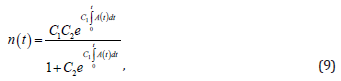
Where 2 C − is the integration constant, and the function

Constant 2 C is found from the initial condition n(0) = n0 . As a result

Substituting solution (9) into (7) now, we come to the following time dependence of the temporal behaviour of healthy cells:

As we can see from obtained solutions (9) and (12), the healthy and diseased cells behave quite adequately. Specifically, if the function is A(t ) = b(t ) −Gc (t ) > 0 , then the disease wins, because

In this case, the function A(t )must be such that condition (13) is fulfilled. As we can see from (9) and (12), in this case

And

At some point of “switching” function A(t ) = b(t ) −Gc (t ) changes sign, that is, it becomes negative ( A(t ) = b(t ) −Gc (t ) < 0 ) and we get the opposite picture. In this case, the disease recedes, and we come to equality

And healthy cells are restored, that is:

Now, as for the functions b(t ) andc (t ) . Due to the fact that the connection with the EM field has the form of a dipole interaction, then, based on purely physical considerations and the correct dimension of the equations, the form of these coefficients can only be as follows:


Where 1,2 a − the linear dimensions of diseased and healthy cells are, respectively, c S − is the contact area along the border line between them, and the dot above the letter means differentiation according to time.

Thus, function (10) of out interest, taking into account (1), (18) and (19), should be the following

Where β − is the angle between the direction of the external constant field and vectorE ′(t ) .

As we can see from (20), when changing the direction of the polarity of the fields, function A(t ) must change the sign to the opposite. By doing this, we achieve the opposite effect, that is, following the theory proposed above, there will have to be the destruction of diseased cells and the growth of healthy ones.

In this case, we may express function E′(t ) , for example, in the form of a linear time dependence, that is

Where γ − is a certain growth coefficient.

The Main Idea of the Work

The general physical considerations outlined above concerning possible methods of combating malignant tumours (and not only them, but many other similar diseases), allow, in our opinion, to select such variable fields and their amplitudes, which, in the process of reversal magnetization of their directions, will certainly lead to a positive therapeutic result.

Conclusion

Concluding this message, it is worth paying attention to two important points. The influence of variable EM fields on the reproduction of malignant tumours has been studied; It is shown that the proposed model quite adequately describes both the growth and disappearance of cancer cells, that is, the recovery of the body.

For more Articles on: https://biomedres01.blogspot.com/

Thursday, June 15, 2023

Invent a Removable Orthosis with the Ability to Prevent Foot Drop and Venous Thrombosis and Maintain Muscle Contraction


Invent a Removable Orthosis with the Ability to Prevent Foot Drop and Venous Thrombosis and Maintain Muscle Contraction

Introduction

From the perspective of the World Health Organization (WHO), health is the science and technology of disease prevention, providing medical services for immediate diagnosis, treatment and development to address problems. Safety is one of the most important aspects of health care systems [1]. Increasing the quality of work, consequently, increases the accuracy and improvement of the treatment process, the speed of the treatment process and also patient satisfaction [2]. Intensive care includes taking care of patients with life-threatening diseases, under the supervision of the most skilled personal, with advanced equipment and facilities, which includes all sensitive care related to the patient’s life [3,4]. A significant part of the treatment system’s effort is focused on being able to provide the best services to its patients in the shortest time and at the lowest cost. Intensive care units (ICUs) have an important place due to the high human and economic costs that they can incur for the health care system [5,6].

Patients with problems such as diabetic ketoacidosis, hypertensive emergency, non-accidental self-poisoning, heart failure, ischemic heart disease and cerebrovascular disease and respiratory conditions can be mentioned [7-9]. Numerous studies have been performed to identify the increasing mortality factors of patients admitted to the ICU. Infectious shock, age, smoking and nosocomial infections are among the factors that increase mortality in ICU wards [10,11]. A comprehensive study of UK hospitals showed that the mortality rate of patients admitted to ICU wards is about 20.6% [12] and that of Singapore public hospitals is 9.4% [13]. Complications of hospitalization in the Intensive Care Unit include foot drop, deep vein thrombosis[14], muscle atrophy [15] and so on. Venous thromboembolism (VTE), including deep vein thrombosis(DVT) and pulmonary thromboendarterectomy (PTE), is a public health problem that results in 250000 hospitalizations per year in the United States [16,17]. On the other hand, one hundred to fifty thousand people are hospitalized every year due to a benign and treatable disease, but die due to pulmonary embolism, while with prevention, the death of these patients can be prevented [18].

Risk factors for this disease are immobility, cancers, myocardial infarction, respiratory failure, surgery, trauma, obesity, use of female hormones and inherited coagulation disorders [19]. Another complication of hospitalization is “foot drop”. Foot drop or foot prolapse is a condition in which a person is unable to perform the dorsiflexion joint function properly due to weakness or paralysis of the anterior tibialis muscle or other muscles originating in the peroneal nerve. Foot drop can be unilateral or bilateral. Symptoms such as pain, weakness, and numbness are sometimes seen with this complication [20]. Diagnosis of this complication is easily possible by physical examination, but the use of imaging techniques and electromyography can also help to examine this complication more closely [21]. Today, there are various treatments to control foot drop, which considering the cause of this complication, the appropriate treatment method is adopted. Among the available treatments for prevention and correction of foot drop, physiotherapy, electrical stimulation and teaching the most common treatments using ankle foot orthosis [22].

Other complications of hospitalization in the intensive care unit include muscle atrophy [15], bed sores, etc. At present, according to the subject and results of research, one of the ways to prevent and reduce complications in hospital wards is to produce and use a mobile orthosis with the ability to prevent foot prolapse and venous thrombosis and maintain muscle contraction. In the following, we will talk about its structure. This idea has been registered in the Patent Organization of Iran under invention number 103769.

Methods

This project started with a new researcher idea Then by searching in the scientific content of articles and book, the title and proposal the project was compiled with opinion of the supervisor. Then model of the device was designed in collaboration with graphic & mechanical engineers Fianccy. The main tool was invented by the researcher. By mechanic engineer and solid work the design of this tool with professional software with academic-theorical proposal researchers (Mahmoudi and Mohammadbeik) about making a tool for the purpose of preventing foot drop, deformity of toes, muscles atrophy, improvement of blood return from distal to proximal organs and keep the function of vainvalves, assessment of vital signs and keep the power of foot solid muscles and prevention from muscels atrophy. This idea is designed by softwares and then the file of this designing was transferred to laboratory by researcher and work mechanic engineer in order to hardware designing and maquette. The structure of orthosis was discussed in terms of all angle’s morphology, measurement of figure based on standards and again after changing s figures and ,measurement was redesigned by software expert.

biomedres-openaccess-journal-bjstr

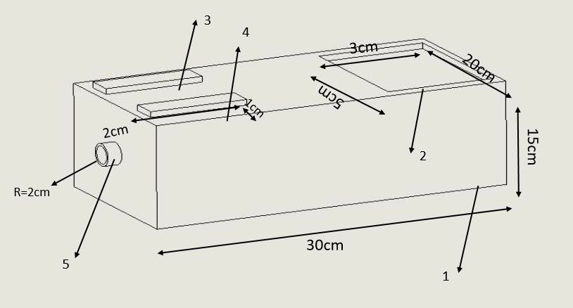
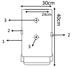
Figure 1: According to Figure 1

1. Orthotic body

2. Mechanical lock

3. Electric leads.

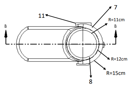
This mobile orthosis with the ability to prevent foot drop and keep the function of vain valve and assessing the vital signs is composed of (mechanical lock: 3×2cm, body: 40 cm length , pulse sensor: 0.5× 0.5 cm, pulse oximeter sensor: 0.5× 1 cm, inflatable inner layer R: 12cm, outer layer R: 15cm, electrical message transmitter leads: R: 1cm, insole: 25cm, removale hinge R: 2cm, connector between inner laye and air pump R:2cm. Orthosis is made up of upper and lower sections which after right locating(positioning) of foot in it, two sections will be connected to each other by mechanical lock (according to Figure 1, number 2). for motion of foot in its rang of motion (ROM), first removable hinge(according to Figure 7, number 11) is connected to monitor screen which is seprated from orthosis by a wire. all normative motion for foot’s motion(foot’s dorsal flexion, foot’s sole flexion,invertion , overtion) will be done by a program which is designed on screen and by choosing each motion, message will be transferred to removable hing(according to Figure 2, number 11) by a wire and by moving the orthosis, the foot moves in the desired direction and according to instruction given to program , the foot remains at desired posture for specified period of time (5s) and returns to its initial posture again.

biomedres-openaccess-journal-bjstr

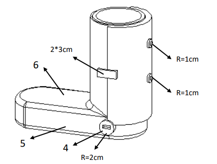
Figure 2: According to Figure 2

1. 7- outer layer of orthosis

2. 8- inner layer of orthosis

3. 11- Removable hinges.

biomedres-openaccess-journal-bjstr

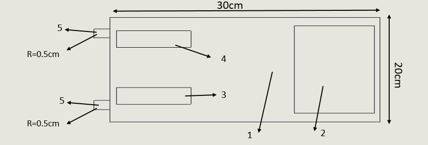
Figure 3: According to Figure 3

1. Air pump

2. LCD

3. Power on

4. Power off

5. Junction with interface.

biomedres-openaccess-journal-bjstr

Figure 4: According to Figure 4

1. 4- Connector

2. 5- Orthotics insole

3. 6-Upper part of orthosis.

biomedres-openaccess-journal-bjstr

Figure 5: According to Figure 5

1. Tens devices

2. LCD

3. Power on

4. Power off

5. Wire connection position.

Four foot motion (foot’s dorsal flexion, foot’s sole flexion, invertion and overtion) is done through a program which is programmed on the screen memory via removable hing, the duration of doing this motion, number of times, the amount of foot’s rest until next move. For preventing deep vein thrombosis (DVT) and maintaining the one way valve function, first the portable small air pump (according to Figure 3, number 1) which is available in the market is connected to the connector between air pump (according to Figure 4,number 4) and inflatable inner layer (according to Figure 2, number 8 ) by an air tube through transfering air from pump to inflatable inner layer the action of inflating and emptying takes place. Of course the number of times, intensity of wind pressure is defined by screen (according to Figure 4, number 6) available on pump’s body(according to Figure 3, number 1) and applied on foot, which this action causes standard pressure(17 mmhg) on one way valve and it causes continuity and helping blood circulation and prevents from the reduction of one way valve function.

biomedres-openaccess-journal-bjstr

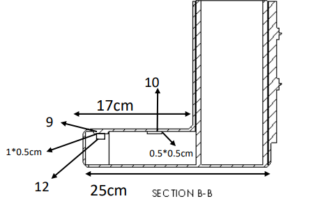
Figure 6: According to Figure 6

1. 9- pulse Oximeter sensor

2. 10- Pulse Sensor.

Also electrical leads (according to Figure 1, number 3) are connected to the desired position (according to Figure 5, number5) by a wire and after connecting to tennes machine (impulse maker machine) by making impulses causes electrical stimulation of muscles and prevents muscles failure and atrophy that voltage intensity, duration of muscle stimulation, number of stimulation are controlled and done by screen (according to Figure 5, number 2) and planning is done. According to location of the sensor in the upper part of orthosis(according to Figure 6 , number 10) number of dorsal pedis pulse is measured and shown on the screen. Also by placing pulse oximeter sensor on the upper part of orthosis (according to Figure 6, number9), the amount of oxygen saturation is shown on the screen. By making movable orthosis and due to structure and muscle electrical stimulation prevents reduction of muscle contraction, atrophy and foot drop and causes maintaining valve function and the amount of oxygen saturation is studied through dorsal pedis pulse sensor and pulse oximeter sensor and can prevent the accurance of many diseases and the death caused by them.

Discussion

This research has been done in the field of medicine and medical engineering. Due to the extent of the foot drop and Deep vein thrombosis when providing medical care to the drug, the use of preventive equipment is felt to prevent this problem. Due to the innovative design of this design, Used to prevent ankle foot drop, deformity of the toes, muscle atrophy, improve blood flow from the lower to the upper and improve blood flow from the lower to the upper and monitor vital signs monitor vital signs and maintain the strength of the leg muscles. The advantages of this plan include moving the foot in four directions (flexion of the back of the foot and flexion of the sole of the foot and inversion and oversight), the ability to plan the number and time of movements, check vital signs in the lower limb, prevent reduction of contraction Muscles, preventing blood stasis and thrombosis in the lower extremities, preventing atrophy and muscles mass. Also, according to the research of Esfandiari et al (2017), a study entitled Literature Review of the Effect of Ankle-Foot Orthosis on Gait Parameters After Stroke [23] and Alnajar et al (2020), with Title Advances in neuroprosthetic management of foot drop [24] and prenton et al (2018), with Title FUNCTIONAL ELECTRICAL STIMULATION AND ANKLE FOOT ORTHOSES PROVIDE EQUIVALENT THERAPEUTIC EFFECTS ON FOOT DROP [25], and this research, the use of preventive equipment to prevent Ankle foot drop, toe deformity, muscle atrophy, improving blood flow from the lower to the upper and maintaining pigeonhole valve function and monitoring vital signs and maintaining leg muscle strength is suggested.

Results

Results of this study and other surveys can be one of the most effective ways to prevent prevent ankle foot drop, deformity of the toes, muscle atrophy, improve blood flow from the lower to the upper and monitor vital signs and maintain the strength of the foot muscles. Also using orothesis can decrease it significantly. Besides, modifying the preventive policies such as producing instruments like Removable orthosis is recommended. We are ready to work with all institutions and individuals in the fields of economics, education, research and health and startups for this idea. If you wish, please contact us via masoudmahmoudi515@ymail.com email.

For more Articles on: https://biomedres01.blogspot.com/

Tuesday, June 13, 2023

Screening of Cervical Cancer in the Dakar Region by Cervico-Vaginal Smear: Epidemiological and Cytological Aspect


Screening of Cervical Cancer in the Dakar Region by Cervico-Vaginal Smear: Epidemiological and Cytological Aspect

Introduction

The cervical smear (CSF) or pap smear is a collection of cells from the cervix for the early detection of any cellular abnormality that may suggest precancerous or cancerous lesions of the uterus [1]. Cervical cancer is one of the most common cancers in the world, particularly in women. Indeed, it is the 3rd most common cancer in women worldwide and one of the most common in our regions [2]. More than 80% of uterine carcinomas are found in developing countries [3]. Cancerous and pre-cancerous lesions of the cervix are therefore a real public health problem in many African countries like Senegal [4]. Cervical cancer is preceded by a long latency phase characterised microscopically by a broad spectrum of events ranging from cellular atypia to various degrees of dysplasia or cervical intraepithelial neoplasia (CIN) before progression to invasive cancer.

Method

This is a retrospective study of 2391 cases of significant cervical smears performed in the period from 01 August 2014 to 01 June 2016. This work was carried out at the Laboratory of Cytology, Cytogenetics and Reproductive Biology of the University Hospital of Dantec in Dakar. First, patients were registered in a register with an identification number, surname, first name, age, origin and telephone number. The interview is carried out before the sample is taken and focuses on marital status as well as gynecological and obstetric complaints and history. The sample is taken from a woman in a gynecological position by inserting a suitable speculum which exposes the cervix then use a cytobrush to take the actual smear by scraping the ectocervix and then the endocervix. Finally, two slides marked exo and endo respectively and the patient’s ID number are spread out. The slides were then fixed with a fixative, dried and stained using the Papanicolaou method. After the staining step, the slides were mounted and then read under the microscope. We wrote the reports in the computer. We used the computer and the register for data processing and were able to collect cases of smears that could be interpreted. All women with inflammatory and atrophic smears of menopausal origin were excluded from the study. The data were stored and analysed in Excel.

Result

We collected 2391 files. The mean age of the patients was 47.72 years with a standard deviation of 11.37 years. The most represented age (mode) was 40 years. Referrals from level 1 and 2 hospitals were 65%. The rest were referred by private doctors’ surgeries, by the pension institute and by some clinics. Patients referred for routine check-up without apparent clinical manifestation were 43.16%. The other requests were due to various clinical manifestations: metrorrhagia, primary or secondary amenorrhea, pelvic pain, dysmenorrhea’s majority of patients (51%) were married in a monogamous union. single women accounted for 14% of the total. The remaining 35% were in polygamous households. Patients with intraepithelial lesions were 7.27% and of these highgrade lesions (HSIL) represented 2.63%.

Discussion

The systematic analysis of the results of our study has provided us with important information on the epidemiological data of cervico-vaginal smears in Senegal. Indeed, after 22 months of data collection, the average age of the patients in our series was 47.72 years with extremes between 15 and 88 years. In Ethiopia, Mesele and his team found the same average age in 2010 (47.7 years), after 6 months of study [5]. This age generally corresponds to the period of genital activity in most women. Epidemiological studies have shown a strong correlation between sexual age and certain infections, particularly HPV [6]. Patients were sometimes referred by various public (more than 65%) and private health structures. This can be explained by the relatively affordable cost of this examination in our center (less than 10 euros) compared to private facilities that perform the same examination. Our countries have very little universal health coverage, in contrast to several countries in the North, where the uptake of screening is greater but limited in some areas by disparities [7]. Patients living in the outskirts of Dakar were 53.47%.

The departments of Pikine and Guédiawaye account for more than 50% of the population of Dakar [8]. We therefore believe that increasing the number of public screening facilities in the suburbs of Dakar would probably increase adherence to routine screening. These results are in contrast to those obtained by Diallo and his team who showed 20 years ago that in the absence of functional urogenital signs, women rarely consulted health facilities in Senegal for early detection of cervical lesions [4]. In France, the Haute Autorité de Santé recommends systematic screening for precancerous and cancerous cervical lesions by cervico-vaginal smear every 3 years in women aged between 25 and 65 years [9]. We thus note that education, information and communication about cervical-uterine diseases are fundamental to their prevention. Of the patients screened who were in a monogamous union, 51% were in polygamous households compared to 35%. Polygamy is a major feature of matrimonial systems in sub-Saharan Africa. It is underpinned by cultural and religious perceptions [10].

Studies have shown a major preponderance of HPV infections in non-monogamous spouses, even in developed countries [5,6]. Our study also looked at the parity of women. Indeed, 8.67% of them had more than 5 children. High parity appears to be a very important risk factor for the occurrence of cervical cancer. Studies in Ethiopia, Costa Rica and Thailand found a higher incidence of cervical cancer in women who had more than five children. Weakening of the cervical mucosa, which reduces the spontaneous elimination of HPV, could explain this link. Other studies in Denmark and Manchester found no association between parity and cervical cancer incidence [5,11,12]. Patients with dyskaryotic cyto-morphological abnormalities of the cervical intraepithelial neoplastic type were 7.27% with 4.64% low-grade intraepithelial lesions (LSIL) and 2.63% high-grade intraepithelial lesions (HSIL). In 20 years, we have seen a very significant reduction in the rates of dysplastic lesions, which were estimated to be 20.54%, of which 17.56% were low Bethesda smears and 3.36% were high-grade smears [4]. Studies have shown that 12% of LSIL can develop into invasive cancer, compared with 1% of HSIL [13]. However, only histology after colposcopy of the lesions could confirm its cytological abnormalities.

Conclusion

Thanks to the work undertaken by Papanicolaou since 1928, gynecological cytology has proved to be of prime importance in the fight against and treatment of dysplastic lesions, and in raising the awareness of health workers, government officials and the education of women in Senegal.


For more Articles on: https://biomedres01.blogspot.com/

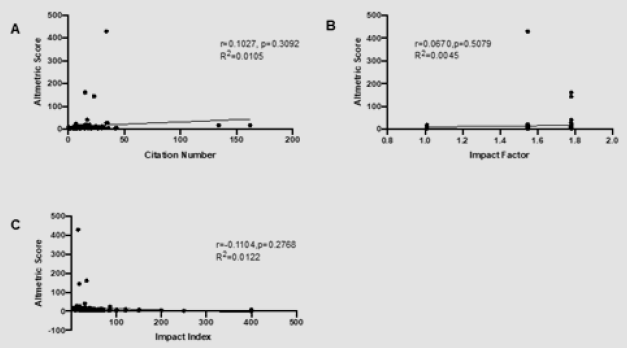
The Correlation between Altmetric Score and Citations in Pediatric Orthopaedic Journal Articles

  The Correlation between Altmetric Score and Citations in Pediatric Orthopaedic Journal Articles Introduction In scientific research, it is...