Thursday, July 30, 2026

Awareness and Attitudes to COVID-19 Vaccines: A Cross-Sectional Study


Awareness and Attitudes to COVID-19 Vaccines: A Cross-Sectional Study

Introduction

The virus SARS-CoV-2 is genetically related to the previous generation of coronaviruses causing the SARS epidemic in 2003 [1]. The challenges created by COVID-19 have affected the wellbeing of all individuals in all communities irrespective of rich-poor, literate illiterate, rural-urban directly or indirectly [2-4]. Preventive measures such as physical distancing, avoiding social gatherings, enforcing masks as mandatory, hand sanitizing, and many others have become a daily routine from the beginning of national-wide lockdown. How- ever, the impact of the second wave has brought the importance of vaccination to the fore [5]. Since the emergence of a new epidemic, the whole human community anticipated effective pharmaceutical management either as medication or vaccine. Globally, more than 15 vaccines have been approved, and many have yet to prove their efficacy in trials. Despite the Government efforts, the hesitancy towards vaccines by the general public is concerning [6-7].

In general, vaccine preparation requires many years, while the fast-tracking of the vaccines against COVID-19 raised concerns among the public regarding vaccine safety and efficacy. The theory of planned behavior suggests that every per- son with particular behavior in taking the COVID-19 vaccine would be influenced by major factors such as an individual’s attitude towards a vaccine and perceived behavioral control regarding taking the vaccine [5]. Vaccines have been the most effective and reliable public health intervention for decades, saving millions of people from deadly infectious diseases [8]. Vaccination is one of the most effective ways to help reduce and eliminate viral infection and its spread [9]. Since the beginning of the Universal Immunization Program (UIP) [6]. Even the best vaccine against any infection may go unfruitful if it is less used or unused [9].

Therefore, the aim of this study was to investigate the acceptance of COVID-19 vaccines and their determinants, as well as public perceptions of these vaccines in Sindh population. General participants from Sindh were surveyed on the acceptability of COVID-19 vaccines using a cross-sectional and self-administered questionnaire.

Materials and Methods

The aim of the cross-sectional study is to assess the possibility of evaluating the levels of healthiness literateness assistances about vaccination in the Sindh general population; through a questionnaire from October to December 2021 a total of 500 participants shall complete the survey. The attitudes towards COVID-19 vaccination were collected via questionnaire and analyzed using descriptive and inferential statistics. The questionnaire were prepared, distributed, and collected by ‘based surveys and shared through other services. The sampling approach was convenient and nonprobability. Participants were given complete freedom to complete the survey and were encouraged to forward and share it with others. On the first page, participants were given information on the survey’s rationale and scope, which included gathering perceptions as well as methods and abilities for collecting, understanding, and using information about vaccination, including possible COVID-19 vaccines. They were assured that moving on to the second page of the survey and filling out the questionnaire meant they had given their consent.

Statistical Analysis

Statistical analysis was performed by using SPSS (version 20, SPSS, Chicago, IL, USA).

Results

biomedres-openaccess-journal-bjstr

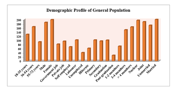
Figure 1: Demographic Profile of General Population.

biomedres-openaccess-journal-bjstr

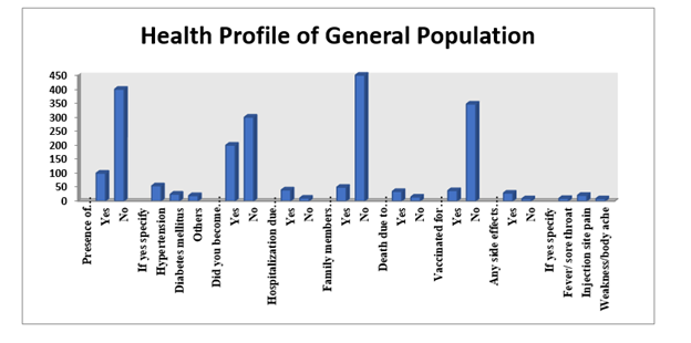
Figure 2: Health Profile of General Population.

biomedres-openaccess-journal-bjstr

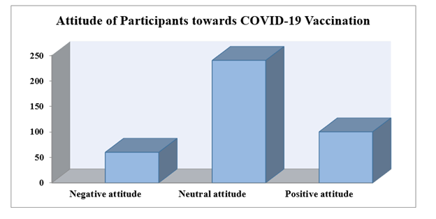
Figure 3: Attitude of Participants towards COVID-19 Vaccination.

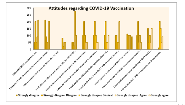
The sample size consisted of 500. More than half of the participants were female. Comorbidities were reported in the general population (Figures 1 & 2). Many participants had a neutral to positive attitude, and very few had a negative attitude towards COVID-19 vaccination (Figure 3); COVID-19 vaccination and mortality among friends and relatives were significantly associated with their attitude towards COVID-19 vaccination. However, participant occupation and number of family members were significantly associated with their attitude towards COVID-19 vaccination. It was also noted that the presence of comorbidity and hospitalization history was not associated with participants’ attitudes from the general population (Figure 4). Almost one-third of participants strongly agreed regarding the safety and efficacy of COVID-19 vaccination. Furthermore, more than half of the participants strongly agreed that the pharmaceutical companies’ rules and regulations in manufacturing the COVID-19 vaccination as per the government norms. However, only about a third of participants agreed to advise their relatives and friends to take the COVID-19 vaccination. Furthermore, less than a third of reported neutral behavior towards the effectiveness of COVID-19 vaccination in preventing virus mutation, while a majority of participants demonstrated a neutral attitude towards the side effects of COVID-19 vaccination on their pre-existing disease conditions (Figure 4).

biomedres-openaccess-journal-bjstr

Figure 4: Attitudes regarding COVID-19 Vaccination.

Discussion

In this cross-sectional study, most participants had a neutral attitude towards COVID-19 vaccination in the general population. The present study findings suggest that the general population shows some hesitancy to-wards the COVID-19 vaccination drive. A global survey study involving 13,426 participants from 19 countries targeting the acceptance of COVID 19 vaccinations in the general population reported China with the highest (88.6%) and lowest (54.8%) in Russia [10]. Moreover, middle-income countries, such as Brazil and South Africa, also show positive public acceptance [11]. How- ever, vaccine acceptance is more or less in harmony with the initial planning in developing countries [12] An Ethiopian study reported that one-fourth of participants (24.2%) had a positive attitude towards COVID-19 vaccination, and around (40.8%) respondents were aware of COVID-19 vaccination [13]. A similar study from Jordan revealed that less than half (37.4%) of respondents showed a positive attitude towards COVID-19 vaccination, and around (26.3%) of respondents are still unsure about vaccination.

The main concern of the general public refused to take vaccination fearing of side-effects of newly launched vaccines against COVID-19 but agreed to take after the licensing of pharmaceutical companies with the proper establishment of favorable effects of vaccines [14]. A study from Bangladesh revealed that more than half (74.5%) of the general population showed a positive attitude towards COVID-19 vaccination with a mean attitude score of 9.34 (2.39), and quite a few (8.5%) still showed some amount of hesitancy towards vaccination. It was more amongst the geriatric population, low literacy level, comorbidities, and less confidence in its healthcare system [15]. In the United Kingdom, it was found that only a few respondents exhibited high levels of uncertainty about vaccines and had a negative attitude towards COVID-19 vaccination, it was seen higher among individuals from ethnic groups, education level, monthly income, and poor knowledge regarding the high level of mutation of this deadly disease among the general population [16].

Another study from Malta reported that half of the participants had a positive attitude towards COVID-19 vaccination and were willing to take the vaccination. Vaccine hesitancy was a major setback in public opinion as one-third of participants were still in a dilemma towards vaccination, and some of them were not in favor of COVID-19 vaccination, and they refused to take it even after robust safety trials [5] The result was incongruent with the study done on the general population of Pakistan and found that most of the respondents showed a positive attitude towards vaccination and are willing for COVID-19 vaccination as soon as their chance will come and agreed to recommend their family and friends [17]. The present study results also suggested no relation of demographic variables with attitude scores. However, there is a significant association of the history of COVID-19 positive status in family and friends. Another study showed that participants more than 45 years of age and socio-economic status were significantly associated with attitude scores.

The willingness to pay for the vaccine was also significantly positively associated with socio-economic status, and the willingness to recommend the vaccine to family and friends was found to be significantly associated with place of residence [17]. A study done in Kuwait showed a significant association of gender with attitude scores as the male population was more willing to accept a COVID-19 vaccine than females and participants who previously received an influenza vaccine were more likely to accept a COVID-19 vaccine. In contrast, participants who were suffering from comorbidities were less willing to accept vaccination [18]. During the initial stages of the pandemic, the rural areas were the least affected compared to urban sectors. However, in the second wave, there was a significant rise in rural areas. The fundamental evidence for concern on vaccine drive is logistical constraints such as poor infrastructure, unskilled workers, and the lack of resources [19].

The survey was conducted when vaccination phase II started in the general population aged above 45 years; resulting uncertainty was more prevalent younger age groups. The vaccine motivation campaign was not active during the data collection period affecting the study findings. The current study’s recommendations suggest that a community-focused approach is required to deal with people’s mentality and mindset. Furthermore, the findings recommend interventional studies compared to rural and urban to attain more accuracy in the results.

Conclusion

The COVID-19 pandemic was, in some aspects, the worst pandemic in history, causing substantial mortality and morbidity rates, but the introduction of the COVID-19 vaccine offered a ray of hope for a better future. Negative attitudes towards vaccination and hesitancy or unwillingness regarding vaccination are the major concerns that need to be addressed. People in Pakistan currently have mainly neutral attitude regarding vaccination, requiring more authentic, reliable, and adequate information to assist them in decision-making. Positive attitudes and perceived usefulness of vaccination in the general population is crucial for a successful vaccination plan and prevention of new epidemics waves in the future.


Follow us for more Articles on: https://biomedres01.blogspot.com/

https://scholar.google.com/citations?user=OFtUOZYAAAAJ&hl=en
https://www.base-search.net/Search/Results?lookfor=Biomed+Journal+of+Scientific+%26+Technical+Research&l=en&refid=dcsuggesten
https://citefactor.org/journal/2574-1241/biomedical-journal-of-scientific-technical-research
https://ideas.repec.org/s/abf/journl.html
https://econpapers.repec.org/article/abfjournl/
https://www.ncbi.nlm.nih.gov/nlmcatalog/101723284
https://bjstr.org/
https://bjstr.org/about.php
https://biomedres.us/reprints.php
https://biomedres.net/submit-manuscript.php
https://independent.academia.edu/BJSTRAngelaRoy
Wednesday, July 29, 2026

Synthesis of a Coordination Polymer of Cu (II), FTIR Spectrum, Single- Crystal X-Ray Crystallography, HOMO – LUMO Analysis, MEP and Biological Investigation In Vitro and Molecular Docking


Synthesis of a Coordination Polymer of Cu (II), FTIR Spectrum, Single- Crystal X-Ray Crystallography, HOMO – LUMO Analysis, MEP and Biological Investigation In Vitro and Molecular Docking

Introduction

Metal-organic polymers are one of the most interesting materials [1]. These compounds have different properties and can be applied as a catalyst in coupling reactions [2], pollution control [3], and anionic polyelectrolyte hydrogels [4]. Polymers supported metals are a significant group of polymers that consist of the coordination of polymeric ligands with transition metal ions [5]. Nowadays, the polymer-supported metal is synthesized with modified structures as various monomers and variant properties [6]. This modified polymer as a ligand has high chemical stability due to its different properties and interesting behavior and is very important for the composition of metal ions. These compounds with hydrophilic properties were investigated in a polymer containing immunodiastates [7]. Polymer-copper (II) complex was studied as a catalyzed conversion of aldehydes to primary amides [8]. These polymers have been applied with various metals such as poly (maleic acid) poly metal complexes with transition metals just as Cu (II), Co (II), Ni (II), and Zn (II) [9]. It is attributed to the formation of stable mononuclear copper (II) complexes with hydroxyl groups of Poly (acrylic acid) is also studied [10,11]. Poly [4-Imino (N-4- ethyl benzoate) benzene p-styrenesulphonate] and the ability of the polymer to release the drug have been investigated [12]. These results prompted us to synthesize and evaluate (1) in a new method. In our previous research, we reported 4-acetylaminobenzene -p-styrene sulphonate and its polymer as a prodrug in controlled release technique [13] and polymer-supported amino acid for alkylation of arenes [14]. On the other hand, metal complexes have special properties such as antimicrobial effects and biological activity [15,16]. Copper is a necessary element in the human body that is a noticed cofactor for many enzymes and extracellular proteins. The deficiency of copper ions in the human body has an effect on the production of white blood cells. In adult humans, the net absorption of dietary copper is approximately 1 mg/d [17]. Copper has long been known to have antimicrobial activity and is used in water drinking refineries. In 2008, it has been recognized by the US Environmental Protection Agency as the first metallic antimicrobial agent. Antimicrobial applications of copper have been reported [18]. Recently, the synthesis of metal-based drugs has been considered. Then, studying the interaction of metal complexes and HSA is very attractive for researchers. In this work, we investigated the interaction of monomeric (1) with HSA and 2JVU using molecular docking methods to evaluate the medicinal properties of this compound as a biocompatible material and an antibacterial agent.

Experimental

Material and Methods

Pyridine-2, 6-dicarboxylic acid, NaOH, Cu (NO3)2⋅3H2O were obtained from commercial sources and used as received. The water was distilled and deionized. Elemental analyses (C, H, and N) were measured on Perkin-Elmer 2400 II elemental analyzer. The FT-IR spectrum was measured as KBr discs in the region of 4000-400 cm-1 on a Jasco FT/IR-430 spectrometer. Crystallography data and structure refinement of (1) were collected on a STOE-IPDS 2T diffractometer equipped with a graphite monochromator and Mo Kα radiation (λ=0.71069 Ã…)

Preparation of [Cu (dipic) (H2O)2]n (1)

dipic is an abbreviation pyridine-2, 6-dicarboxylic acid (0.084 g, 0.5 mmol) was deprotonated using the aqueous solution (5 mL) of NaOH (0.04 g, 1 mmol) and stirred for 20 min, at room temperature. A high-density ultrasonic probe with a power of 80 W was immersed directly into the above solution. Then, an aqueous solution of Cu (NO3)2⋅3H2O (0. 121 g, 0.5 mmol) was added dropwise to this solution. The product was sonicated for 30 min at room temperature. After the solution was slowly evaporated and blue crystals of 1, was appeared. This mixture was filtered and washed with ethanol (3 × 10 mL). This crystal was dried in the air. Then, Single crystals for X-ray crystallography were obtained (Yield 3.72 g, 54.0%). IR (KBr, Cm-1): 3595(νH2Oas), 3080(νCH),3058(νCH), 1682(νC=O), 1588(νC=O), 1574(σH2O), 1304(νΦ + δCH), 1241(νPh), 1122(νPh), 1117(δCH + δ Ph), 1113( δ Ph + δCH), 1044(δCH + δ Ph ), 1003(δCH + δ Ph ), 991(δCH + δ Ph ), 924(γCH), Elemental analysis Calcd (%) for (1), C7H7CuNO6: C, 31.72; H, 2.64; N, 5.72. Found: C, 31.64; H, 2.70; N, 5.65. Other methods are described in Ref [19].

Computational Details

The monomer of (1) was calculated using Gaussian 09 software [20]. The computation including geometry optimization (optfreq), MEP, FT-IR spectrum, and HOMO – LUMO analysis were applied using B3LYP/6-311G (2d, 2p) [21]. A modeling and docking tool was performed to predict the activity of the monomer [22]. As mentioned before, the monomer of (1) as a compound was optimized by using the B3LYP method 6-311G (2d, 2 p) basis set and prepared for docking. The structure of Escherichia Coli and HAS (human serum albumin) were obtained from Protein Data Bank, respectively, (PDB ID: 2jvu, 1AO6). Auto Dock software is a new tool to get an insight into ligand-receptor interaction and screen molecules for the binding affinities against a receptor. These molecular docking computations were performed on Auto Dock software. The most popular algorithm, the Lamarckian Genetic Algorithm (LGA), in Auto Dock for this docking was employed [23,24].

Antibacterial Assays

The minimal inhibitory concentration (MIC) is determined by preparing solutions of the chemical in vitro at increasing concentrations, incubating the solutions with separate batches of cultured bacteria, and measuring the results using agar dilution. It is used to measure the Minimum Inhibitory Concentration [MIC] of an antimicrobial agent, which is the lowest concentration of antimicrobial agent which will prevent the growth of microbes. The MIC of the (1) as an antibacterial agent against Escherichia coli will be reported in the next section. The (1) was tested for its antibacterial activity Escherichia coli as Gram-negative bacteria by the disk diffusion method [25].

Preparation of Nutrient-Agar Medium

For this work, 3.80 g of Nutrient-Agar (NA) medium was dissolved in 100 mL of distilled water. This solution was sterilized at 120 °C for 20 min in an autoclave. Then, 20 mL of this solution was solidified in a Petri plate.

Result and Discussion

Synthesis and Characterization

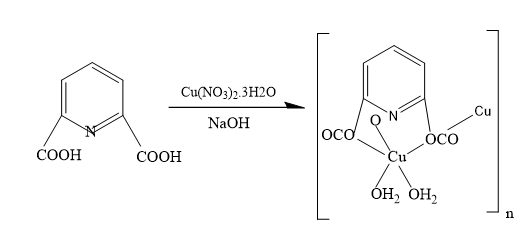
The synthetic route for the preparation of (1), as explained in the experimental section, is schematically represented in Scheme 1.

Molecular Structural

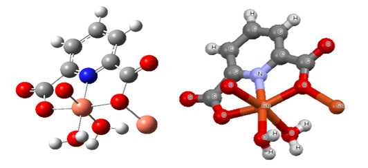
The calculated optimized geometry of monomer (opt-freq) using B3LYP/6-311G (2d, 2p) of monomer and crystal structure are shown in Figure 1.

Spectroscopic Characterization of (1)

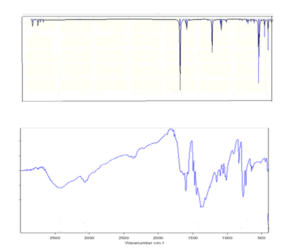
The experimental FT- IR of (1) is reported in the experimental section (Figure 2). The C–H stretching frequencies of aromatic can be observed in the range of 3100–3000 cm-1 is shown at 3106 cm- 1. The C= C stretching vibration in the range of 1650-1430 cm-1 and the C–H bending bands have appeared in the regions 1275-1000 cm-1 (in-plane C-H bend) and 900-690 cm-1 (out-of-plane C-H bend). In the following discussion.

Molecular Electrostatic Potential

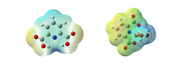
Predicting electrophilic and nucleophilic attacks for biological interactions by using MEP (Molecular electrostatic potential) as an important tool was shown [26,27]. The geometry of dipic (pyridine-2, 6-dicarboxylic acid) and monomer of (1) were optimized using the B3LYP method 6-311G (2d, 2p). As in Figure 3 can be observed, the different colors are indicated different values of the electrophilic and nucleophilic potential. Red< orange < yellow < green< blue. The blue illustrates the strongest attraction, and this area (positive area) is located around CH groups. The negative area (red color) is related to C=O and O-H groups over the electronegative oxygen atoms. These regions of negative potential are associated with the lone pair of electronegative atoms. The nitrogen of pyridine and oxygen of hydroxyl anion is coordinated to Cu and lead to form (1).

biomedres-openaccess-journal-bjstr

Scheme 1: The reaction involving synthesis of (1).

biomedres-openaccess-journal-bjstr

Figure 1: Calculated optimized geometry of monomer (in the left) and its Crystal structure (in the right).

biomedres-openaccess-journal-bjstr

Figure 2: The experimental FT-IR spectra of (1) (bottom) and theoretical FT-IR spectra of the monomer (top).

biomedres-openaccess-journal-bjstr

Figure 3: MEP plot dipic (in the left) and monomer of (1) (in the right).

Frontier Molecular Orbital Analysis

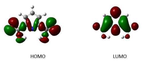
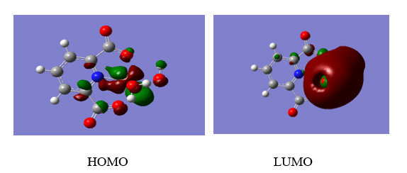
Analyzing the highest occupied molecular orbital (HOMO) and lowest unoccupied molecular orbital (LUMO) is important in a molecule as a compound. Soft systems are large and highly polarizable, while hard systems are relatively small and much less polarizable. For the comprehension of various aspects of drug design and their properties, several new chemical reactivity descriptors have been proposed [28]. The LUMO energy explains the ability to accept an electron and the HOMO energy is related to the ability to donate an electron. Both the HOMO and the LUMO, play a significant role in the electrical properties and chemical activities in the compound [29,30]. The HOMO and the LUMO orbital energy are important parameters to predict the chemical properties of a compound. The HOMO and the LUMO orbital energy of dipic (2) and an aqueous solution of [Cu (dipic) (H2O)2] (3) are calculated at the B3LYP method 6-311G (2d, 2p) basis set. The energy values (2) and (3) are, EHOMO-1 = -0.107 and -0.155 EHOMO = - 0.369 and -0.265 eV, respectively. The energy difference (gap) between the HOMO and the LUMO is 5.796 and 3.468 eV, for (2) and (3), respectively. These energies of HOMO and the LUMO orbitals of compounds are negative showing that these compounds are stable and do not decompose spontaneously into their elements. According to Parr, et al. [31]. A molecule with a high energy gap is less polarizable and is termed a hard molecule. The softness (strongly polarizable) can be explained by the deformation of electron cloud and polarization of chemical systems during the chemical process. By using the HOMO and the LUMO orbital energies, the ionization energy and electron affinity can be calculated as: I = - EHOMO =0.107 and 0.155 eV for (2) and (3), respectively, and A = - ELUMO = 5.796 and 3.468eV for (2) and (3), respectively. The global hardness η and chemical potential μ are given by using the relation η = 2.898 and 1.734 eV and μ =5.502 and 5.483 eV global electrophilicity = μ 2/2 η = 5.226 and 8.668 eV for (2) and (3), respectively. The atomic orbital components of the frontier molecular orbital are shown in Figures 4 & 5.

biomedres-openaccess-journal-bjstr

Figure 4: HOMO and LUMO plots of Pyridine-2, 6-dicarboxylic acid (2).

biomedres-openaccess-journal-bjstr

Figure 5: HOMO and LUMO plots of the monomer (3).

The Disk Diffusion Method

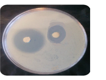
A microbial suspension (1 mL) of Escherichia coli was spread over the surface of an agar plate, which was incubated for 24 h at 37°C in an autoclave. Inhibitory zone values (diameter of inhibition) from disk diffusion tests and growth inhibition ring for cu complex were reported in Table 1 and Figure 6.

biomedres-openaccess-journal-bjstr

Table 1: Inhibitory zone values (diameter of inhibition) from disk diffusion tests.

biomedres-openaccess-journal-bjstr

Figure 6: The growth inhibition ring observed for ligand in E. coli.

Molecular Docking Studies

biomedres-openaccess-journal-bjstr

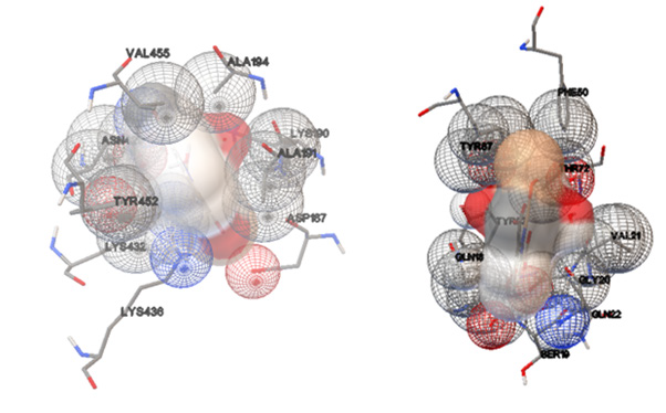
Figure 7: Left) View of the HSA binding site for (1) and its selected cluster. Right) View of the 2JVU binding site for (1) and its selected cluster.

The study of molecular docking is a significant tool for the prediction of the ligand-receptor interactions [32] and was performed to indicate the HSA and 2JVU-binding site for the cu complex. The crystal structures of HSA and Escherichia coli (PDB ID: 1AO6, 2JVU) were taken from the Protein Data Bank, respectively. These crystals were prepared for the docking system. Flexible ligand docking was carried out by Auto Dock 4.2.5.1 molecular docking program using the implemented empirical free energy function and the Lamarckian Genetic Algorithm. We decided to show this compound has an antimicrobial property in the human body. The cu complex as a ligand was prepared for docking by using the B3LYP method 6-311G (2p, 2d) basis set. In the first step, the docking of the cu complex with 1AO6 and 2JVU, a blind docking with 126 lattice points along X, Y, and Z axes was performed to find the binding site of the complex on these crystals with a grid point spacing of 0.375 Ã…, to allow the complex to rotate freely. In the next step, the second docking was performed using a cubic box with 60×60×60 Ã… dimensions. Among the docked conformations, the best-scored conformation predicted by the Auto Dock scoring function was visualized for complex-HSA and complex-2JVU interactions in Auto Dock software. The resulting docking for the obtained molecular docking in which the Cu complex binds into HSA and 2JVU, as a receptor is illustrated in Figure 7. The HSA creates one hydrogen bond with the (1), which includes LYS436. The 2JVU forms one hydrogen bond, which is UNK1. There are hydrophobic contacts between Cu complex with HSA (VAL 455, ALA194, LYS190, ALA191, ASP187, LYS436, LYS432, TYR452, ASN429) and 2JVU (TYR87, PHE50, GLN18, THR72, VAL21, TYR23, SER19, GLN22, GLY20), respectively. The binding free energies (ΔG°) of -6.55and -5.94 kcal mol-1 were predicted for HSA and 2JVU in the best conformation of the ligand.

Conclusion

We report that (1) as a new polymer-supported metal has been synthesized for the first time from the reaction of Pyridine-2, 6-dicarboxylic acid, and Cu (NO3)2⋅3H2O solution under ultrasonic irradiation in the mild condition. (1) was confirmed using FTIR spectrum and single-crystal X-ray crystallography. Molecular electrostatic potential and frontier molecular orbital analysis were indicated polymer-supported cu due to special property that can be designed for a new drug. This study leads us to the conclusion that this polymer has antibacterial properties. These results of biological research confirmed that (1) can be used to design and synthesize new based-drug materials.


Follow us for more Articles on: https://biomedres01.blogspot.com/

https://scholar.google.com/citations?user=OFtUOZYAAAAJ&hl=en
https://www.base-search.net/Search/Results?lookfor=Biomed+Journal+of+Scientific+%26+Technical+Research&l=en&refid=dcsuggesten
https://citefactor.org/journal/2574-1241/biomedical-journal-of-scientific-technical-research
https://ideas.repec.org/s/abf/journl.html
https://econpapers.repec.org/article/abfjournl/
https://www.ncbi.nlm.nih.gov/nlmcatalog/101723284
https://bjstr.org/
https://bjstr.org/about.php
https://biomedres.us/reprints.php
https://biomedres.net/submit-manuscript.php
https://independent.academia.edu/BJSTRAngelaRoy
Rare Case of Streptococcal Meningitis in a Child with Sars Cov2 Infection


Rare Case of Streptococcal Meningitis in a Child with Sars Cov2 Infection

Introduction

Group A Beta-hemolytic Streptococcus remains a rare cause of childhood meningitis (0.2-1% of all bacterial meningitis) despite the increasing trend of invasive streptococcal infections in recent years [1,2]. From the experience of the 3-year pandemic caused by SARSCoV- 2, it is well known that there are varying degrees of neurological damage in a strictly viral context in some patients, but data in the literature are still insufficient to explain the pathophysiology of the phenomenon. However, similar to influenza viruses, it causes a certain degree of immunosuppression that may predispose previously healthy patients to contracting bacterial infections. In November 2020, the male child B.A., aged 13, with no known personal pathological antecedents, was admitted to the Clinical University Infectious Diseases Hospital “Saint Parascheva” from Iasi, being transferred from “Saint Mary” Pediatric Hospital in Iasi, for fever (38 degrees C), nausea, food vomiting, frontal headache, colicky abdominal pain and semi-constant diarrheal stools, in the context of a positive SARS-COV-2 RT-PCR test. The objective clinical examination at admission showed a moderately influenced general condition, afebrile, circling facies, sabural tongue, tachycardia, absence of meningeal contraction syndrome, the clinical picture being dominated by frontal headache and semi-constant stools. The paraclinical investigations objectified the presence of the biological syndrome of inflammation and leukocytosis with neutrophilia, without other major changes. Twelve hours after admission, the patient’s condition worsens, the fever reappears, the patient presents drowsiness, bradylalia, bradypsychia, psychomotor agitation, complains of photophobia and develops a motor deficit in the left upper limb. Upon resuming the clinical examination, the presence of neck stiffness and the Kernig-1 sign is noted, for which reason a lumbar puncture is performed, with the extraction of an opalescent, hypertensive cerebrospinal fluid, with increased cellularity (240 ecn/mmc) and negative latex agglutination (Table 1).

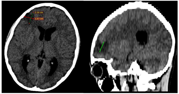
Also, it was extremely important for the therapeutic orientation to exclude a meningeal injury caused by the SARS COV2 virus (PCR RNA SARS COV 2 negative in CSF). Empiric intravenous antibiotic therapy with Ceftriaxone 3g/24 hours, drugs against cerebral edema, dexamethasone and B-group vitamin therapy are initiated. The evolution under treatment is unfavourable and on the 3rd day of hospitalization the child presents tonic-clonic convulsions in the left hemibody and peripheral paresis of left facial nerve, for which neurological and neurosurgical consultation as well as a cranio cerebral computer tomography with contrast substance are requested. The CT showed moderate cerebral edema, hydrocephalus and the presence of a hypo-dense area located on the right frontal lobe with a diameter of 27 mm, interpreted as a brain abscess in the pre-suppurative phase (Figure 1). Antibiotic therapy is escalated to Meropenem 3g/24 hours in association with Vancomycin 1g/24 hours, administrated intravenously. The following day, the result of the CSF cultures confirms the etiology of the meningoencephalitis, by isolating the Group A Beta hemolytic Streptococcus. As a result, we decided to gradually deescalate the antibiotic treatment with Ceftriaxone to a total duration of 28 days. The clinical and paraclinical evolution was slowly favourable, with remission of the fever, the convulsive syndrome, the peripheral paresis of the left facial nerve, as well as the complete recovery of the upper limb mobility. The follow-up cerebral CT performed 30 days after the first one confirms the regression of the frontal abscess, the hydrocephalus and the cerebral edema.

biomedres-openaccess-journal-bjstr

Figure 1: Cerebral CT-scan.

biomedres-openaccess-journal-bjstr

Table 1: Cytological and biochemical examination of CSF in evolution.

Discussion

The SARS COV 2 infection recorded a benign evolution in children, in most cases [3,4]. However, if we refer to the prognostic significance of a viral infection that occurs during an epidemic/ pandemic in a previously healthy organism, we can state that virally induced anergy is the explanatory key to the septic complications deriving thereof. Thus, Raul O. Ruvinsky et al. mentions, in a 2020 study, that the invasive infection with SBHA is favored by the coinfection with the influenza virus [2]. In the case presented, the nasopharyngeal exudate for influenza viruses was negative, but the presence of the infection with the SARS-CoV-2 virus seems to have been the predisposing factor. However, the medium or short-term impact of this virus on immunity is not fully known.

Apart from the correlations mentioned above, Streptococcus pyogenes is not per se a frequent cause of meningitis in children. In a retrospective study from the English literature carried out in 2005, a number of 25 cases registered during 25 years is mentioned [3]. Another study shows that meningitis caused by Streptococcus pyogenes usually occurs through dissemination from a neighbourhood outbreak [2].

Thus, the apparent absence of a portal of entry in the case discussed is a particularity of the case worth mentioning. Cases of streptococcal co-infection (pharyngitis, sepsis) in SARS-CoV-2 positive patients, but not meningitis with S. pyogenes in children evolving to brain abscess, have been described in recent literature [4]. So far, in our hospital there have been no cases of association of SARS-CoV-2 infection to bacterial meningitis.

Despite the early institution of first-line antibiotic therapy (which later proved to be targeted and in accordance with the results of the antibiogram) [5], the initial evolution is unfavourable, with the formation of a brain abscess in a short time. Thus, the escalation of antibiotic therapy was necessary over a period of 7 days, even after confirmation of the antibiotic susceptability of the etiological agent [6]. However, the adequate therapeutic response of the patient, as well as the absence of surgical indications during hospitalization, indicate that the medical intervention was prompt, early in the abscess formation phase. Also, the reduced initial inflammatory reaction for a bacterial meningitis constitutes an additional argumentative element in support of this hypothesis [7].

Conclusion

The association between COVID-19 and CNS infections remains a rarity, especially in pediatric patients. The case presented is not an edifying one in terms of the potential severity of the SARS COV 2 infection, but rather in outlining the role of a turntable for the bacterial superinfections that can evolve in this context. Although it is a rare cause of meningitis in children, the mortality rate in the case of meningitis caused by Group A Betahemolytic Streptococcus is high. However, provided early diagnosis is established and the microorganism’s susceptibility to beta-lactam antibiotics (especially to Penicillin) is preserved, the prognosis tends to be favourable.


Follow us for more Articles on: https://biomedres01.blogspot.com/

https://scholar.google.com/citations?user=OFtUOZYAAAAJ&hl=en
https://www.base-search.net/Search/Results?lookfor=Biomed+Journal+of+Scientific+%26+Technical+Research&l=en&refid=dcsuggesten
https://citefactor.org/journal/2574-1241/biomedical-journal-of-scientific-technical-research
https://ideas.repec.org/s/abf/journl.html
https://econpapers.repec.org/article/abfjournl/
https://www.ncbi.nlm.nih.gov/nlmcatalog/101723284
https://bjstr.org/
https://bjstr.org/about.php
https://biomedres.us/reprints.php
https://biomedres.net/submit-manuscript.php
https://independent.academia.edu/BJSTRAngelaRoy
Monday, July 27, 2026

Bridging the Gap Between COVID-19 and Subarachnoid Hemorrhage: A Case Report and Literature Review


Bridging the Gap Between COVID-19 and Subarachnoid Hemorrhage: A Case Report and Literature Review

Introduction

The novel coronavirus was reported in December 2019 during a pneumonia epidemic in Wuhan, China. It surged rapidly and the World Health Organization designated it a pandemic on March 11, 2020 [1]. COVID-19 was the term given to the disease induced by the virus known as severe acute respiratory syndrome coronavirus 2 (SARS-CoV-2). Over 559 million confirmed cases and 6.3 million fatalities had been recorded globally as of July 17, 2022 [2]. SARSCoV- 2 infects host cells via Angiotensin Converting Enzyme-2 (ACE-2) receptors, ACE-2 is found on the apical membranes of polarized cells of the testis, cardiovascular epithelium, cardiac myocytes, cardiac fibroblasts, kidney, liver, intestine, brain, and lung epithelial cells, resulting in coronavirus disease (COVID-19)- related pneumonia as well as acute myocardial injury and long-term cardiovascular damage [3,4]. SARS-CoV-2 can potentially infect the nervous system, skeletal muscles, and respiratory tract. Neurologic involvement is increased in patients with severe infections, including acute cerebrovascular disorders, altered consciousness, and skeletal muscle damage [5]. The probable processes behind covid-19 involvement, stroke, and subarachnoid hemorrhage (SAH) are yet unknown. However, according to numerous studies, covid-19 can increase intracranial pressure, increasing the likelihood of an intracranial aneurysm [6]. At the moment, there seems to be minimal information available on the SAH following COVID-19. An overt or covert link between the incidence of spontaneous SAH and COVID-19 has yet to be established. It is crucial to identify parameters for the diagnosis and therapy of patients with COVID-19 infection and SAH. We herein report a case of a non-comorbid young woman infected with SARS-CoV-2 presenting with severe cough and headache and eventually saccular aneurysmal SAH, who recovered with conservative management and a literature review exploring links between SAH and SARS-CoV-2.

Case Report

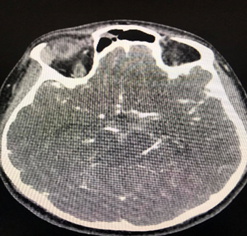
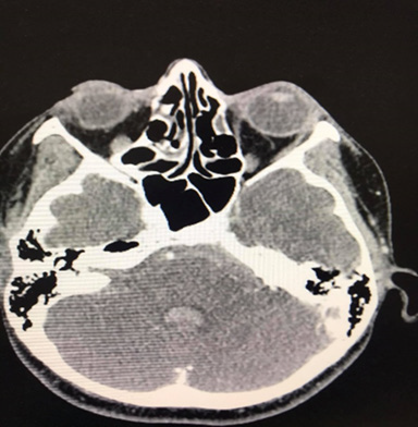
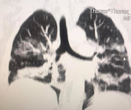
A 35-year-old Caucasian woman, 178cm, 72kg, presented with nausea, vomiting, dry cough, fever, anosmia, and headache for 5 days. Comorbidities or prior neurological episodes were not reported. The patient was alert and oriented upon admission to the emergency room. The patient denied any family history of brain aneurysms or past medical history of any neurological deficits. The likelihood of infection by the novel coronavirus was raised after reports of cough, fever, and anosmia in the preceding 5 days. As a result, throat swab samples were collected for 2019-CoV RNA RT-PCR, which confirmed SARS-CoV-2 infection. The patient was admitted to ICU to receive hospital care. Within 48 hours of hospital admission, the patient was disoriented and confused and developed acute respiratory failure. After clinical stabilization, a cranial CT scan was performed, and a Fisher score II SAH was visualized (Figures 1 & 2). The patient was placed on ventilation and had suffered a subarachnoid intraventricular hemorrhage. Upon further observation, the dynamic condition did not improve according to neuroimaging, later the patient’s consciousness was suppressed, and her Glasgow Coma Scale (GCS) score was 3. The patient was transferred to Caucasus Medical Center, Evex hospitals, Tbilisi, Georgia, to receive specialist care. Upon admission, the patient was in a comatose state, while her photoreaction and corneal reflexes were sluggish. The patient underwent further testing and a chest CT scan was performed and it demonstrated bilateral glass opacities, suggestive of COVID-19 pneumonia (Figure 3).

A Selective digital cerebral angiography was performed - an aneurysm of a complex configuration, a two-chamber, left posterior cerebral artery aneurysm, with a wide neck saccular aneurysm was detected. A spasm of the basilar artery was also detected as shown in Figures 4 & 5. Based on the angioarchitecture of the aneurysm, and to prevent its re- rupture, endovascular closure was performed via balloon-assisted embolization, and complete occlusion of the aneurysm was achieved. Pharmaco-angioplasty for correction of pronounced vasospasm was performed using Percutaneous Transluminal Angioplasty (PTA) of the basilar artery using Nimodipine I/A infusion (Figures 6 & 7). Based on the angioarchitecture of the aneurysm, and to prevent its rerupture, endovascular closure was performed via balloon-assisted embolization, and complete occlusion of the aneurysm was achieved. Pharmaco-angioplasty for correction of pronounced vasospasm was performed using Percutaneous Transluminal Angioplasty (PTA) of the basilar artery using Nimodipine I/A infusion (Figures 6 & 7). The post-operative period proceeded with positive dynamics, without complications. The patient became more active and the headaches decreased. Neurologically at discharge, the patient experienced clear consciousness, adequate orientation in time and environment, and relatively fewer headaches. The patient was discharged after 24 days, with no neurological disabilities. The patient was advised to rest for 2-3 weeks and begin neurorehabilitation.

biomedres-openaccess-journal-bjstr

Figure 1: Brain CT-scan showing Fisher score II SAH.

biomedres-openaccess-journal-bjstr

Figure 2: Brain CT-scan showing Fisher score II SAH.

biomedres-openaccess-journal-bjstr

Figure 3: A chest CT scan in coronal section reveals ground-glass opacities, indicating lung infection by the new coronavirus

biomedres-openaccess-journal-bjstr

Figure 4: Left Posterior Cerebral Artery Aneurysm.

biomedres-openaccess-journal-bjstr

Figure 5: Coiled Aneurysm.

biomedres-openaccess-journal-bjstr

Figure 6: Basilar artery.

biomedres-openaccess-journal-bjstr

Figure 7: Basilar artery post percutaneous transluminal angioplasty with nimodipine I/A infusion.

Epidemiology

Subarachnoid hemorrhage was the 53rd leading cause of death worldwide in 2019, with 11.2 million cases and 373,000 fatalities and a gross mortality rate of 3.33 percent. Furthermore, 240,000 deaths in patients aged over 60 was noted, compared to 103,000 deaths from patients aged between 40 to 59 [7]. Following the study conducted by Sepide Kashefiolasl et al, 10 dated March 21st, 2022, it discussed the prevalence of COVID-19 and Subarachnoid Hemorrhage concerning the age of the patients. In their study, they retrospectively analyzed some subsamples of patients that independently suffered from COVID-19 and SAH and the other group of patients that shared the homogeneity of both diseases. To make their conclusion more referable they underlined the prepandemic era as well. It was then thereby found that there were 12 out of 56 (21%) patients aged less than 50 with a diagnosis of aSAH whereas in comparison to the pre-covid era there were 19 out of 84 (23%) patients aged less than 50 with a diagnosis of aSAH. Concerning this data there was a conclusion that SAH interlinked with COVID-19 was much more prevalent in patients of young age, to emphasize more on the findings they used multivariable analysis to verify significant factors for a favorable outcome (mRS ≤ 0–2) after aSAH during the COVID pandemic [8]. In another similar study conducted by Qureshi AL et al, 282,718 patients were evaluated to assess the risk of SAH development in patients with COVID-19. It was concluded that 0.3% to 1.2% of the patients develop Sub arachnoid Hemorrhage as a complication of Covid-19 [9]. In a gender-based study of the incidence of SAH in association with Covid, male patients were found to be younger than older female patients. The evident causes of younger age group predominance were cigarette smoking in males and hypercholesterolemia in females [10].

Pathophysiology

SARS-COV-2 is a member of the coronavirus family, namely the Beta coronavirus. Beta coronaviruses are known to frequently infiltrate the central nervous system. This behavior has also been observed in other coronaviruses such as SARS, MERS as well as porcine hemagglutinating encephalomyelitis [11]. SARS-COV-2 infects host cells by entering via angiotensin-converting enzyme-2 receptors (ACE-2). ACE-2 receptors are omnipresent within the human body, particularly overexpressed on intestinal epithelial cells, endothelial and smooth cells of blood vessels, heart, lung, renal tubular epithelial cells [12], and in cerebrovascular endothelium which interacts with the viral “s” protein (homotrimer spike glycoprotein) [13]. A potential risk factor that causes intracranial hemorrhage (ICH) is intracerebral capillaries bursting as a result of direct endothelial toxicity. Additionally, endothelial damage may initiate a series of events involving the coagulation cascade, the complement system, and proinflammatory cytokines. Ultimately, this causes increased permeability, breakdown of cellular tight junctions, and the disruption of the blood-brain barrier leading to ICH. In addition, the renin-angiotensin-aldosterone pathway is disrupted by SARS-Cov-2 suppressing the expression of the ACE- 2 receptor. As a result, the autoregulation of cerebral blood flow is compromised, and the local endothelium accumulates high amounts of angiotensin II. This subsequently results in uncontrolled hypertension, significantly raising the risk of subarachnoid hemorrhage (SAH) and ICH [13].

In microvascular lesions of cerebral hemorrhage, increased D-dimer level enhances fibrinolytic function and plasmin generation, which may result in the hypercoagulable conditions. Elevated D-dimer levels are associated with a hyper-fibrinolytic, pro-inflammatory state in SARS-Cov-2 patients, and these levels have been shown to correlate with the severity of the illness and may raise the risk of intracranial bleeding [14]. Innate and acquired immune responses that are elicited by the virus are the main defense mechanisms that protect the host organisms from the crippling effects of SARS-Cov-2 infections. Identification of pathogen-associated molecular patterns (PAMPs) and induction of antigen-specific adaptive immunity is of prime importance to cause these responses. The body’s response to viral infections is influenced by the secretion of cytokines, chemokines, leukotrienes, proteases, reactive oxygen species, and the rate of viral clearance [15]. Furthermore, SARS-Cov-2 causes systemic inflammation as a result of cytokine down-regulation leading to septic shock [16]. Inflammation associated with SARS-CoV-2 and aneurysmal rupture is both heavily influenced by macrophages. The generation of IL- 1, IL-6, and tumor necrosis factors by macrophages have a role in SARS-CoV-2 infection and cerebral aneurysm rupture [17]. Studies have shown males had higher rates of severe Sars-Cov-2 symptoms necessitating ICU admission and worse ischemic stroke outcomes upon hospital release while females had higher rates of milder symptoms [18].

Comorbidities Associated with Covid

Patients with Subarachnoid hemorrhage plus SARS-COV2 had a greater chance of mortality than patients with subarachnoid hemorrhage without SARS-COV2 or patients with SARS-COV2 without subarachnoid hemorrhage. These individuals had a higher incidence of systemic comorbidities which served as a factor in the elevated risk. Patients with COVID-19 and subarachnoid hemorrhage had higher rates of pneumonia, pulmonary embolism, urinary tract infection, acute kidney damage, liver failure, Heart failure, acute MI, septic shock, and respiratory failure [9]. A study evaluating Patients hospitalized with COVID-19 disease reports a significant and high risk of thrombotic events including stroke and SAH. 52% of Covid -19 patients present SAH, while 95% of them have a history of hypertension and 60% have a history of diabetes Mellitus, 40% have cardio embolism and 5% have vessel disease, and 35% cryptogenic [19]. Hypertension (HTN) and chronic heart disease were the two most common vascular comorbidities, as documented in various studies on ICH in COVID-19 patients [13]. Due to the resulting autonomic imbalance, more individuals are developing cardiac arrhythmias as the coronavirus 2019 pandemic advances relentlessly. The most frequent rhythm disturbance in COVID-19 infection patients is sinus tachycardia, which can be brought on by fever, hypoxia, and hemodynamic compromise. Transient sinus bradycardia is also a potential sign of COVID-19, according to a retrospective series of 4 patients. Numerous ventricular arrhythmias can happen as a result of triggers such as COVID-19 infection-induced systemic inflammation and severe respiratory insufficiency. Additionally, many medications used to treat COVID-19 infection have the potential to affect the cardiac system by lengthening the QT interval and inducing polymorphic Ventricular Tachycardia (VT) in the form of Torsade de pointes (TdP) [20].

A hemorrhage was found in a group of covid 19 patients 67 years old where the majority of comorbidities diseases were hypertension, diabetes, and obesity. SARS-CoV-2 may raise blood pressure and increase the risk of cerebral hemorrhage once it enters the bloodstream, especially in people who already have high blood pressure [21]. When compared to controls, patients with spontaneous ICH or SAH and concurrent COVID infection were more likely to be members of racial or ethnic minorities, diabetics, and obese, and they also had higher mortality rates and longer hospital lengths of stay [22]. Diabetic patients were more likely to be admitted to the intensive care unit (ICU) during the SARS epidemic at a rate that was higher than that of non-diabetic patients. Research and Reviews proved that diabetic patients have a significantly lower absolute lymphocyte count than those who do not have diabetes. Clinical studies revealed that once diabetic individuals contracted SARS-CoV2 due to a variety of causes such as decreased physical activity and irregular nutrition, the insulin dose rose and blood glucose control became challenging. Additionally, as the virus employs the human angiotensin-converting enzyme (ACE2) as a receptor for cellular entrance, which enhances personto- person transmission, the increased infectivity and virulence of SARS-CoV2 in diabetes is explained. Noting that the reninangiotensin system (RAS) family, which has been linked to diabetes, contains the key component ACE2 [23].

Additionally, a review of the international Health Outcome Predictive Evaluation for SARS-COV2 registry evaluating the effect of renal function on admission and mortality with SARS-CoV-2 infection found that 30% of patients had kidney dysfunction upon admission and that CKD was prevalent in 8.5 % of infected patients [24]. In COVID-19, AKI is linked to a greater risk of death. A case in point is sepsis. Microvascular dysfunction, a rise in vascular permeability, and tissue damage are its defining characteristics. Cardiomyopathy, viral myocarditis, and left ventricular failure are additional conditions that can cause hemodynamic abnormalities. Additionally, cytotoxic reactions harm podocytes and tubules and cause hematuria, proteinuria, and AKI. Moreover, collapsing glomerulopathy is an extra consequence that is brought on by a direct viral effect, the presence of increased cytokines from the systemic inflammatory response, or both [25].

Risk Factors for Covid and SAH

Risk factors of high-rate mortality are based on a cohort study, Elevated INR, severe pulmonary symptoms, and spontaneous hemorrhagic presentation [26]. Cigarette smoking, cocaine use, hypertension, low body mass index, first-degree relatives with hemorrhagic stroke, caffeine in pharmaceutical products, lower educational attainment, and nicotine in pharmaceutical products are all risk factors for SAH that can be modified. Age, sex, and race are further risk variables that cannot be changed [27]. To be more specific hypertension can classify as the first and the most notable risk factor caused by SAH in human beings regardless of age and sex which’s followed by the second risk factor in young males is cigarette smoking and hypercholesterolemia in the older woman [10]. There is a strong association between a higher risk of Intracranial Aneurysm (IA) and aneurysmal Subarachnoid Hemorrhage (aSAH) and genetic propensity to smoking, sleeplessness, and high blood pressure. The risk of IA and aSAH may also be influenced by factors like physical activity, body mass index, triglyceride levels, and lowdensity lipoprotein cholesterol levels. These findings support the triangulation of information regarding IA and aSAH risk factors and call for additional research in future extensive MR and other epidemiological studies. The biggest risk factors for IA and aSAH, according to the current MR study, include smoking and high blood pressure. Additionally, this study discovered data that suggests insomnia may be a brand-new risk factor for IA and aSAH [28].

In the indirect way of SAH hemodynamic stress, chronic inflammation and vascular wall remodeling are the most common causes of the ruptured Cerebral aneurysm which leads by the reactive oxygen species through the activation of nuclear factor Kappa-B to the endothelial dysfunction. Hemodynamic stress is brought on by the activation of the renin-angiotensin system, which causes hypertension. Unlimited consumption of (alcohol, antioxidants vitamins, B vitamins, flavonoids, and n-3 fatty acids) leads to increased blood pressure which increases the risk of the rupture of cerebral aneurysm and causes SAH [29]. The most important risk factors of SAH based on studies are Smoking, hypertension, and excessive alcohol consumption, Besides, there are many other uncertain factors such as nonwhite ethnicity, HRT, hypercholesterolemia, and diabetes in the etiology [30,31]. Cigarette smoking and hypertension increase the risk of cerebrovascular diseases which are the main causes of SAH. However, cigarette smoking cessation doesn’t decrease this risk [32].

Regarding covid 19, The Elderly COVID-19 patients, who had a higher risk of hospitalization, mechanical ventilation, and mortality, demonstrated dyspnea as a risk factor. Additionally, because of weakened immunity, patients with cancer and hematologic malignancies are more susceptible to SARS-CoV-2 infection. An Individual may be more susceptible to COVID-19 infection if they are pregnant in addition to these risk factors. This may be due to physiological changes in the immune system and placental immaturity during the first trimester [33]. One of the most important complications of Covid 19 disease is thrombosis, while preventing this thrombotic complication by using antithrombotic agents, there are important side effects, from the use of these agents, such as hemorrhagic stroke, subarachnoid hemorrhage, and intracranial bleeding [34].

Complications

Major Complication Associated with SARS-CoV-19

Pneumonia: The Lower respiratory tract’s innate and acquired defense systems produce an inflammatory response after infection, as in the case of SARS-CoV-19. The cytokines TNF-a, IL-8, and IL-1 released by the resident alveolar macrophage in the lungs attract inflammatory cells like neutrophils to the parenchyma thereby generating clinically symptomatic pneumonia [35,36]. Additionally, the macrophages function as Antigen-presenting T-cells (APC)s, activating a plethora of immune responses, cell-mediated, humoral, complement-activated, and antibody-production. As a result, the lung parenchyma becomes inflamed, and capillary leakage leads to fluid-filled alveolar sacs, thus producing the fundamental pathogenesis of pneumonia [36]. Patients experience productive cough (greenish, yellow, or bloody mucus), tachycardia, tachypnea, fever with chills, malaise, loss of appetite, and myalgia. A fraction experience altered mental status, abdominal pain, stabbing chest pain and further systemic findings. Physical examination common findings include crackles, dullness on percussion and egophony and tactile fremitus (both suggestive of consolidation) [35,36].

Acute Respiratory Distress Syndrome (ARDS)

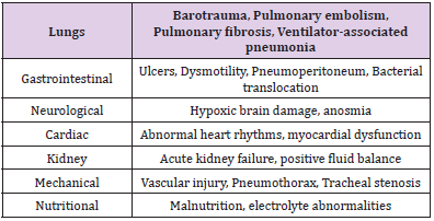
ARDS is a cause of respiratory failure in critically ill patients and is the acute onset of pulmonary edema (noncardiogenic), atelectasis, hypoxemia and the need of mechanical ventilation. ARDS is marked by increased permeability to fluid and protein across the lung endothelium leading to edema in the lung interstitium. Damaged tight junctions between type I and II alveoli, the edematous fluid translocate into the alveoli space. Typical hallmarks of ARDS include increased permeability to fluid, proteins, neutrophils and RBCs, leading to their accumulation within alveolar space [37]. Vascular hypoxemia seen is caused by ventilation-to-perfusion disbalance as well as right-to-left intrapulmonary shunting. Furthermore, hindered carbon dioxide excretion is a major component of respiratory failure, resulting in increased pulmonary dead space with elevated ventilation [37]. Increasing pulmonary dead space and decreasing respiratory compliance are predictors of ARDS mortality [37]. Diffuse alveolar damage (DAD) is the main pathologic finding and is characterized by the destruction of both alveolar type I and II cells. This ultimately leads to pulmonary edema as the destroyed alveolar type II cannot remove leaked fluid, this then is followed by hyaline membrane depository on the alveolar walls. During this time, surfactant produced by alveolar type II cells cannot reduce surface tension and inflate the alveoli for respiration. During the recovery process, collagen deposited may not be completely reabsorbed, limiting alveolar expansion and thus respiration [37,38]. A plethora of problems arises from ARDS which can be seen in the Table 1 below [37,38].

biomedres-openaccess-journal-bjstr

Table 1: Further systemic complications of ARDS. It can be seen that ARDS in its own right is a complication, yet it can bring additional problems that can ultimately lead to death.

Multi-Organ Failure and Death

Whilst common cold symptoms are observed in mild forms of COVID-19. In severe cases, there is the involvement of different organs that can quite frequently lead to death, often characterized by acute lung failure, liver failure, kidney injury and cardiovascular disease, and a spectrum of hematological and neurological abnormalities [39,40]. It is suggested that the main cause of multi-organ failure is the cytokine storm, which is induced by inflammatory mediators, endothelial dysfunction, coagulation abnormalities, and inflammatory cell organ infiltration [40]. Along with the plethora of complications previously mentioned, death can ultimately occur and can be a result of multi-organ failure or any other complication such as ARDS or Acute heart failure [39,40].

Common Complications Pertaining SAH and SARS-CoV-19.

In comparison to COVID-19, some patients with the condition: SAH (Subarachnoid Hemorrhage) will survive and make a satisfactory recovery. However, a significant number of individuals still do develop serious complications which further decline their health and can potentially lead to death. Complications can be classified into intracranial or extracranial where, according to Hall and O’Kane (2018), intracranial consequences are factored in the initial treatment while the latter is closely monitored [41]. Furthermore, according to Daniere et al., complications can also vary in severity, from acute, subacute, and chronic listing the following in respective order: hydrocephalus, vasospasms, and cognitive disorders [42]. In many instances, the complications can be closely related to the activation of the sympathetic nervous system, catecholamines, and the systemic inflammatory response syndrome (SIRs) (Garg and Bar, 2017) [43]. Data supported by Hammer et al., (2020), has shown that the following are statistically significant complications found after SAH: pneumonia, sepsis, hydrocephalus, and delayed cerebral ischemia [44]. Apart from the neurological complications seen in SAH, a patient may also develop secondary non-neurological conditions, such as neurocardiogenic injury, pulmonary edema, and hyperglycemia (Chen, et al. [45]). From these non-neurological conditions, it is common to observe cardiac and pulmonary manifestations following the complication of SAH [38].

Conclusion

This case study and literature analysis on COVID-19 and SAH established that patients with a prior COVID-19 infection have a higher likelihood of developing SAH as opposed to patients who have not acquired COVID-19. The primary etiology of SAH during COVID-19 infection was proven to be thrombotic vascular events, which caused microvascular lesions of cerebral hemorrhage and disrupted the angioarchitecture. Additionally, these lesions were most frequently seen in patients with a history of comorbidities like diabetes mellitus and hypertension resulting in higher mortality rates. Regardless of the patient’s age or comorbidities, the ratio of major respiratory complications such as pneumonia and ARDS were significantly higher. The major determinants of higher mortality in patients with SAH and COVID-19 were elevated INR, insidious pulmonary presentations (effusion, ARDS, pneumonia), and spontaneous hemorrhagic conditions. It was further noted that the strokes were typically initiated after the COVID-19- associated lung infection had completely subsided in the body. Furthermore, this led to coagulation in the arteriovenous system of the brain, resulting in the individual becoming more prone to brain hemorrhages. Although there were generalized reviews stating that COVID-19 infection caused an increase in the intracranial pressure while simultaneously surging the risk of aneurysms, there was ambiguity in the data that determined the underlying mechanism of the aforementioned. Consequently, thorough research in this area would be beneficial in determining factual data.


Follow us for more Articles on: https://biomedres01.blogspot.com/

https://scholar.google.com/citations?user=OFtUOZYAAAAJ&hl=en
https://www.base-search.net/Search/Results?lookfor=Biomed+Journal+of+Scientific+%26+Technical+Research&l=en&refid=dcsuggesten
https://citefactor.org/journal/2574-1241/biomedical-journal-of-scientific-technical-research
https://ideas.repec.org/s/abf/journl.html
https://econpapers.repec.org/article/abfjournl/
https://www.ncbi.nlm.nih.gov/nlmcatalog/101723284
https://bjstr.org/
https://bjstr.org/about.php
https://biomedres.us/reprints.php
https://biomedres.net/submit-manuscript.php
https://independent.academia.edu/BJSTRAngelaRoy

Antimalarial Aloe Compounds

  Antimalarial Aloe Compounds Introduction Among the most prevalent diseases caused by protozoan parasites, malaria is caused by parasites o...