The Use of Ionic Implantation for The Medical Materials Modifying
Introduction
Modern medicine is not possible without the use of steel tools and implants. Usually, in order to meet the needs of physicians, manufacturers of tools use chrome and chromium-nickel corrosion-proof alloy steels 30Cr13, Cr13 40, 95Cr18 [1,2], AISI 301 (similar to 07Cr16Ni6), AISI 316L (analogue 03Cr17Ni14Mo3) [3,4] for multiple uses and special alloys [5] and steel ShKh15 (analogue 52100) [2] for disposable products. To produce materials that are in contact with biological media also often stainless steels [6] are used. But in this case steel is required much corrosion resistance and wear resistance [7]. While the key issue is the biocompatibility of the material of the implant and the environment in which it works [8]. One of the ways to succeed in creating such a modern medical material is surface modifying already existing metals and alloys [9]. A promising direction in the synthesis of new materials using the modifying surface is ion-plasma technologies, in particular, the method of ionic implantation [10-14]. The essence of ionic implantation, as technology of impact on the properties of materials, is the introduction of high-energy target ions in the surface layer and their accumulation with the subsequent creation of a surface film. Thanks to this the new distribution of surface energy and change of mechanical, physical-chemical, optical, and other characteristics of the processed sample are achieved. Therefore, the purpose of the work was to investigate the properties of stainless steel and steel ShKh15 as the materials that are widely used in medicine, after the ionic implantation.
Synthesis of Samples and Methods of Investigation
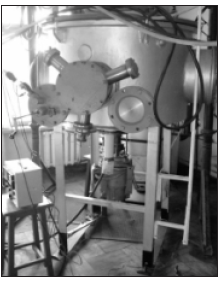
Treatment of the samples was carried out by titanium ions in nitrogen plasma using installation of ionic implantation (Figure 1). Mode of implantation corresponded to fluence near 5·1017 cm-2. The energy of the ions was about 20 keV. It allows to introduce them to a depth of up to 1 micron. The pressure in the vacuum chamber, which provided the ion jet did not exceed 10-4 Pa. Research of the surface hardness were carried out using the device “NOVOTEST T UD”. Sclerometric analysis of adhesive properties was performed with the help of a microhardness meter “PMT-3”. The state of the surface was shown wits using optical microscopy (microscope “MIM- 7”, additionally equipped with digital camera “Kodak Easy Share C1013”). Corrosion resistance was tested for long-term exposure to solutions of sulfuric acid and with subsequent gravimetric analysis.
Results and Discussion
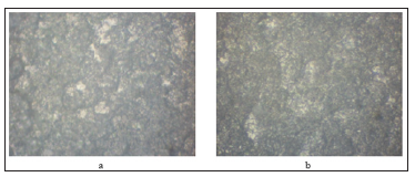
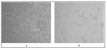
As a result of the studies a substantial increase in the hardness and strength of the surface for steel 12Cr18Ni10Тi and steel ShKh15 was received. In particular, the hardness for steel 12Cr18Ni10Тi increased from 220 HB to 280 HB. The hardness of treated steel ShKh15 sample was changed from 2 GPa to 4 GPa. This data indicate about the strengthening of surface layer and increase the durability of the material in terms of friction. Mechanical strength of the surface layer increased too (Table 1). One of the reasons for strengthening the surface is changing of the material structure. The results of optical studies (Figures 2 & 3) indicate about changing in the roughness of the surface in the direction of smoothing. Since stainless steel is intended for reusable medical materials, it must maintain a long term contact with the biological environment. Therefore, studies on corrosion stamina were conducted. Endurance stainless steel sample (12Cr18Ni10Тi) for 500 hours showed that depth of the dissolved layer for untreated material is about 400 μm/year. Instead, steel with titanium ions had a value of no more than 12 μm/year.
Figure 2: Microphotographs of steel ShKh15 structure:
a) Initial sample
b) Sample after implantation with an optical magnification x500.

Figure 3: Microphotographs of steel 12Cr18Ni10Тi structure:
a) Initial sample
b) Sample after implantation with an optical magnification x1500.

Conclusion
Consequently, studies have shown significant prospects for the use of ionic implantation technology for the production of metal medical materials. The increased hardness and strength of the surface layer will ensure the use of treated steel as a material for tools. Mechanical properties in a heap with increased corrosion resistance indicate the potential for application of samples in the function of implants.
Obesity: Modulating Adiponectin Levels Through Diet-https://biomedres01.blogspot.com/2020/07/obesity-modulating-adiponectin-levels.html
More BJSTR Articles : https://biomedres01.blogspot.com


No comments:
Post a Comment
Note: Only a member of this blog may post a comment.