Traumatic Bilateral Globe and Optic Nerve Avulsion: Rare Case Report
Introduction
Optic nerve avulsion (ONA) is a condition that usually results from severe trauma of the orbit and face [1]. ONA can be divided as a partial avulsion, where partial visual recovery can be achieved and complete avulsion usually leading to total vision loss [2]. There is a wide range of causes that lead to the globe avulsion and optic nerve avulsion. Globe avulsion (GA) may occur from finger pricking during sports trauma, from severe facial and orbital fractures after vehicle accidents [3] or after a rare animal attack or physical assault [4,5]. In children, ocular trauma is leading cause of vision loss [6] where ONA can occur following the blunt or penetrating ocular trauma [7]. Furthermore, in every maxillofacial trauma associated with globe protrusion, there should be suspicion for the ONA [8]. Some unusual cases like auto-enucleation have been reported in psychiatric literature [9] and more rarely there is a case representing accidentally self-inflicted ONA [10]. Although the spectrum of globe injures is wide, bilateral ONA following complete globe protrusion is very rare [1]. Herein, we present a case with complete bilateral globe and optic nerve avulsion following tractor accident.
Case Report
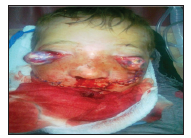
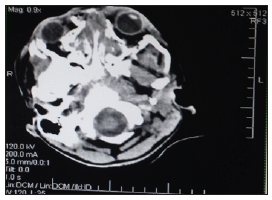
We present a case of 12-year old boy, admitted to emergency unit after an accident where the tractor falling over him causing se vere injuries. Physical examinations revealed unconsciousness, oedematous and bruised face with facial lacerations and both globes were luxated anteriorly from the orbital sockets (Figure 1). There were no direct or indirect light reflexes and no eye movements. Computed tomography (CT) showed multiple fractures of maxillary and nasal bones, including lamina cribrosa of ethmoid bone, fracture of the orbit medial wall and zygomatic arch of both sides. Both globes were protruded outside the orbits, with suspicion of the ONA. Neurosurgery consultation revealed no sign of systemic trauma except minor frontal lobe contusion (Figure 2). After evaluation in the emergency unit, the patient was transferred in operation room where he underwent surgery under general anaesthesia.
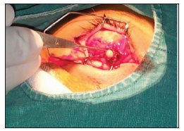
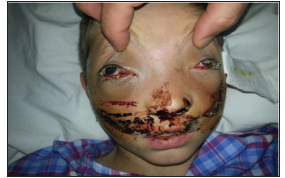
The orbital soft tissue and Tenon’s capsule were attached to the globe. During the exploration, all the extraocular muscles and optic nerve of both eyes were found to be ruptured, presumably because of fractured bony segments, but there were no retrobulbar haemorrhage. Optic nerve stump in both eyes were found (Figure 3). The stumps of lateral and inferior rectus muscle on the right eye were found and the stumps of the superior and lateral rectus muscle of the left eye were also found. After repairing the muscles in both eyes, the globes were repositioned. The facial fractures were immobilized with plates and screws. After the operation, the patient was observed in the intensive care unit for 2 weeks. The patient was re-evaluated six months after surgery and there was hypotony of eyes and no signs of facial deformity (Figure 4).
Discussion
Globe protrusion with ONA, is a rare condition resulting from severe trauma of the maxillofacial area [11]. The globes and the optic nerve are usually resistant to mild and moderate trauma. However, high energy trauma can cause multiple fractures reducing the orbital volume, promoting protrusion of the globe from its socket [11]. Moreover, Morris et al., have described three potential mechanisms: [1] protrusion of the globe, after an elongated object enters the medial orbit; [2] a wedge-shaped object enters the orbit medially, displacing globe anteriorly; [3] a penetrating object transects the optic nerve directly [12]. In some reported literature, optic nerve transection is a major cause of globe subluxation [1,13]. If there are also disruptions of extraocular muscles, total luxation of the ocular globe will occur [12]. In our case, it is supposed that multiple fractures of the orbital walls caused transection of the optic nerve and globe protrusion. Although we were not able to explain why the extraocular muscles were detached from globes. To evaluate the extent of orbital, ocular and intracranial injuries before the surgery, brain and orbital imaging are advisable. Although, it has been reported a normal CT images with presence of optic nerve avulsion [14]. In presented case, ONA was detected during exploration of the orbits, because there was no suggestion from the CT before surgery. The primary goal in complete GA, is prevention of the visual impairment. If the optic nerve is partially avulsed, partial recovery of visual function can be achieved [2].
However, in total transacted optic nerves with absence of visual function, only cosmetic concerns may become important. In these patients with no visual functions, the management is controversial. Some authors recommend primary enucleation because there is no visual recovery [15], others suggest repositioning of the globe, even if the patient had no light perception, presuming that saving the eye reduces the psychological stress after the abruptly traumatic loss of vision [16]. Although, even if we have the repositioning of the globe there is always a risk of the bulbar atrophy due to ischemia [17]. Taking in consideration the possible prognosis of these severe injuries and their management dilemma, it is necessary to have a good management plan which can help to optimize outcomes. Therefore, knowing that there is no treatment for optic nerve avulsion [18] and it can adopt different intraocular courses and outcomes depending the visual equity after the injury [19], the globe preservation can have great psychological impact and also ease the cosmetic rehabilitation. Goal would be to have satisfactory results as represented in some cases [20]. In our case, both globes were repositioned after repairing the extraocular muscles and performing a lateral canthoplasty. Facial bone fractures were repaired with plates and screws. After sixth months, evaluation revealed that there was no sign of bulbar atrophy, which could be possible due to survival sources of the blood supply in the soft tissue attachments.
Conclusion
ONA with complete globe protrusion is very rare complication of maxillofacial trauma. However, in every facial trauma the assessment of optic nerve is crucial. Depending on the degree of the visual acuity after the injury there can be different outcomes because of the intraocular morphological changes. However, optic nerve avulsion causes nerve damage leading to the permanent visual acuity impairment. Therefore, the best approach for these patients is organized plan management for globe preservation aiming for the best outcome which can help having better psychological impact and cosmetic rehabilitation.
Thermal Studying Dry Chemicals as Hygienically Safe Extinguishing Substances-https://biomedres01.blogspot.com/2020/09/thermal-studying-dry-chemicals-as.html
More BJSTR Articles : https://biomedres01.blogspot.com


No comments:
Post a Comment
Note: Only a member of this blog may post a comment.