Evaluation of Aqueous Extract and Fractions from theSeed of Magonia Pubescens in Sleep Models, Anxiety and Depression in Mice
Introduction
The “tingui” - Magonia pubescens, is a plant distributed predominantly in Brazilian Cerrado, in the states of Goiás, Mato Grosso, Mato Grosso do Sul and Minas Gerais. This tree has been used in civil construction, such as rafters and slats, in the manufacture of frames, portals, firewood and charcoal. JOLY; Barroso [1]; SCHVARTSMAN; Lorenzi [2]. The plant is used for cleaning animal ulcers, as a soothing and in fishing by means of water poisoning. Its fruits and seeds are still used in the manufacture of homemade soap and dry floral arrangements Joly et al. [3]; Silva & Lorenzi [4]. Bioactive compounds extracted from Tingui were mainly studied for their larvicidal activity against Aedes aegypti Arruda et al. [5]; Da Silva. Ethanol extracts from the stem bark showed high levels of tannins Silva et al. [6]. Tannins are widely distributed in nature, being generally the active principles of plants used in traditional medicine. Condensed tannins have a great ability to interact with metal ions and macromolecules and form soluble complexes with electron donor groups, such as those found in alkaloids and proteins. This may be one of the reasons related to its toxicity towards different organisms including insects, fungi and bacteria. The morphological changes caused by these compounds on the midgut epithelium of A. aegypti larvae resemble those recorded for tannic acid Rey et al. [7].
Several extracts of plants such as Magnolia officinalis, Zizipus spinosa and Valeriana walichii have sedative action, as sleep inducers or on mood disorders such as anxiety and depression, because they have bioactive components capable of interacting as monoamines and their receptors of the central nervous system Koetter et al. [8]; Sahu et al. [9]. Benzodiazepines are the drugs most often prescribed due to their anxiolytic properties or treatment of sleep disorders such as insomnia. However, these medications have limited benefits with obvious side effects, such as impairment of cognitive function, memory, and overall daytime performance Thomas et al. [6]. In addition, the long-term results of the administration of such drugs cause tolerance and dependence Gyllenhaal et al. [4]. Sleep disorders, anxiety and depression, or using in a generalist the term behavioral disorders, present increasing number of cases. Projections indicate that depression will be the most prevalent disease in the world by 2020 Murray et al. [10]. In many cases, the diagnosis of patients with such morbidities is not done correctly, or inadequately treated, leading to conditions of functional decline, decreased productivity and increased mortality of these patients Belmake et al. [11].
The relationship between inflammatory processes and behavioral changes has been widely studied in recent years. Patients with depression have high levels of inflammatory markers, I is also reported that patients with depressive behavior have elevated levels of some pro-inflammatory cytokines (such as TNF- α, IL-1β, IL-6, INFα) and C-reactive protein Maes et al. [12]. Phytotherapeutic drugs are the subject of further studies because of their ability to interact with various compounds and receptors, making it a potential therapeutic target. Despite the enormous diversity of biomes and plants in Brazil, there are few studies that investigate the potential of biomolecules in these species. About 80 species of plants are described in the Phytotherapic Form of the Brazilian Pharmacopoeia, and of these only seven species have reports on sedative or anxiolytic actions. In view of the above, this work intends to evaluate the ability of Magonia pubescens extract to alter sleep pattern in treated animals, as well as to test the effects of this extract on locomotion tests in healthy animals and with LPS - induced peritonitis.
Methods
Animals
For the execution of the work Swiss female mice weighing between 30 and 35 g were obtained from the bioterrorism department of the Biophysics and Pharmacology Department of the Federal University of Rio Grande do Norte. The animals were housed in polyethylene boxes with free access to food and water and kept in a 12-hour dark-light cycle (6:00 a.m. to 8:00 p.m.) in an air-conditioned environment. The Ethics Committee on the Use of Animals (CEUA) of the Federal University of Rio Grande do Norte (n º 033/2014) approved all the experimental procedures of this work plan. Eight animals were used per group. At the end of the behavioral tests the animals were euthanized with a thiopental solution at a dose higher than 100 mg / kg (volume 10 ml/kg) and after confirming the death, the cadavers were stored in a freezer at -20 ° C until collected by company specialized.
Drugs and Drug Treatments
Drugs administered were thiopental and aqueous extract of MP, all in the volume of 10 ml / kg. Thiopental (Laboratory of Pharmaceuticals Cristália, Brazil) is a sedative-hypnotic substance of the fast acting barbiturates class whose mechanism of action is characterized by the potentiation of the inhibitory effect of gammaaminobutyric acid (GABA) in the central nervous system and will be used in order to induce sedation in animals. This drug was solubilised in saline and administered intraperitoneally (ip) at the dose of 50 mg / kg, since previous studies have shown that this dose is effective in inducing sedative effects by ip administration in mice. Since there are no studies characterizing the effect of aqueous extract of M. pubescens on animals, it was administered ip, in a dose range delimited according to the toxicity test of the extracts (Hemolysis Test / Cell Viability in Macrophages), using three non-toxic concentrations with maximum values around 10 μg / ml, considering that hemolysis curves were performed in order to characterize the concentrations used in which there is no hemolysis, and thus, without toxicity. Doses of 10 μg / ml, 1 μg / ml and 0.1 μg / ml will be used.
Extraction, Fractionation and Characterization of Extracts
Extraction: After collection, the seeds were separated, cleaned and pulverized, 17 g of seed meal was homogenized in 170 ml of distilled water ratio 1:10, kept for 4 hours on a magnetic stirrer. After the time elapsed, the mixture was centrifuged for 30 min at 4 ° C, 8000 rpm, then the supernatant called crude extract and the precipitate was discarded. The supernatant was lyophilized to obtain the aqueous extract.
Chemical Characterization: The total sugars were quantified by the phenol / sulfuric acid method according to Dubois et al., using the monosaccharide galactose as standard. The readings were performed at 490nm. Protein content was determined by the method of Bradford, using Coomassie Blue Brilliant reagent and bovine albumin as standard, and the reading performed at 595nm.
Electrophoresis: The samples were subjected to Agarose electrophoresis gel, SDS-PAGE. The color of the gels was made by Comassie.
FPLC Fractionation: The extracts were clarified and subjected to low pressure molecular exclusion chromatography on AKTA purifier chromatograph with Tricorn or Hiprep columns and fixed phase of Sephacryl or Superdex with particle sizes appropriate to the composition of each sample.
Assessment of Toxicity of extracts
Hemolysis Test: In order to evaluate the toxicity of the extracts they were incubated at different concentrations (1000 μg / ml, 100 μg / ml, 10 μg / ml, 1 μg / ml and 0.1 μg / ml) with human erythrocytes obtained from bag donations by HEMOCENTRO-RN. These bags were out of the recommended shelf life for transfusions. In order to evaluate the hemolytic effect of the extracts on human erythrocytes, human red blood cells were separated from the plasma by sedimentation and washed three times with 0.01M Tris- HCl buffer pH 7.4 containing 0.15M NaCl. The same buffer was used to prepare a 1% (v / v) suspension of red blood cells and solubilize the samples. In 1.5 mL tubes, 100 μL of the red blood cell suspension was incubated with 100 μL of the sample for 60 minutes at room temperature. The 100% and 0% hemolysis references were made by incubating 100 μL of the RBC suspension with 100 μl 1% (v / v) Triton X-100 or Tris buffer, respectively. After incubation, the tubes were centrifuged at 3000 x g for 2 minutes and 100 μl aliquots of the supernatants were transferred to 96-well microtiter plates and analyzed in reading at 405 nm.
Cell Viability Assessment: In order to evaluate the cytotoxicity of the extracts obtained from the seeds of M. pubescens, they were incubated at different concentrations (100 μg / ml, 10 μg / ml, 1 μg / ml and 0.1 μg / ml), with a cell proliferation inducer (LPS) and using LPS in synergy with the different concentrations of the extract (MP) in macrophages obtained by primary culture. The MTT method (3- [4,5-dimethylthiazol-2-yl] -2,5-diphenyltetrazolium bromide) is a cell viability assay used to determine cytotoxicity after exposure of the cells to test substances. The cytotoxicity of extracts of M. pubescens (MP) was measured as previously described by Mosmann [13]. Cells were exposed to different concentrations of MP (in triplicate) and incubated at 37 ° C for 24 h. After incubation, 100 μl DMEM medium containing MTT (final concentration 5 mg / ml) was added to each well, and the plates were incubated at 37 ° C for 4 hours. After the time the supernatant was removed and 100 μL of P.A. ethanol was added to each well to solubilize the formazan crystals. After homogenization, the reading was carried out at 570 nm.
Behavioral Models
Thiopental-Induced Sleep Test: In order to evaluate the sedative effect of the aqueous extract of M. pubescens, the animals will undergo the thiopental-induced sleep test. For this, the animals were given aqueous extract of M. pubescens 30 min prior to the administration of thiopental (50 mg / kg, ip). Immediately after receiving thiopental, the animals were placed individually in boxes to record sleep-onset latency and sleep duration. The latency for sleep onset corresponds to the time elapsed since the administration of thiopental until the animal loses the righting reflex when placed in the position of dorsal decubitus. The duration of sleep corresponds to the time elapsed from the loss, until the recovery of the righting reflex.
Open Field: The locomotor activity of the mice was measured using the open field. This test was performed in a square black formica box containing four open fields measuring 40 cm x 40 cm x 40 cm each, so that four animals are filmed and evaluated simultaneously. Each mouse has been placed in the center of one of the open fields and will be allowed to freely explore the device. The distance traveled every 5 minutes was recorded automatically by software (Any-maze, Stoelting, USA) for 15 minutes. After the behavioral evaluation of each mouse, each open field was cleaned with a 5% ethanol solution. The test environment was quiet and under controlled lighting.
Rota-Rod: The evaluation of the motor coordination of the mice was performed by means of the rota-rod test (AVS Projetos, Ribeirão Preto, SP, Brazil) as previously described by Rosa’s, the apparatus consists of a bar 3 cm in diameter that rotates clockwise at different speeds, measured in revolutions per minute (rpm). The bar is raised from the support platform by 22 cm. The rotating bar is divided into 5 compartments so as to allow testing of more than one animal simultaneously. During the behavioral tests, the mice were placed on the rotating bar of the device under acceleration conditions of 35 rpm and the number of times the animal was dropped from the device in one minute. All experiments were performed between 13:00 and 17:00.
Statistical Analysis
The data presented in this paper will be reported as mean ± standard error. Comparisons between the treated and control groups were performed using the one-way ANOVA. Values of p <0.05 will be considered significant. These analyzes were done using the SPSS program, version 16.0.
Results
Obtaining the Aqueous Extract of the Seeds of Magonia Pubescens
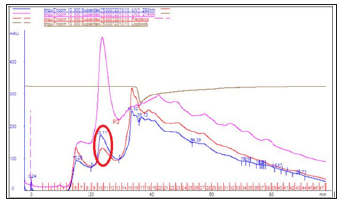
The partial chemical characterization of the extract obtained, showed that it does not have carbohydrates, being composed mainly by molecules of protein origin, as shown in Table 1. After extraction, the obtained extract was fractionated by chromatography by molecular exclusion in the FPLC system. Peaks at 280 nm related to the presence of proteins demonstrate that they are two protein populations, called P1 and P2. According to the spectrum profile, it can be inferred that such populations have amino acids with hydrophobic characteristics in their composition (Figure 1).
Table 1:Total sugar and protein contents in extract obtained from M.P.

Figure 1:Chromatography of molecular exclusion Superdex 75 column in FPLC system.
Electrophoretic Profile of P1 and P2
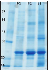
The electrophoretic profile reveals a characteristic bank in the crude extract and fractions 0-30% of ammonium sulphate and 30- 60% of ammonium sulphate. Most of the proteins present in both P1, P2 and crude extract (EB) have molecular weight around 25KDa (Figure 2).
Figure 2: Polyacrylamide gel electrophoresis, Tris-Glycine running buffer with 1% SDS. About 50μg of each fraction were submitted to electrophoresis. The development was done with 0.01% toluidine blue.
Evaluation of MP Toxicity in Hemolysis Tests
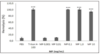
According to the applied methodology, it was demonstrated that MP has cytotoxic action in erythrocytes at doses above 0.1 μg / mL, shown by the presence of significant hemolysis. At the concentrations of 0.001 μg / mL and 0.1 μg / mL, no hemolysis was detected, thus, at these concentrations PM does not cause damage to the membranes of these cells (Figure 3).
Figure 3:PEvaluation of the percentage of hemolysis caused by MP. Level of significance in relation to control (PBS): (***) p <0.001. Values are means ± standard deviation (n = 3).
Evaluation of Cell Viability of Mp in Macrophages
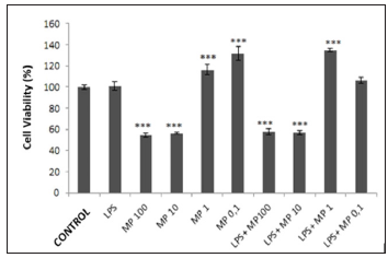
The cell viability test was performed in the laboratory of Profª. Drª. Janeusa Trindade, Department of Immunology, UFRN. Cell viability was verified in macrophages through the MTT test, which evaluates the mitochondrial function of viable cells through the conversion of this salt to its derivative, formazan. In Figure 4, it can be observed that in the test, the LPS had no significant proliferative action, compared to the cell type tested. MP showed antiproliferative effect at the concentrations of 100 μg / ml (54.3 ± 2.1%) and 10 μg / ml (56.3 ± 0.3), however, in none of the doses tested reached the LD50.
Figure 4: Evaluation of MP cytotoxicity in macrophages obtained by primary culture and MP in synergy with LPS. Level of significance in relation to control: (***) p <0.001. Values are means ± standard deviation (n = 3).
Dosage of IL-6 and TNF-α secreted by RAW lineage cells incubated with different concentrations of MP
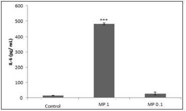
After incubation of the RAW 264.7 macrophages with different concentrations of MP, the culture medium was collected in order to perform the IL-6 and TNF-α dosing through ELISA kits. The dosages showed that incubation with MP does not alter the secretion of TNF-α, however it increases the secretion of IL-6 by approximately 34 times in the cells incubated with 1 μg / mL MP (Figure 5).
Figure 5: Increased IL-6 secretion in macrophages incubated with 1 and 0.1 μg / mL MP. Values expressed as mean ± SD (n = 3) (P <0.01, ANOVA).
Behavioral Models
Evaluation of the Sedative Effects of PM through the Thiopental-Induced Sleep Test
The mice were randomly divided into the following groups:
a) MP 1 μg / ml (ip) + thiopental 50 mg / kg (i.p.) n = 8
b) MP 0.1 μg / kg (ip) + thiopental 50 mg / kg (i.p.) - n = 8
c) Saline (ip) + thiopental solution 50 mg / kg (i.p.) - n = 8
d) MP 1 μg / ml (ip)
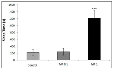
Thirty minutes after the treatment, each mouse was submitted to the sleep test. The results presented for the groups showed that there was no significant statistical difference between the animals of groups 2 (240.5 ± 100.2 s) and group 3 (219.1 ± 80.9 s), P> 0.05. However, when comparing group 1 (1217 ± 223s) and group 3, there was a significant increase in sleep time in animals treated with MP 1 μg / ml, P≤0.01 (Figure 6). The animals in group 4 did not sleep.
Figure 6:Analysis of the effect of MP extract on a sleep model induced by thiopental in mice (P <0.01, ANOVA).
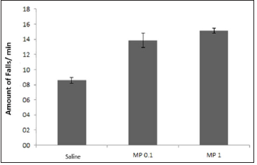
Evaluation of MP Effects in Animals Submitted to Open Field: To evaluate the effect of MP on the immobility of animals, the animals were dosed at 1 and 0.1 μg / ml (ip), and 30 minutes later they underwent the open field test. The distance traveled by the animals through the Anymaze Software was evaluated (Figure 7). There was no statistically significant difference between groups. In this stage, the effect of the substance on motor coordination was evaluated through the rota-rod test. For this, the mice previously submitted to the open field test were used for this test, after one week of the open field. No significant difference was observed in the motor coordination of control animals (saline) and those treated with different MP doses (Figure 8).
Figure 7:Analysis of the effect of MP extract on a sleep model induced by thiopental in mice (P <0.01, ANOVA).
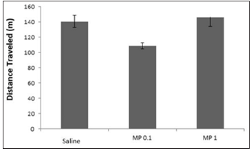
Figure 8: Analysis of the effect of MP extract on the motor coordination of mice submitted to the Rod Route (P> 0.05, ANOVA).
Discussion
In general, depression is a chronic illness that results in functional impairment and often a lifetime physical and social disability. Patients diagnosed with depression often have frequent recurrences due to incomplete recovery and residual symptoms Lopez et al. [14]. In addition to psychosocial issues, patients may also have physical health problems, such as heart disease, type 2 diabetes, autoimmune diseases and cancer Leonard [15]; McIntyre et al. [16]. Recent studies have shown that disturbances in the endocrine-immune axis could be indicative of signals of depression-related behaviors, directly relating mental disorders to physical symptoms Leonard [17]. Research on depressive and non-depressive patients has shown that depressives exhibit cardinal signs of the inflammatory process, including an increase in inflammatory cytokines and their receptors in the peripheral blood and cerebrospinal fluid Zorilla et al. [18]. The literature reports an association between the inflammatory pattern and time-specific symptoms of depression, such as fatigue, cognitive dysfunction and compromised sleep Meyers, et al. [19]; Bower.
Both in patients with inflammatory diseases and in patients with depression, sleep dysfunctions have been associated with increased levels of Interleukin-6 (IL-6), as well as activation of nuclear factor kappa β (NF-κB), this transcription factor is classically activated at initiation of the inflammatory response Motivala et al. [20]; Irwin et al. Thus, the use of a drug with direct actions on sleep, psychic disorders and inflammatory process, may be a promising therapeutic target. The aqueous extract of M. pubescens has been used in popular culture as fish narcotics to facilitate fishing in breeding sites, our results demonstrated that the form of absorption and the metabolism in mammals is quite different. Two routes of administration were tested intraperitoneally and orally for animals submitted to the thiopental sleep induction test. The animals receiving oral thiopental + MP had no difference in sleep time relative to the control group (thiopental), indicating that because it is a compound of protein origin, it undergoes digestion in the digestive tract of the animal and its metabolites do not have sedative action.
Animals receiving MP intraperitoneally had their sleep time increased at 1 μg / ml, however the MP extract per se had no sedative potential. Thus, it is inferred that the sedative action of MP is due to its capacity to potentiate the sedative effect of thiopental. These results are corroborated by the absence of the MP action in the verification of the locomotion of the animals in the open field and in the tests of motor coordination by the Rota Rod, which are complementary tests to evaluate the sedative activity. Despite the modification in IL-6 inflammatory cytokine patterns in vitro, it can be observed in the results that this pattern in vivo may not be repeated, since the literature reports the direct influence of these cytokines on the modifications of sleep patterns Motivala et al. [20]; Irwin et al.
Conclusion
From the results presented, we can conclude the aqueous extract of M. pubescens is formed by molecules of protein origin that potentiate the effects generated by i.p. of thiopental at the dose of 50 mg / kg, increased by about 6 times the sleep time in mice receiving MP 1 μg / ml (ip) + thiopental 50 mg / kg [21-32]. The doses tested had no toxicity in cultured cells and increased about 34 fold the secretion of IL-6 in macrophages incubated with 1 μg / ml MP.
Artificial Intelligence and Virtual Environment for Microalgal Source for Production of Nutraceuticals-https://biomedres01.blogspot.com/2021/01/artificial-intelligence-and-virtual.html
More BJSTR Articles : s://biomedres01.blogspot.com


No comments:
Post a Comment
Note: Only a member of this blog may post a comment.