Saturday, February 27, 2021

Questionnaire Based Study about Association between Blood Oxygen Level and Mysophobia

Questionnaire Based Study about Association between Blood Oxygen Level and Mysophobia


Introduction
Optimum level of arterial oxygen in a healthy individual ranges from 75-100 millimeter of mercury (mm Hg). Any individual with oxygen level under 60 mm Hg is suffering from hypoxemia. Hypoxemia is related to low oxygen level in blood especially in arteries, that results in trouble breathing and nausea. Blood oxygen level can be measured by Arterial-Blood Gas Test (ABG-Test). ABG test measures the level of oxygen and carbon dioxide by taking blood sample from arteries. Sometimes the ABG- test can be difficult to do at home therefore, a small device known as pulse oximeter is commonly used. Pulse oximeter is a small clip like device that does not require blood samples, it measures the oxygen level with special sensors when attached to fingertip, toe or ear lobe [1]. Although pulse oximeter is easy to use and very convenient but it is less accurate than ABG as it measure the saturation of peripheral oxygen (SpO2) from extremities, results may differ due to external factors e.g. poor circulation, dirty fingers, nail polish and standing position [2]. Normal SpO2 level measured by pulse oximeter ranges from 95-100%, while individuals with less than 90% are considered with lower oxygen level or suffering from hypoxemia [3].

Mysophobia sometimes also referred as germophobia, verminophobia and germaphobia is the distress about germs and contamination. Germophobia falls at fifth position in the Diagnostic and Statistical Manual of Mental Disorder (DSM) [4]. In 1879, William A. Hammond first time used the terminology germophobia while observing obsessive-complex disorder (OCD) of washing hands among his patients [5]. While being a germaphobe not certainly mean that individual is suffering from OCD until diagnosed with excessive and unreasonable standards of hygienic environment [6]. An individual is regarded as germaphobe if DSM-5 lasts for a period of 6 months or more. Social Phobia Inventory (SPIN) questionnaire is used to identify phobias [7]. Symptoms related to germophobia includes intensive fear of germs and being contaminated that disturb psychological heath [8]. Behavioral symptoms include excessively washing hands, use of sanitizers/antibiotics and avoiding crowded places perceived to fear of germs exposure [9].

Common physical symptoms examined in an individual suffering from germophobia are rapid heartbeat, difficulty in breathing, muscle tension and sometime chills. Genetic linkage of family history, environment issues, brain health and many other factors are believed to be the main sources of germophobia [10]. Purpose of this study was to evaluate the effect of germophobia on normal oxygen level (SpO2). To check the correlation between germophobia and saturation of peripheral oxygen that will help in estimating the body response against germophobia.

Materials and Methods
Project Design
A questionnaire was designed to check the correlation between germophobia and blood oxygen level. 200 individuals from IMBB (Institute of Molecular Biology & biotechnology) department of Bahauddin Zakariya University were questioned either they are suffering from germophobia or not. Their blood oxygen level was measured by pulse oximeter device, it checks the saturation of peripheral oxygen (SpO2 ) from fingertip. SpO2 level was examine in both male and female individuals.

Measurement of Peripheral Oxygen Saturation
Saturation of peripheral oxygen (SpO2 ) was measured in the blood by the pulse oximeter. It can be calculated with pulse oximetry according to the following formula
HbO2 is oxygenated hemoglobin and Hb is deoxygenated hemoglobin

Statistical Analysis
M Stat software was used to analyze the results. Student’s t-test was performed. p<0.05 was considered as significant.

Result and Discussion
The correlation between peripheral oxygen saturation and germophobia is shown in Table 1. T-test was performed to check the impact of mysophobia on blood oxygen level. 200 individuals were questioned while the p-value less than 0.05 was considered as significant. Analysis of collected data presents that there is no impact of blood oxygen level on the germophobia. p-value of the collected data for both male and female was greater than 0.05, which characterizes that it is non-significant. Out of 200 individuals there were 60 males and 140 females. Fifty male out of 60 individuals were germaphobe while 10 were not suffering from germophobia. 91 female out of 140 had fear of germs, while 49 were non-germaphobe. Among 200 individuals, 141 have germophobia while 59 were normal. 83.3% males and 65% females said that they are germaphobe, while 16.7% male and 35% female said that they are non germaphobe. Out of 100, total 70.5% individuals said they are germaphobe while 59% are not. p-value for male, female and both was 0.13, 0.46 and 0.65 respectively which is greater than significant value of 0.05, hereafter shows that results are non-significant for both male and female categories. Percentage of mysophobic and non-mysophobic individuals is shown in Table 2.

Table 1: Association between blood oxygen level (Mean ± S.D) and mysophobia.
biomedres-openaccess-journal-bjstr
Note: Non-significant (where p>0.05)

Table 2: Percentage of mysophobic and non-mysophobic individuals.
biomedres-openaccess-journal-bjstr
Note: Non-significant (where p>0.05).

Conclusions
In this study a correlation between mysophobia and blood oxygen level was measured. 200 individuals were questioned either they have germophobia or not, while their oxygen level was measured by pulse oximeter. Analysis and prediction of collected data shows that there is no significant association between germophobia and blood oxygen level. It shows that blood oxygen level have no clear impact on the germophobia as the calculated p-value was greater than 0.05, a standard value of significance.

Relation Between Normal Blood Pressure and Half Fried Eggs Lovers-https://biomedres01.blogspot.com/2021/02/relation-between-normal-blood-pressure.html

More BJSTR Articles : https://biomedres01.blogspot.com

Relation Between Normal Blood Pressure and Half Fried Eggs Lovers

Relation Between Normal Blood Pressure and Half Fried Eggs Lovers


Introduction
Rake air atmospheric insistency is the force that movement parentage through our circulatory system. Blood line atmospheric imperativeness is also vital because it delivers white stock cells and antibodies for granting immunity and hormones. Blood pressure is synchronized by baroreceptors which spot by way of the smartness to control nervous and endocrine systems. Blood pressure that is too low is called hypotension, and pressure that is constantly is high is hypertension. Blood pressure readings consist of two numbers first is systolic and the second is diastolic blood pressure. The systolic pressure is the higher pressure caused by the hearts muscle contraction, while the diastolic identification number is the lower pressure in the arteries during the brief resting menses between heartbeats. The device used to measure blood pressure is a sphygmomanometer and is measured in millimeters of atomic number 80mmHg, it consist of a rubber armband the manacle that is inflated by hired man or machine pump. Half fried ballock are very tasty cocked dish. These are amazing or wonderful protein in factory. Most of the people eat this cocked dish in breakfast or tiffin. Half fried testicles are tastier than omelet. These eggs are cocking in petroleum or vegetable ghee. Children’s are very fond to eat it. Idea of the existing study was to correlate the relation between normal blood pressure and half fried eggs lovers [1-10].

Data and Technique
200 objects were involved in this research or project. We were used sphygmomanometer for measuring normal blood pressure. All these objects or students were my classmate

Estimate Style: We arranged a feedback form on the subject of the relation between normal blood pressure and half fried eggs lovers.
Analysis of Statistics
a. Processing of Examining Data: M State was performed by using Statistical analysis.

Calculation and Debate
200 objects were involved in this research or project. We were used sphygmomanometer for measuring normal blood pressure. All these objects or students were my classmate. We were used Ms excel for taking the result. After knowing the blood pressure of every men and women in class room we layout these result on the page. Then we were found the value of lovers or hated half fried eggs and discussed in Table 1.

Table 1: Relation between normal blood pressure and half fried eggs lovers (Means ±SD).
biomedres-openaccess-journal-bjstr
Conclusions
We were concluded that numbers of half fried eggs lovers greater as compare to the hated half fried eggs.

Endothelial Progenitor Cells: Novel Biological Marker for Risk Stratification in Arterial Hypertension?-https://biomedres01.blogspot.com/2021/02/endothelial-progenitor-cells-novel.html

More BJSTR Articles : https://biomedres01.blogspot.com

Endothelial Progenitor Cells: Novel Biological Marker for Risk Stratification in Arterial Hypertension?

Endothelial Progenitor Cells: Novel Biological Marker for Risk Stratification in Arterial Hypertension?


Introduction
Endothelial progenitor cells (EPC) are defined as CD34(+) precursors of mature endothelial cells originated from pre-endothelial resident cells or having mononuclear origin [1]. EPCs are involved in maintaining vascular integrity, endothelial function, vascular repair and angiogenesis [2]. There is a large body of evidence regarding the pivotal role of EPCs in developing cardiovascular (CV) diseases and CV events [3-6]. For instance, nature evolution of vast range of CV diseases were associated with deficiency of circulating number of EPCs as well as weak function of endothelial precursors including mobbing, proliferation, differentiation and survival, which were incorporated into term EPC dysfunction [7]. In patients with pre-hypertension and hypertension lowered number and declined function of EPCs were identified and EPC dysfunction appeared to be a biomarker of endothelial dysfunction and arterial stiffness [8]. However, there are serious controversies in understanding whether EPCs dysfunction is causative factor for arterial hypertension or it appears to be just whiteness of nature evolution of endothelial dysfunction. Indeed, impaired endothelial repair capacity of early EPCs was found in pre-hypertensive in significantly relation to conventional CV risk factors [9,10]. However, an accelerating senescence of early EPCs was being an attribute of aging, some CV risk factors (smoking, abdominal obesity, insulin resistance, dyslipidemia), co-existing metabolic conditions including diabetes mellitus, hyperuricemia, hyperthyroidism, atherosclerosis [11].

In fact, number and function of pro-angiogenic EPCs with immune phenotypes CD34+CD133+, CD34+CD133+VEGFR2+, and CD34+CD133+VEGFR2+Tei2+ are reduced in hypertensive patients and in hypertensive disorders of pregnancy and strong correlate to severity of endothelial dysfunction. Nevertheless, clear characterization of dysfunctional phenotype of circulating EPCs in pre-hypertensive patients includes shaping of colonies and ability to trans-differentiation into mature endothelial cells and smooth muscle vascular cells were found in close relation to co-morbidity conditions, while similar association was not confirmed in arterial hypertension patients [12,13]. Moreover, there was no evidence between impaired vascular integrity evaluated as EPCs dysfunction and excess CV morbidity and mortality in patients with pre-hypertension, but for hypertensive patients including gestational hypertensive female the evidence was received [14,15]. The exact molecular mechanisms, which lead to EPC dysfunction in pre-hy-pertensive and hypertensive patients, are not fully clear. Because EPCs are able to enhance neovascularization and support vascular function through the JAK2/STAT3 signaling pathways, wide spectrum factors contributing to expression on the surface of EPC appropriate Tie2+ receptors could be embedded in this interacting.

Yet, epigenetic impact on the Lnk gene in EPCs, which are crucial for proliferation, migration, and tubule-like formation and for angiogenesis overall, is considered a core element for shaping incompetence of EPCs. There are wide ranges of stimuli that are candidates for co-regulators of epigenetic influences and receptor-targeting effectors result in both decreasing number of EPCs in peripheral blood and weak their function, i.e. inflammatory cytokines, increased fasting glucose, hyperinsulinemia, components of oxidative stress such as oxidized low-density protein cholesterol, galectines, free radicals, as well as growth factors (growth factor-bets), catecholamines, and hormones (renin, angiotensin-II, aldosterone, endothelin-1) [4,7,10]. Unfortunately, all these findings do not explain why some patients without CV risk factors demonstrate EPC dysfunction before hypertension manifestation. For instance, in early pregnancy female at risk of preeclampsia and with established preeclampsia the number of circulating pro-angiogenic EPCs was lower in comparison to pregnancy female in the absence of preeclampsia risk [16]. Additionally, EPC colony formation, as well as differentiation and migration abilities were also impaired in the gestational hypertensive female [17]. It has been speculated that vascular health in early pregnancy could be altered in women with aberrant numbers of EPCs with pro-angiogenic immune phenotypes (CD34+CD133+VEGFR+ or CD34+CD133+VEGFR+Tie2+) and might represent significant CV maladaptation contributing to an increased risk of preeclampsia. The majority of data received by several investigators supported a hypothesis that EPCs dysfunction reflects an endothelial alteration that accompanies arterial hypertension and that the extent of endothelial damage does not appear to be associated with the severity of the disease [18].

Additionally, in pulmonary hypertension colony forming ability of EPCs was rather associated with right ventricular (RV) remodeling and RV dilation than pulmonary vascular obstruction and disease severity [19]. However, EPC dysfunction is novel biological marker of endothelial dysfunction and vascular reparation in hypertension with predictive value requires to be investigated in large clinical trials. In conclusion, hypertensive patients present greater apoptosis and impaired repair capacity EPCs that they were found in healthy individuals, but perhaps EPCs dysfunction is not cause of hypertension, while impaired EPC function may contribute to target organ damage manifestation and appearing CV complications.

Resveratrol Play as Anti-Heat Stressor-https://biomedres01.blogspot.com/2021/02/resveratrol-play-as-anti-heat-stressor.html

More BJSTR Articles : https://biomedres01.blogspot.com

Friday, February 26, 2021

Resveratrol Play as Anti-Heat Stressor

Resveratrol Play as Anti-Heat Stressor


Opinion
A natural botanical polyphenolic compound, resveratrol (3,5,4’-trihydroxy-trans- stilbene) was reported to have anti-oxidation, anti-tumor, anti-inflammatory, neuro-protection and cardiovascular protection effects [1] Numerous studies have shown that resveratrol effectively eliminated reactive oxygen species (ROS), reduced DNA fragmentation, and enhanced the regulatory effects on growth factors and anti-inflammatory cytokines [1]. Resveratrol was considered to be important bioactive compound in monomers, and it has shown strong antioxidant capacity including scavenging free oxygen and lipid radicals [2]. Resveratrol is able to inhibit the formation of glutathione disulfide and keep glutathione in a reduced state, thereby inhibiting free radicals [3]. It has also been shown that resveratrol and its derivatives have activities of antimicrobial (bacterial), and anti-proliferation of cancer cells [4]. In recent study, resveratrol was shown to protect DNA damage against oxidative damage [5].
It has been reported that some plant polyphenols can also be used as anti-stress additives of livestock and poultry to alleviate tissue damage caused by heat stress [6,7]. Heat stress causes an imbalance in physiological status, a general reduction of the antioxidant system, and a reduction in the immunological function of chickens [8]. The research team leading by Dr. Jianhua He conducted a series of trial with broiler chicken to verify the anti-heat stress effect of resveratrol [1,9,10]. The resveratrol (≥98% purity) they used to be extracted from Polygonum cuspidatum at Hunan Engineering and Technology Center for Natural Products. He [9] reported that Resveratrol supplementation improved average daily gain (p = 0.001) and decreased (p < 0.05) rectal temperature from d 3 when compared with heat-stressed (maintain room temperature at 34℃ for 8h from 9:00 to 17:00) birds fed diet without resveratrol. In addition, supplementation with resveratrol at 350 or 500 mg/kg lowered (p < 0.05) the contents of corticosterone, adrenocorticotropic hormone (which are highly related to heat stress); it also decreased the content of triglycerides, uric acid, malonaldehyde, and activities of aspartate aminotransferase, alanine aminotransferase, and lactate dehydrogenase, increased (p < 0.05) the levels of triiodothyronine, the ratio of triiodothyronine to thyroxine, total protein, glutathione, and activities of alkaline phosphatase, total superoxide dismutase, catalase, and glutathione peroxidase, the above results suggested that dietary resveratrol supplementation could improve the growth performance by positively regulating serum metabolic parameters and alleviating tissue oxidant damage of broilers under heat stress. The results provided by Liu [10] also showed that dietary supplemented with resveratrol improved growth performance, feed intake and body weight gain of blackboned chickens during heat stress period, while feed conversion ratio was decreased. And it reduced oxidative stress in heat-stressed black-boned chickens by increasing serum growth hormone concentrations and modulating the genes expression of heat shock protein in organs of the immune system. She also found that dietary supplementation with 400mg/kg of resveratrol improved the villus morphology, increased the numbers of goblet cells and lymphocytes, attenuated the mRNA overexpression of HSP70, HSP90 and NF-κB on the 6th, 10th and 15th days of heat stress (maintain room temperature at 34℃ for 8h from 9:00 to 17:00)(P <0.05), activated the expression of EGF (P <0.05) in the jejunal mucosa.
Resveratrol reduced protein expression of HSP70, HSP90 and NF-κB in the jejunal villus after 15-days heat stress, increased EGF expression from the lamina propria toward the epithelial cells of the villus. These results suggest that dietary resveratrol offers a potential nutritional strategy to improve intestinal morphology and alleviate jejunum mucosa injuries by modulating the mRNA and protein expression of HSPs, epithelial growth factor and transcription factor in black-boned chickens subjected to circular heat stress. Liu [1] found that Resveratrol attenuated the heat stress-induced overexpression of Hsp27, Hsp70, and Hsp90 mRNA in the bursa of Fabricius and spleen and increased the low expression of Hsp27 and Hsp90 mRNA in thymus upon heat stress. Preliminary studies also have indicated some benefits of resveratrol supplementation to alleviate negative effect of heat stress, and increased SOD, GSHPx and CAT enzyme activities in mice [11], quail [7] during heat stress exposure. Das [11] also observed that resveratrol is able to resist the body organ dysfunction induced by heat stress. Sahin [7] reported that resveratrol can inhibit the high-levels of HSP70/90, NF-κB proteins in quail liver during heat stress. In conclusion, dietary supplementation of resveratrol could alleviate the heat-stress effect of broiler by reduced oxidative stress and its recovery effect of intestinal damage caused by heat stress. It was suggested that eat resveratrol containing herbs or food may play benefit effect for Human in tropic area.

Correlation of Normal Peripheral Oxygen Saturation with Tea Likeliness-https://biomedres01.blogspot.com/2021/02/correlation-of-normal-peripheral-oxygen.html

More BJSTR Articles : https://biomedres01.blogspot.com

Correlation of Normal Peripheral Oxygen Saturation with Tea Likeliness

Correlation of Normal Peripheral Oxygen Saturation with Tea Likeliness

Introduction

Blood oxygen level represents the fraction of oxygen i.e. saturation or unsaturation in haemoglobin of Erythrocytes. Blood should maintain normal levels of oxygen for aerobic metabolism which helps in Breathing. Human body needs specific oxygen level in blood and normal oxygen level in blood of human beings should be 97 percent and it can fluctuate during exercise. Haemoglobin of red blood cells collects oxygen in the lungs by using respiratory system and distributes it to the whole body. Oxygen level below 90 percent (low partial pressures of oxygen) leads to hypoxemia because hemoglobin is deoxygenated. The symptoms of low blood oxygen level include changes in the color of nails and skin i.e. bluish color appears. Blood oxygen level below 40 percent leads to compromise the function of Brain and Heart and Blood oxygen level below 20 percent leads to comma and ultimately, it causes death. Continue low levels of Blood oxygen causes many serious problems like cardiac, respiratory and neurological problems. A pulse Oximeter is an instrument which indicates the oxygen level in our blood. Oxygen therapy can be used to raise blood oxygen levels.

In ancient years, Tea was used as an aromatic beverage for many medicinal problems because it helps in improving our health i.e. it contains a chemical known as Nicotine. Now a days, Tea is also used to regulate the blood level of human beings because it has boosting antioxidants like catechins and ingredients which helps in the activation of human Brain and releases tension of the persons. Tea is simply prepared by the addition of teabags and milk in kettles of boiling water with a bit sugar according to one’s taste. It is considered as White tea and it is the simplest tea. Tea is also considered as a lifestyle for the present generation. Many types of tea have been discovered yet i.e. White tea, Black tea, Green tea and Kashmiri tea etc. Black tea is considered as a strong tea because it contains potential ingredients in it and it is taken mostly in Europe. Green tea has its own great importance because it is taken as a diet tea i.e. it is helpful in the dieting. Kashmiri tea is used mainly in gatherings and it is used as a trend in Asian countries to serve Kashmiri chaye to guests. In short, Different types of tea have different nutritional and health values. By addition of Fruit flavors to the tea, we can get natural health benefits. We can also add the flavors of different things like chocolate. Tea has given us many benefits like it can reduce cancer related diseases and heart diseases. Furthermore, it gives us energy and refreshes our mind but excess of everything is bad. Therefore, we should not take more than 4 cups of tea per day. The aim of the recent study was to interlink the Blood oxygen level with Tea likeliness [1-5].

Materials and Methods

Approximately 200 participants took part in this research and Blood oxygen level of every participant was checked by using Oximeter. To interlink Peripheral oxygen saturation with Tea likeliness, Questionnaire was prepared to estimate the likeliness and Dislikeliness among participants.

Estimation of Blood Oxygen Level

Peripheral Oxygen Saturation represents the oxygen levels in blood, and it can be measured by using a device named Pulse Oximeter which clips to the finger or earlobe, light sensitive sensors in the device measure absorption of red and infrared light and it calculates the Blood Oxygen Level by finding the difference between oxygenated and deoxygenated hemoglobin levels in blood. By using a Formula, we can calculate the Blood Oxygen Level with Pulse Oximeter as follows:

Spo2 = Hbo2/Hbo2 +Hb

Where HbO2 = Oxygenated Hemoglobin

Hb= Deoxygenated Hemoglobin

Statistical Analysis

Statistical Analysis was done by using state software. To analyze the results, t-Test was done, and p-value was observed by considering p<0.05 as significant value.

Results and Discussion

t-Test was used to analyze the Correlation of peripheral oxygen saturation with Tea likeliness. In Table 1, Correlation of Peripheral Oxygen Saturation (Mean±SD) with Tea likeliness among males, females and Both (Males and Females) is given as follows in which p-value is calculated by t-Test is greater than 0.05 i.e. it is non- significant (p-value smaller than 0.05 is referred as significant while p-value larger than 0.05 is referred as non- significant) and it means that there is no correlation of Peripheral Oxygen saturation with Tea likeliness [6-8]. This Questionnaire based studies will give great importance to the latest researches because no research has done before this about correlation of normal blood oxygen level with Tea likeliness. Such type of researches show advancement in the scientific era and give us broad idea to relate different variables with different Blood factors. In this way, we can also know about the diagnosis of different diseases related to human beings.

biomedres-openaccess-journal-bjstr

Table 1: Co-rrelation of Peripheral Oxygen Saturation (Mean±SD) with Tea likeliness among Males, Females and Both (Males and Females).

Note: Results were non-significant ( p<0.05).

Conclusion

By Statistical Analysis, it was interpreted from the recent research that Peripheral Oxygen Saturation with Tea likeliness does not correlate in human beings because p-value was larger than significant value which was calculated by using t-Test (p<0.05).


Active Probe Emitting HF Electromagnetic Fields and Piezoelectric Emissions in Association with Local Carbon Nanotubes : Preliminary Tests in C6 Glioma Cell Death Induction: A Possible Application in Glioblastoma Therapeutics ?-https://biomedres01.blogspot.com/2021/02/active-probe-emitting-hf.html


More BJSTR Articles : https://biomedres01.blogspot.com

Active Probe Emitting HF Electromagnetic Fields and Piezoelectric Emissions in Association with Local Carbon Nanotubes : Preliminary Tests in C6 Glioma Cell Death Induction: A Possible Application in Glioblastoma Therapeutics ?

Active Probe Emitting HF Electromagnetic Fields and Piezoelectric Emissions in Association with Local Carbon Nanotubes : Preliminary Tests in C6 Glioma Cell Death Induction: A Possible Application in Glioblastoma Therapeutics ?

Introduction

Malignant Glioma (MGs) variants, Glioblastoma (GBM) is the most common malignant tumor in the central nervous system. His prevalence is about 1/100 000, with a survival time post-diagnosis of 15 months and a mortality rate exceeding 95% [1]. Common treatments for GBM include surgical resection, radiotherapy, chemotherapy, or combinations of these three modalities [2,3]. Nevertheless, there is no general agreement on the radiation pro tocol to use. If fractionation seems to be the most appropriate, the choice mainly depends on the radiotherapy options available to the treating physician. In case of recurrent glioblastoma, the treatment is then focused on the use of chemotherapy. Numerous studies have demonstrated the safety and efficiency of various agents, both alone and in combination [4-8]. GBM have an extremely low survival rate due to the high infiltrating properties of the glioma cells. Typically, these cells could infiltrate up to 2 cm exceeding the volume of visible tumor, making them difficult to detect and treat. Treatment of GBM is also limited by several other factors such as, the insufficient delivery of chemotherapy drugs caused by the blood-brain-barrier, the radio resistance of the cells and the need to preserve functional parenchyma.

Despite the existence of different therapeutic strategy treatments, the evolution of the post-diagnosis median survival only increased from 13 to 15 month, highlighting the need of curative approaches. In the recent past, some new innovative researches have produced promising results or are the subject of ongoing investigations [9-16]. Among them, we can find biophysics approaches using electromagnetic fields or nanoparticles. The results of Kirson’s group highlighted the first point. This team have shown a decrease of the malignant cell proliferation with no or minimal side effects in animal tumor models and human brain tumors [11.12] by using an alternative electric field (also called TT Fields for Tumor Treating Field). According to the authors, the mechanism involved is related to the disintegration of the mitotic spindle [11]. NovoTTF therapy is a new therapy using this observation and has undertaken the phase III clinical trial [13].

Another Group (Magforce AG) is using iron nanoparticles combined with an alternative magnetic field (100 kHz). This field induces nanoparticle’s vibrations and then, locally increases the temperature leading to the tumor cells death. The main drawback of such approach is the lack of specificity. Even if nanoparticles are addressed to malignant cells, the increase in temperature destroys all the cells located in the vicinity of the particle. However, this therapy seems efficient and safe with no or moderate side effects [14-16]. We propose an alternative project using these two different aspects by using both nanoparticles and electromagnetic field without inducing any thermal effect. This new approach is inspired by recent works [17] showing a strong interaction between Carbon Nanotubes (CNTs) and electromagnetic field.

According to this observation, a synergic toxic effect between electromagnetic fields (RF) and CNTs could be expected. CNTs could increase the toxicity of the RF by focusing them and inducing an electromagnetic field increase. The RF could also increase the toxicity of the CNTs by a direct helping of their dispersion and penetration into the cells. The intrinsic characteristics of CNTs make them specifically sensitive to electromagnetic field. Due to theirs high length compared to their diameter, they could be at the origin of a strong point effect (like a lightning rod) inducing a local increase of the fields’ strength. Both CNTs and electromagnetic field are major public health issues and many studies tried to determinate their toxicity [18-21].

The drawbacks of both Kirson and Magforce AG studies was that the field emitter (electrodes on the skin / external magnetic emitter) were located outside the body, i.e. far away from the target. As field power density generally decreases with the square of the distance, using an intra cerebral emitter, combined with CNTs induced local magnification of the field would be promising. Such mechanisms would clearly differ from those of Magforce AG team, since neither tissue heating nor thermal reaction occur in the former case. Final therapeutic hope would use a combination of the convective infusion technique [22,23] together with the inclusion of the RF emitter in the catheter used for intra-tumoral injection. The aim of this study was to evaluate the effects of a co-exposure to carbon nanotubes and combined electromagnetic field/ultrasounds on an in vitro glioma model.

Materials and Methods

Cell Culture

The C6 cells (Rat Glioma) were obtained from the ATCC cell base (ATCC® CCL-107™). The cells were maintained in exponential growth in a 5% CO2 humidified incubator held at 37°C. The C6 cells were placed in cell culture flasks (75cm²) and were grown with 12 ml of DMEM, Glutamax supplemented with 10 % heat-inactivated fÅ“tal bovine serum and antibiotics (100 U/ml penicillin and 100 µg/ml streptomycin).

Synthesis and Particles Characterisation

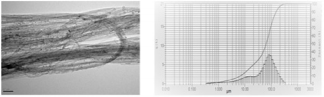
Single wall carbon nanotubes (CNTs, quoted here CNTs, Figure 1) produced by Catalytic Chemical Vapor Deposition were purchased by SIGMA-Aldrich, St Quentin Fallavier, France. The CNTs diameter distribution was ranged from 0.7 to 1.3nm, and length between 450 and 2300nm, although bundles may be much longer (up to 100µm at least) with a density of 1.7-1.9 g/cm3 at 25°C.

biomedres-openaccess-journal-bjstr

Figure 1: Left : Electron microscope of CNTs in suspension in aqueous medium after dispersion Left : relative distribution of CNTs aggregates in suspension.

Particle size distribution of the stock solution used for cells exposure was analysed with a Laser Scattering Particle Size Distribution Analyser, LA-950V2 (HORIBA) with DMEM as solvent. Particles dispersion in cell culture medium and cells exposure : Stock solution of 5 mg/ml CNTs was prepared by dispersion of sterilised particles in DMEM containing 25 % FBS. Previous study has reported that serum produced particle suspensions were observed with the fewest large agglomerates [24]. The nanoparticles were homogeneously dispersed by vortexing for 30 seconds followed by sonication in a water bath (Al 04-02, Advantage Lab) for 5 min. This procedure was repeated five times. A “Sham Stock Solution” of sterile DMEM containing 25 % FBS was prepared and sonicated at the same time for use as control.

The stock solutions were kept at 4°C. The stock solution of CNTs was resuspended by vortexing for 30s followed by sonication for 5 min and diluted in cell culture medium to the required concentration (50 µg/ml, 120µl per flask). An equal volume of “Sham Stock Solution” was added to cell culture medium for control cells. Before any experiment, CNTs size were analyzed using a Laser Scattering Particle Size Distribution Analyser, LA-950V2 (HORIBA) with DMEM as solvent (10%) and a dilution at one centesimal (150µl in 15ml). The size of the CNTs measured by light scattering was defined as the equivalent spherical diameter and could not be related to the exact particle size but represent a relative size of nanotubes which are long cylinders. This led to identify different particle sizes in solution : 100µm diameter aggregates (35%), a minor component of intermediate aggregates (1µm-30µ). Sizes below 1µ were considered as unbounded carbon nanotubes.

Temperature Control

The absence of temperature increase was controlled over 16Hrs exposure under the RF/CNTs conditions with probe immersed in the culture flask, using a FLIR A320 Temp screen camera. No significant variation was detected within the observation period.

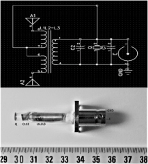
Measurement of the electromagnetic field : The probe was composed of three parts (electric, magnetic and ultrasonic with a piezoelectric component, CNTs) realizing the emission of an electromagnetic signal and a piezoelectric operating phase. Electromagnetic fields were then applied by connecting the wires to an amplifier operated by a signal generator with frequency and amplitude controls. The probe was powered by a square wave modulated signal working at 100 kHz (low-intermediate frequency emission) from an Agilent 3312DA generator, peak to peak tension of 10Vpp, and duty cycle DC=50%. This first prototype version is presented Figure 2.

biomedres-openaccess-journal-bjstr

Figure 2: Top: Electronic wiring of the probe; Bottom: The probe, as connected with BNC connectors. GND: Ground; E: RF input via BNC connector; Q: 100KHz resonating piezo; C1,C2 tune capacitors; L1=1mH, L2=L3=16mH; L2L3-L1transformator; A1, A2: antennas.

Field estimations were attempted with a PM03 field meter and a H/E fieldmeter ESM-100, Meshket. As the exposure conditions (inside the flask) were obviously in reactive nearfield conditions, E values were extremely heterogenous (5dB , with a maximum of 80V/m in close vicinity of the probe extremity ) while H variations were found less important ( 3dB , maximum 90nT in close vicinity of the probe extremity). The 100kHz resonating piezo was a PRYY0073 from PICERAMIC, Germany. The electric field was evaluated with the help of a PMOR 03 field meter. In order to isolate the exposed flask, the electromagnetic exposed flask was placed into an absorbing chamber allowing an attenuation of the signal by 12 dB.

In vitro Experimental Setup

Each flask was randomly assigned to one of the four different groups: Sham / Sham, Sham / Nano exposed, Sham / Field exposed, and Nano / Field exposed (CNTs/RF+piezo, quoted CNTs/RF in the followings). All the cultures were realized in triplicate under the same conditions as the CNTs/RF group except that the generator was turned off, with the probe into the flask. The initial C6 cell population was 0.2* 106 cells per flask. 72h exposure starts immediately after the cells have been passaged. Sham / Sham and Sham / Field groups received 120µl of the Stock Sham solution (without CNTs), the double exposed and the Sham / Nano groups received 120µl of the Stock solution containing the CNTs. After exposure, in order to eliminate the SWCNTs in solution, each flask was rinsed 3 times with Phosphate Buffered Saline (PBS). The cells were chemically detached from the flask using trypsin EDTA. C6 cells were centrifuged 5 min at 300G. Cell pellets were resuspended in 1 ml cell culture medium. 50 µl of C6 cells solution were added to 50 µl of Trypan Blue. C6 cells were counted by a TC20TM automatic cell counter. Results were expressed as means ± Standard Deviation (S.D.)

Results

Cells Count and Viability

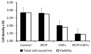
In these experiments, cell counts were performed after 72h culture with/without RF exposure and/or in the presence or not of CNTs (see Figure 3). By comparison with reference sham values measured at 72h (2,84 106/mL) and also after 72Hr RF exposure (2,8 106/mL) an important reduction in absolute values of cells counts was found when CNTs were present alone (2.106 /mL) and especially when CNTs were associated with RF exposure (1,46 106/mL) . These results are all the more evident when expressed in percentage to reference sham values and showed an important decrease in the number of the cells compared to the control group (51% for the Nano / Field group; 75% for the Sham / Nano group; 98% for the Sham / Field group). Conversely, the survival rate of the cells within any group was only slightly decreased whatever the exposition factors considered : hence, the survival rate is maxima (100%) for the Sham / Sham control group and slowly decreases with the exposition of CNTs and electromagnetic field (97% survival rate for the Sham / Field group, 95% survival rate for the Sham / Nano group and 94% survival rate for the Nano / Field group. As this step it is worth to note than rinsing process (see methods) induced an averaging over the whole flask, this resulting in a smoothing of any possible local drastic effect. Morphological control had thus to be performed.

biomedres-openaccess-journal-bjstr

Figure 3: Cell count (cells/ml 106 related to sham / sham group (percentage) and survival rate distribution in C6 cell population with exposition of SWCNTs or electromagnetic field according to their respective group. The cells were exposed for 72h right after injection of the cells into the flask.

Cell Morphologic Observation

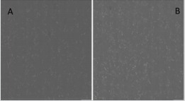
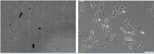
As mentioned above, microscopic observation (Figure 4) clearly shows the reduction of cell density in the flask, even like here after the rinsing process between CNTs/RF and sham groups. Moreover, the absence of morphological change was also in full agreement with the minor differences in cell viability between groups. Here differences appear between CNTs and CNTs/+RF groups. As noted in the methods section, CNTs spontaneously selforganized in µ-sized aggregates, with only a partial contribution of dispersed nanoparticles. This was observed on the Figure 5A, especially before rinsing of the flasks, where these aggregates are visible even at low magnification (x4). Such a distribution appeared partially overcome (or really dispersed) due to rinsing, as shown on Figure 5B, where higher magnification allows to detect smaller particles among the C6 cells, without specific contact or geometric distribution.

biomedres-openaccess-journal-bjstr

Figure 4: A: Sham / Sham group cells after 72h exposure time (X4 magnification); B: CNTs + RF group cells after 72h exposure time and rising of the flask (X4 magnification).

biomedres-openaccess-journal-bjstr

Figure 5: Sham / Nano group cells after 72h exposure time with nanotubes aggregates, before the rinsing process, with X4 magnification (left) and x10 magnification (right).

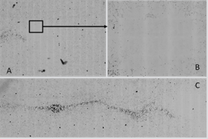
Besides, no evidence for cell distribution, i.e. heterogeneity around the probe (not emitting, sham) was observed. Such was not the case in the combined CNTs/RF samples (Figure 6). Several CNTs aggregates were still present whereas more numerous and of smaller size, in an area close to the extremity of the probe. This feature was not reasonable to quantify - a factor of 2 to 5 ? - due to the important heterogeneity in the half centimeter around the probe wall. However, a true “no cell area” was truly present in the several millimeters around the probe. Furthermore, linear arrangements of CNTs were observed (Figure 6) in the closest vicinity of the antennas (wires inside the probe while aligned along its main axis) , this revealing true interactions between CNTs and RF and/or ultrasound coming from the piezo.

biomedres-openaccess-journal-bjstr

Figure 6: CNTs + RF group after 72h exposure time showing a cell depleted area close to the probe extremity; longitudinal CNTs orientation in the CNTS+RF group after 72h exposure time before rinsing resulting homogenization.

Discussion

The aim of the preliminary work was to evaluate the effects of a co-exposition from carbon nanotubes and electromagnetic field on an in vitro glioma model. The Sham / Field group cell count showed a limited decrease of the C6 cells population compared to the Sham / Sham group and a similar survival rate between the two groups, 99% and 100% respectively. Compared to Kirson’s results several differences arise (9,11) :in the present work, the frequency was fixed at 100 kHz while Kirson’s group used a 100-300 kHz frequency system, with an emitting power twice as ours. The Sham / CNTs group cell count showed a 25% decrease of the C6 cells population compared to the Sham / Sham group with a 97% survival rate. CNTs alone have a strong effect on C6 cells. This could be related to their own direct or indirect toxicity related with the ability of CNTs to trap growing factors in a culture medium [18-21]. The most interesting results were to find a synergic effect of the CNTs with the electromagnetic field. The Nano + RF cell count showed decrease to 51% of the C6 cells population compared to the Sham / Sham group with a 95% survival rate. As an hypothesis, the SWCNTs could induce an increase of the local field’s strength and a disruption of the field direction. According to the mechanism of action of TTFields described by Kirson, TTField efficacy must be a function of the angle between the field and axis of division of the cells; when the two are parallel it has a maximal effect and when one is perpendicular to the other, it has a minimal effect [11].

In our case, the carbon nanotubes could solve this problem as they might cause a local reorientation of the field, overcoming the previous problem. In addition, the effect of the piezoelectric component might be useful to open the blood-brain-barrier and increase the dispersion of the CNTs into the tumor after a local injection using the convective infusion technique [23]. The results showed a strong effect associated with an important decrease of the C6 population subjected to the double exposure. However, the survival rate of the cells is still high and this combination tends to prove that the effect of the co-exposition is not on the destruction of the cells herself but by an inhibition of the cell proliferation [11].

At this step it is not possible to ensure any specific effect due to the several bias of the study. First of all, the studied population of cell wasn’t enough to draw conclusion. More experimentation should be performed in order to obtain a strong statistical analysis. In addition, most of the dead cells are eliminated during the rising process because they can’t bind the flask anymore once they lost their structure. In order to include these cells into the final calculation, centrifugation of the rising solvent might be a good idea to retrieve dead cells. Another possibility would be gel phase culture and direct final count by optical density lecture , for instance. Besides, the specific effect of each component separately or combined with one or all the others should be addressed, for instance separate piezo and RF contribution and association with CNTs. Finally, the particle exposure could also be improved.

The particle size distribution showed that the dispersion of the carbon nanotubes was neither homogenous not ideally dispersed. Due to their strong hydrophobic properties, CNTs bind each other and form large aggregates. This could diminish the efficacy of the CNTs on the cells and hide a potential effect. The CNTs concentration used here (50µg/ml) may be at the origin of the problem and using a lower concentration could increase the dispersion of the carbon nanotubes. This concentration was chosen based on the previous studies on the CNTs toxicity [18,21,27]. The CNTs concentration isn’t the only parameter that could affect the results. The previous pictures were obtained using a phase contrast technique that could not distinguish apoptotic cells and necrotic cells (especially at the beginning of an apoptotic process).

Flow cytometry imaging could solve this problem, with a Hoechst stain to visualize the cell nucleus or even a fluorescent stain using PKH [28,29]. From a mechanistic point of view, different cell types should be tested to ensure (or not) the existence of aspecific mechanisms at mitotic bundle destructuration, as mentioned by Kirson. From this , and by designing the probe small enough to be implanted in living systems, in vivo experimentation procedures would be envisaged. These different points are presently addressed, and we plan to set up a “matches-sized” probe, including a coaxial catheter for direct perfusion of CNTs or charged drugs. Hence other drugs could be carried by the convective infusion system and the combination with a special drug carried by CNTs [30]. The idea of an intra tumoral catheter including an RF emitter allowing the distribution of chemotherapy or carbon nanotubes using the convective infusion technique seems promising. This thematic actually plans to in vivo animal experimentations procedure where other questions such as possible blood-brain-barrier opening and safety evaluation of such therapy.


The Effectiveness of Polarized Polychromatic NonCoherent (BIOPTRON) Light in the Management of Acute Lateral Elbow Tendinopathy: A Case Report-https://biomedres01.blogspot.com/2021/02/the-effectiveness-of-polarized.html


More BJSTR Articles : https://biomedres01.blogspot.com

Thursday, February 25, 2021

The Effectiveness of Polarized Polychromatic NonCoherent (BIOPTRON) Light in the Management of Acute Lateral Elbow Tendinopathy: A Case Report

The Effectiveness of Polarized Polychromatic NonCoherent (BIOPTRON) Light in the Management of Acute Lateral Elbow Tendinopathy: A Case Report


Introduction
Lateral elbow tendinopathy (LET) commonly referred to as tennis elbow and / or lateral epicondylitis is the most common tendinopathy in the elbow area. Pain and decreased function are the main symptoms of LET [1]. Diagnosis is simple. The symptoms are reproduced by
a) Gripping activities;
b) Palpation on the site of pain (facet of the lateral epicondyle); and
c) Clinical tests such as resisted middle-finger extension and/ or cozen’s test [1].
The management of LET is usually conservative. A plethora of physiotherapy modalities have been recommended for the management of LET. The effectiveness of the recom mended physiotherapy treatments has been investigated in chronic LET. Chronic LET is failed healing tendon response or degenerative rather than inflammatory [2]. To our knowledge, there have been no studies to find out the effectiveness of physiotherapy modalities in acute LET. More recently, physiotherapists can use a new modality called polarized polychromatic non-coherent (BIOPTRON) light. However, the evidence of the Bioptron light in the management of LET, acute or chronic, is minimal. Therefore, the objective of the present report is to find out the effect of Bioptron light in acute LET.

Case Report
Ms. V., a 23- year-old female complained of acute pain (less than 48 hours) in the lateral aspect of her right elbow, of her dominant hand. The pain was on the facet of the lateral epicondyle. She played tennis for about 4 hours, 36 hours ago. She experienced pain about 6 hours after completing the tennis practice. Since then, she complained of constant pain. She was not able to sleep. She did not receive any NSAIDs. She did not have any other symptoms or any problems in peripheral joints or in the spine. In the cozen’s test (extension of the wrist with the elbow in extension) the power was 1 on the Oxford scale and there was pain over the facet of the lateral epicondyle of the humerus, 9/10 on the VAS. Resisted extension of the middle finger was painful (9/10 on the VAS) on the facet and the power was 1 on the Oxford scale. The reported pain of the patient in the handgrip dynamometer test was 9/10 on the VAS.

All the other movements (passive, active and under resistance movements of the elbow, wrist and neck) were pain free, with full range of motion and strength. Finally, the pain was reproduced by palpation over the common extensor tendon on the facet of the lateral epicondyle of the humerus. The patient received Bioptron light therapy. A Bioptron 2 device administered Bioptron light therapy. Bioptron light therapy was administered to three locations for 6min in each location (i.e. 18 min in total) [3]. The probe of the Bioptron 2 was held at a 90oangle 5-10cm above the clean bare skin of the lateral condyle (1) from the upper surface (anterior) with the elbow in extension and the forearm in supination and (2) from the lateral surface with the elbow in 900 of flexion and the forearm in pronation. In addition, the probe of Bioptron 2 was held at a 900angle 5-10cm above the clean bare skin of the bellies of the extensor’s muscles of the wrist with the elbow in 900 of flexion and the forearm in mid-position of pronation - supination.

The emission of light was administered in 1-min steps and controlled by an integrated soft-start/soft-stop electronic switch. When the treatment with Bioptron 2 was over, there was a characteristic sound (beep tone). Treatment was delivered twice a day (morning and afternoon) for five consecutive days providing ten sessions in total. Function and pain were measured in the present study. The patient was evaluated before and after the treatment period. Pain and function were measured on a visual analogue scale (VAS), a valid and sensitive approach of the LET [4]. In addition, pain-free grip strength was used to measure function as described in previous trial [5]. Finally, pain and function were measured using the Patient-Rated Tennis Elbow Evaluation (PRTEE). The PRTEE questionnaire, provides a very quick (it takes less than 5 min to complete), easy, and standardized quantitative description of pain and functional disability in patients with LET. It has been translated and culturally adapted into Greek [6].

Results
Pain on VAS was 9, function on VAS was 1, pain-free grip strength was 18lb and the score of the PRTEE questionnaire was 94 at the initial evaluation. At the end of the treatment (session 10), there was a decline in pain on VAS of 6 units, a rise in function on VAS of 6 units, a rise in pain-free grip strength of 35 units and a decline in the score of the PRTEE questionnaire of 74 units (Table 1).

Table 1: Pain, function, pain-free grip strength and PRTEE questionnaire over the 24 h before each evaluation.
biomedres-openaccess-journal-bjstr

Discussion
The present case report has looked at the effect of Bioptron light in a patient experiencing acute LET and its findings have demonstrated significant improvements in terms of pain and disability. The results obtained from this case report are novel; as to date, similar studies have not been conducted. The most common physiotherapy treatment for LET is a supervised or in clinic exercise program [2,7]. However, the patient with acute LET was not able to perform an exercise program due to pain. The patient was advised by the clinician to follow an exercise program after completing the Bioptron light treatment. Like Low Level Laser Light (LLLL), Bioptron light is also a low-power light source but differ from LLLL in that Bioptron light is incoherent and polychromatic rather than coherent and monochromatic [8]. Moreover, Bioptron light combines infrared light at a wavelength of 700–3400nm and visible light at a wavelength of 480-700nm [8]. In contrast, LLLL contains either infrared or visible light at one specific wavelength [8].

Several drawbacks have impaired the usefulness of LLLL in comparison to Bioptron light, such as high risk, required user skills, the small diameter of the laser beam, which allows only a limited area to be treated and high cost [8]. Both the infrared and visible parts of the electromagnetic spectrum of Bioptron light can explain its mechanism of action [8]. It is probable that Bioptron light has biostimulative effects improves the blood supply and accelerates the cellular mechanisms [8], but more research is needed to investigate how this occurs. Since pain relief and improvements in function were noted in the present case study on a short term, it is proposed that Bioptron light may potentially have promoted an anti-inflammatory effect in the soft tissues [8]. However, to understand the potential changes to the tissues in response to Bioptron light therapy, future studies should consider employing outcome assessments that can monitor the changes in deeper tissues. The present case study was the first report to examine the effectiveness of Bioptron light on acute LET.

Previous research assessed the effectiveness of Bioptron light on chronic conditions such as LET [3,9] and carpal tunnel syndrome [10,11]. However, acute and chronic conditions are two different conditions and the results are not comparable. In addition, one controlled study assessed the effectiveness of Bioptron light in acute ankle sprains [12], but ankle sprain is a ligament injury whereas LET is a tendon injury. Therefore, ligament injury and tendon injury are two different conditions and the results are not comparable. The previously reported trials found that a course of Bioptron light may improve patients’ symptoms. The findings of these trials encourage the design of future well-designed RCTs that might produce strong evidence for the effectiveness of Bioptron light on sports / musculoskeletal injuries. A course of Bioptron light treatment was applied in the present study based on manufacturers’ claims [13]. It is a dose-response modality and the optimal treatment dose has obviously not yet been discovered.

Future studies are needed to standardize Bioptron light parameters in the management of LET (acute, chronic and calcific) and on other sports / musculoskeletal conditions. Although the positive effects of such a treatment approach in acute LET have been reported in the present study, its study design limits the generalization of these results. Future well-designed trials are needed to confirm the results of this case report establishing the effectiveness of such a treatment approach in the management of LET (acute, chronic and calcific). In addition, structural changes in the tendons related to the treatment interventions are needed to investigate. Further research is needed to establish the cost-effectiveness of such treatment, because reduced cost is an important issue for the recommendation of any given treatment and the possible mechanism of action of this treatment approach [8].

Conclusions
A course of Bioptron light treatment had reduced the pain and improved the function in a patient with acute LET at the end of the treatment. Further well-designed trials to confirm the results of the present case study are needed.

Can Aerobic Training Restore the Vascular Dysfunction Induced by Intrauterine Growth Restriction? Evidences from Experimental Studies-https://biomedres01.blogspot.com/2021/02/can-aerobic-training-restore-vascular.html

More BJSTR Articles : https://biomedres01.blogspot.com

Semaglutide in Obesity and Non-Alcoholic Fatty Liver Disease

  Semaglutide in Obesity and Non-Alcoholic Fatty Liver Disease Introduction Semaglutide injection was introduced in the US as an antidiabeti...