Branching Patterns of the Facial Nerve within the Parotid Gland Among Sudanese
Introduction
The extratemporal component of the facial nerve starts when
the facial nerve exits the stylomastoid foramen [1,2]. In the adult,
it is protected laterally by the mastoid tip, tympanic ring, and
mandibular ramus, whereas in children younger than 2 years it is
relatively superficial [3]. Postauricular incisions in this younger
population must be carefully planned because the trunk of the
facial nerve is a subcutaneous structure at this level [1,4]. After
exiting the stylomastoid foramen, the facial nerve gives off motor
branches to the posterior belly of digastric, stylohyoid, and the
superior auricular, posterior auricular, and occipitalis muscles [5].
The facial nerve then travels along a course anterior to the posterior
belly of the digastric and lateral to the external carotid artery and
styloid process before dividing into its main motor branches at the
posterior edge of the parotid gland. The facial nerve trunk is usually
identified approximately 1 cm deep and just inferior and medial to
the tragal pointer [6].
The parotid and superficial musculoaponeurotic system (SMAS) can then be carefully divided to expose the facial nerve for facial nerve reconstruction [7]. Alternatively, branches of the facial nerve can be identified distally as they exit the anterior border of the parotid gland. Here, the buccal branches of the facial nerve become quite superficial, lying immediately beneath the SMAS. Facial nerve branches are then traced posteriorly to the main facial nerve trunk. Advocates of this technique note that damage to a small branch of the facial nerve during the initial exploration is far less devastating than an inadvertent injury to the entire motor trunk. However, these peripheral branches are more difficult to identify because of their smaller size and a lack of consistent landmarks [8-10]. The barbarization of the extratemporal facial nerve typically begins within the substance of the parotid gland and ultimately gives rise to the cervical, marginal mandibular, buccal, zygomatic, and frontal (or temporal) nerve branches [11]. The aim of this study is to describe variations of the course and main divisions of the facial nerve in Sudanese people -cadaveric study.
Materials and Methods
Study Design
A descriptive, cross-sectional study.
Study Area
Dissection rooms of the Sudanese Medical colleges in Khartoum and North Kordofan states.
Study Duration
In the time between April to June 2017.
Data Collection
Data collection sheet.
Sample Size and Sample Technique
I shall collect 20 random samples of cadavers from about 7-10 medical schools.
Data Analysis
Data analysed by SPSS (Statistical Package for Social Sciences).
Results
Research about a pattern of branching of the Facial nerve within the parotid region was done after dissecting 10 cadavers fixed with formalin bilaterally and one cadaver unilaterally.
The following were observed:
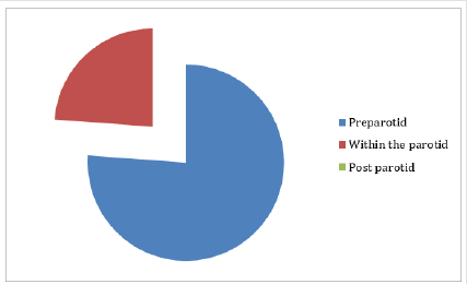
- 16 facial nerves were divided before reaching the parotid gland. Show Table 1 and Figure 1.
- 5 facial nerves were divided within the substance of the gland.
- No post parotid division was observed.
- In one cadaver on the right side the division was pre parotid while on the left side it was within the parotid gland substance.
Relation of the Facial Nerve to Retromandibular Vein
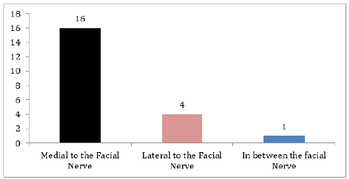
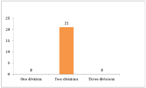
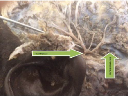
In 7 cadavers the retromandibular vein was medial to the facial nerve, In one cadaver the vein lies in between nerve divisions on the left side and medial to the nerve on the right side. In Two cadavers the retromandibular nerve was lateral to the facial nerve. show Tables 2-5 and Figures 2-4. In all the dissected cadavers the facial nerves were divided into upper and lower trunks only.
Discussion
Knowledge of the main landmark of the facial nerve trunk is crucial for safe and effective surgical outcome in the region of the parotid gland [12]. The branching pattern of the facial nerve within the parotid region shows individual variations [13-15]. This study examines the anatomic relationships and variability of the facial nerve trunk and its branches [16], with emphasis on the site of division in relation to the parotid gland, number of divisions and the position of the retromandibular vein in comparison to the facial nerve [17,18]. Dissections were performed on 11 Sudanese cadavers fixed with formalin; 21 half-heads has bees dissected [19], and the facial nerve trunks and branches were exposed [20]. The facial nerve trunk bifurcated into two main divisions in all cases (100%), and no trifurcation pattern was seen (0%). In 16 cases (76.19%) it bifurcates before the parotid gland, while in 5 cases (23.8%) it bifurcates within the gland. In one cadaver the bifurcation on the right side was before the parotid while on the left side it was within the gland [21,22].
We also investigated the relationship between the facial nerve and the retromandibular vein. In 16 (76.19%) of the cases the retromandibular vein was located on the medial side of the upper and lower trunks of the facial nerve, and in 4 (19.04%), the course of the retromandibular vein was lateral to the lower and upper trunks [23]. In one (4.76%) case the retromandibular vein was medial to the upper trunk and lateral to the lower trunk, the course of the retromandibular vein was different on the right and left sides of the face in the same cadaver [24,25]. A study in Korean cadavers revealed that (86.7%), the facial nerve trunk bifurcated into two main divisions, and a trifurcation pattern was seen in the other cases (13.3%) [26]. Another study in Turkey 50 dissections were performed in 30 cadavers in (90%) of the cases the retromandibular vein was located on the medial side of the upper and lower trunks of the facial nerve [27], and in (10%) the course of the retromandibular vein was lateral to the lower trunk and medial to the upper trunk. also, there is a study done in 79 facial halves from formalin-fixed cadavers in Malaysian subjects .trifurcation was seen in three facial halves (3.8%) [28-30]. For me the absence of trifurcation pattern in Sudanese people compared To those in Turkey and Malaysian could be due to racial and environmental differences [31,32].
In Conclusion
The facial nerve trunk bifurcated into two main divisions in all
cases (100%), in 16 cases it bifurcates before the parotid gland,
5 cases it bifurcates within the gland and only one cadaver the
bifurcation on the right side was before the parotid while on the left
side it was within the gland. In 16 of the cases the retromandibular
vein was located on the medial side of the upper and lower trunks
of the facial nerve, four of them course of the retromandibular
vein was lateral to the lower and upper trunks, In one case the
retromandibular vein was medial to the upper trunk and lateral to
the lower trunk.
For more Articles on : https://biomedres01.blogspot.com/


No comments:
Post a Comment
Note: Only a member of this blog may post a comment.