Minimally Invasive Rehabilitation Techniques for the Endodontically Treated Tooth – A Case Report
Introduction
Today, the options available to restore an endodontically treated tooth with great loss of dental structure are limited. In most cases, the traditional approach is chosen to rehabilitate the remanent with the use of a post and crown. However, it is necessary to prepare the tooth prior to treatment, which reduces the amount of already limited dental structure. The objective of this case report is to describe a minimally invasive rehabilitation treatment option in an endodontically treated anterior tooth (ETT).
Case Report
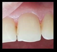
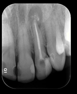
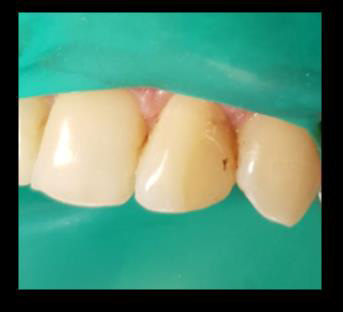
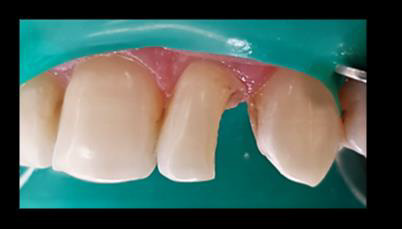
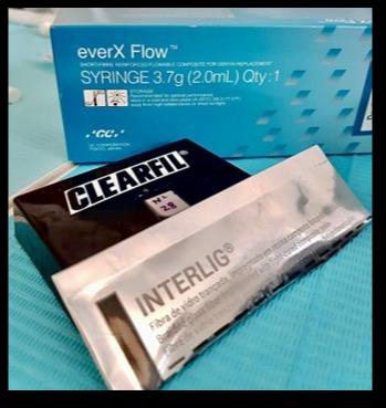
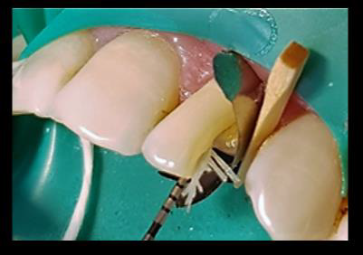
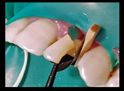
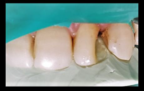
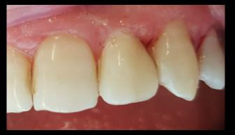
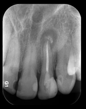
Female patient, 54 years old, was referred for the rehabilitation of ETT 2.2. The tooth was endodontically treated, restored, and double sealed 2 weeks prior.After removing the previous restoration, the Clearfill SE® 2-step self-etching adhesive protocol was used (Figures 1-6). Then, EverX Flow® flow resin with a 2mm x 6mm piece of Interlig® reinforced fiber were used. The coronal area was restored with z350 resin using a silicone key layering technique (Putty speedex®). Control and polishing were later carried out (Figures 7-11).
Discussion
evidence shows that the use of cast or fiber posts to rehabilitate ETTs presents a high risk of medium- to long-term biomechanical failures, due to the great loss of dental structure [1]. In addition, cast posts have a greater tendency to microleakage [2]. Fiber posts are preferred over cast posts owing to their lower risk of catastrophic type fractures [3].Multiple studies reach consensus that the ferrule effect is one of the most important factors in determining the rehabilitative success of endodontically treated teeth, and that the posts material choice does not show statistically significant differences when a ferrule effect exists [4-7].
It has been reported that ETTs treated with reinforced glass fibers can have an increased fracture resistance compared to the ones rehabilitated with fiber posts at different lengths. However, it is not yet possible to guarantee more favorable fracture patterns with the use of reinforced glass fibers over conventional techniques due to the limited amount of evidence available.
Conclusion
1. It is essential that dental professionals are familiar with the different treatment options available in order to carry out the most effective treatment for the case. The judicious use of posts in ETTs should be emphasized in order to preserve healthy dental tissue when possible.
2. The restoration after fiber reconstruction can be either direct or indirect, which will be determined by the clinical situation and the individual characteristics of the case at hand. There is need for more evidence considering rehabilitative alternatives on fiberglass stumps in ETTs.
3. It is advised that long-term studies be conducted evaluating clinical performance of fiber-reinforced ETTs, as well as shortterm ones comparing fracture types in ETTs that have been either fiber-reinforced or rehabilitated with a fiber-glass post.
For more Articles on: https://biomedres01.blogspot.com/



No comments:
Post a Comment
Note: Only a member of this blog may post a comment.