Thursday, February 29, 2024

Role and Relevant Significance of Autonomic Nerves in Esophageal Tumors


Role and Relevant Significance of Autonomic Nerves in Esophageal Tumors

Introduction

Tumor cells continue to proliferate in human body which is a very complex environment. The impact of tumor microenvironment on the occurrence and development of tumors is also one of the key directions of tumor prevention and treatment, and the nervous system, especially the autonomic nervous system, is an important part of the tumor microenvironment. Many studies in recent years had shown that the human autonomic nervous system interacts with tumors and stromal cells to mediate the occurrence and development of a variety of malignant tumors. In addition, new evidence suggests that malignant tumors may reshape autonomic nerves states, thereby maintaining the growth and survival of tumor cells [1]. Therefore, exploring and clarifying the autonomic nerves state of the human body with different tumors is of great significance to the research of tumor diseases. This article will make an exploratory analysis and exposition of autonomic nerves states in esophageal cancer and find ways to slow down the development of the tumor.

The Relationship between the Vagus Nervous System and the Occurrence and Development of Esophageal Cancer

It is well known that the esophagus is innervated by the vagus nerve and the spinal nerve. AS the vagus nerve and the esophagus go together, and anatomical studies had also confirmed that the vagus nerve is the most densely distributed on the esophagus, we need to consider whether the vagus nerve plays a role in esophageal lesions. At the same time, there are phenomena indicating that the vagus nerve in the distal esophagus has low distribution density and small branches, and this area is the high incidence of esophageal cancer. Therefore, some people suspect that vagal denervation may promote the occurrence of esophageal cancer. Ai Qun Wu and some other scholars [2] found that local anastomotic adenosquamous carcinoma was the main cause of recurrent esophageal cancer after surgery, and Esophageal motility disorders such as delayed gastric emptying after surgery were important reasons for the recurrence of this cancer, which suggested that vagal nerve injury may be related to the recurrence of esophageal cancer.

At the same time, Ai Qun Wu et al. believed that the vagus nerve and its transmitter acetylcholine (ACh) can inhibit esophageal cancer by reducing the inflammatory response which leads to oxidative membrane damage and DNA damage. In subsequent cell experiments, acetylcholine and norepinephrine could upregulate DNA repair enzyme expression in the cells of esophageal cancer and promote cell differentiation, which suggested a close relationship between these two neurotransmitters and cancer cell transformation. Except for Ai Qun Wu’s discussion on the relationship between vagus nerve and esophageal cancer, we have not find any other papers on this aspect. This suggests that the decrease of vagus nerve excitation can promote the occurrence of esophageal cancer, which may be just a guess, but not been effectively proved.

The Relationship between the Sympathetic Nervous System and the Occurrence and Development of Esophageal Cancer

As for the relationship between sympathetic nerve and the occurrence and development of esophageal cancer, most of the current studies focus on β receptors. Liu, et al. [3] used β-receptor selectors to regulate esophageal cancer cells and found that β -receptor agonists could promote cell proliferation, while β-receptor blockers significantly inhibited cell proliferation, suggesting that β-adrenergic receptors of the sympathetic nervous system are related to the growth of esophageal cancer. However, β receptor is related to the proliferation of a variety of tumor cells, such as breast cancer, gastric cancer, pancreatic cancer, malignant melanoma, prostate cancer, etc. and studies had shown that the use of β-adrenergic receptor blockers did not improve survival in patients with common cancers [4]. Therefore, whether there is a clear relationship between sympathetic nerve and esophageal cancer still needs to be confirmed.

Relationship between the Occurrence and Development of Esophageal Cancer and Nitric Oxide (NO)

It can be seen from the above studies that the autonomic nervous system, which is mainly composed of the sympathetic nerve and vagus nerve, does not seem to have much influence on the occurrence and development of esophageal cancer. Is the growth of esophageal cancer not affected by the tumor microenvironment constructed by the autonomic nervous state? However, it is not likely to grow independently. Could there be a non-adrenergic, non-cholinergic nerve that plays a role in esophageal cancer? NO is the important inhibitory non-adrenergic, non-cholinergic neurotransmitter, it mainly exists in the tissues of the gastrointestinal tract and is responsible for regulating the movement of the gastrointestinal tract [5]. It is generally believed that the release of NO in gastrointestinal tract is related to nitric oxide synthase (nNOS) expressed on the neuroexpandable membrane of intestinal neurons [6].

There is a large literature showing that nitric oxide (NO) plays a role in the development of esophageal cancer. For example, in the 57 cases of human esophageal squamous cell carcinoma studied by Tanaka et al. [7], high expression of iNOS was detected in 50 cases (87.7%), while the expression of iNOS was weak in esophageal non-neoplastic diseases. McAdam, et al. [8] found that iNOS/NO can induce DNA damage and nuclear factor -κB (NF-κB) signal transduction in esophageal cells, leading to the occurrence of esophageal cancer. Chen, et al. [9] found that selective iNOS inhibitors can significantly inhibit the progression of esophageal cancer by reducing NO production. Chen, et al. [10] found that benzo (a) pyrene in tobacco can increase the expression of iNOS, leading to the occurrence of esophageal cancer. These studies indicate that NO, a non-adrenergic and non-cholinergic neurotransmitter, plays a definite role in the occurrence of esophageal cancer.

The Tumor Mostly Occurs in the Middle and Lower Esophagus, Which May be Related to the Release of NO

Studies have shown that the contraction and relaxation of the lower esophageal sphincter are mainly affected by the action of NO [11-13], so where is the main source of NO in the esophagus? Kuramoto, et al. [14] found that nitric oxide synthase (NOS) in the esophagus is mostly concentrated on Fos neurons, while the distribution of Fos neurons in the esophagus gradually increased from the oral cavity to the gastric end and was highest in the abdominal esophagus. This is consistent with the common clinical phenomenon that esophageal cancer occurs in the middle and lower segments. It was also found that when the esophageal vagus nerve was stimulated, NOS in Fos neurons first showed high expression. Thus, it could release a large amount of NO to inhibit esophageal movement, while Fos neurons only expressed a small amount of acetylcholine transferase [14,15]. Meanwhile, Vazquez, et al. [16] showed that nitric oxide synthase inhibitors can promote the release of acetylcholine in the brain. Leonard, et al. [17] showed that NO can regulate the release of ACh. This can reasonably explain why there may be a relationship between vagus nerve and esophageal cancer mentioned in this paper, but no further research has been conducted.

The Occurrence and Development of Esophageal Cancer is Closely Related to Inos/NO, Especially Squamous Cell Carcinoma

As a physiological messenger, NO is synthesized by three different gene-encoded NO synthases (NOS) in mammals: neuronal NOS (nNOS or NOS-1), inducible NOS (iNOS or NOS- 2) and endothelial,NOS (eNOS or NOS-3). NO regulates a variety of important phy-siological responses, including vasodilation, respiration, cell migration, immune response and apoptosis. All these features are relevant in cancer [18]. It may be related to concentration, location, targets, source and other factors, NO can plays a role in promoting as well as inhibiting tumors [19,20]. In esophageal cancer, a large number of studies have found that iNOS/NO is closely related to the occurrence and development. For example, Kumagai, et al. [21] showed that the expression intensity of iNOS is positively correlated with the depth of tumor invasion of the esophageal wall. This may be due to iNOS and COX- 2 from cancer cells induce angio-genesis from the early stage of esophageal squamous cell carcinoma (ESCC) progression, in which NO produced by iNOS has been reported to enhance the activity of COX-2 [22].

Bednarz-Misa’s research has shown that Esophageal squamous carcinoma might be locally characterized by upregulated expression of genes encoding glucose transporter 1 (GLUT1) as well as inducible nitric oxide synthase (iNOS) and ornithine decarboxylase (ODC) of the L-arginine/nitricoxide (NO)/polyamine pathway. Karakasheva, et al. [23] found that iNOS could induce CD38-expressing in myeloidderived Suppressor in a Murine model of oral-esophageal cancer, thereby promoting tumor Growth. In esophageal adenocarcinoma, the Reflux esophagitis causes Barrett’s metaplasia [24], an abnormal esophageal mucosa predisposed to adenocarcinoma; McAdam’s research has shown that the protein levels of iNOS in esophageal adenocarcinoma was increased and the activity of DNA damage induction and nuclear factor-kappa B (NF-κB) signalling was dependent on iNOS/NO. Vaninetti, et al. [25] found that the expression of iNOS mRNA is higher than induced esophagitis and Barrett’s in esophageal adenocarcinoma, while Ferguson, et al. [26] explored the association Between iNOS polymorphisms and risk of esophageal adenocarcinoma, Barrett’s esophagus, or reflux esophagitis, did not show a positive result in the experiments.

Therefore, the role of iNOS/NO in esophageal adenocarcinoma remains to be further explored. Takala, et al. [27] found that iNOS positivity was more com-monly seen in squamous cell carcinomas than adenocarcinomas in esophageal cancer patients. For other types of NOS, only Chandra, et al. [28] detected NOS-3 expression in tissue samples of 20 patients with esophageal adenocarcinoma. It can be seen from the above studies that the expression of iNOS is more closely related to esophageal squamous cell carcinoma, and we can intervene the development of ESCC by inhibiting the expression of iNOS/NO.

Screening Drugs for Esophageal Cancer through Inos/ NO Pathway, Traditional Chinese Medicine May be the Best Choice

The role of iNOS/NO in the occurrence of esophageal cancer has been found, so what is the effect of relevant inhibitory drug intervention based on this? A study found that L-748706, a selective COX-2 inhibitor, and S,S′-1,4-phenylene-bis(1,2-ethanediyl)bisisothiourea (PBIT), a selective iNOS inhibitor, significantly inhibit NMBA-induced rat esophageal tumorigenesis [9,29]. Chen, et al. [30,31] found lyophilized Strawberries significantly reduced the protein expression level of iNOS and were shown to be effective in the prevention and treatment of esophageal cancer. Many studies have found that traditional Chinese medicine can achieve different therapeutic effects by interfering iNOS/NO. Guo, et al. [32] quickly screened and identified iNOS inhibitors from Ophiopogon japonicus. Chen, et al. [33] screened effective anti-inflammatory drugs by measuring the iNOS/NO inhibitory effect of extracts of 81 Kinds of traditional Chinese medicines. Mei, et al. [34] found two new ingredients from Lonicera Macranthoides, exhibited inhibitory effects on iNOS. Tian, et al. [35] found Berberine attenuates Renovascular hypertension and Sympatholay via the iNOS Pathway.

Such as Viola Yedoensis Makiho [36], Illicium Difengpi [37], Plantanone C [38] and Tribulus Terrestris L [39] can downregulation the expression of iNOS. It can be concluded that screening drugs for the prevention and treatment of esophageal cancer from Chinese herbal medicine through iNOS/NO pathway has great advantages. Qigesan, Shashen Maidong Decoction and other traditional Chinese medicine prescriptions for the treatment of esophageal cancer, as a kind of empirical medicine, it is still used in clinical Chinese medicine and has shown good results. Our previous studies found that Qigesan significantly inhibited the migration and invasion of esophageal cancer cells in vitro [40], and Qigesan inhibits esophageal cancer cell invasion and migration by inhibiting Gas6/Axl-induced epithelial-mesenchymal transition and NF-κB expression [41,42]. In the next step, we can use the iNOS/NO pathway to more accurately screen the active ingredients of related drugs from traditional prescriptions.

Conclusion

In conclusion, during the occurrence and development of esophageal cancer, the human autonomic nervous system releases a large amount of NO and only expresses a small amount of ACh, thus promoting the progression of esophageal cancer. Therefore, we can also treat esophageal cancer by inhibiting NO and increasing ACh. In this respect, the relevant research and clinical experience of traditional Chinese medicine are worth our reference.


For more Articles on: https://biomedres01.blogspot.com/
Wednesday, February 28, 2024

Kidney Injury Associated with Cocaine Use


Kidney Injury Associated with Cocaine Use

Introduction

Cocaine is a drug of abuse, currently considered a public health problem worldwide because of its addiction due to its use for recreational purposes. By 2019, around 20 million people between 15 - 45 years old (0.4% of the world population) have been cocaine users, however, due to its high demand, the trend continues to increase despite the availability of new alternatives to recreational drugs of abuse of synthetic origin [1]. After the isolation of coca paste by the German doctor Albert Nieman in 1865, the interest in this substance in the medicinal field increased, being mainly used as a local anesthetic in ocular surgery due to its vasoconstrictive properties; which led to a plethora of uses in medicine. Sigmund Freud extensively described its psycho-stimulant properties, and he used it as a treatment for some psychiatric disorders in particular those related to heroin abuse. Subsequently, recreational use popularized, particularly in the United States and Europe, the first cases of complications due to its massive consumption began to be reported in the 1980s. In 1982, it was reported the first myocardial infarction that was associated directly with cocaine 2 consumption [2]. The effects at the systemic level are widely documented [3], at low or moderate doses in the central nervous system it causes an increased mood, reduces appetite, causes insomnia, improves attention, and produces a surge in motor activity; in the cardiovascular system, it acts as a class I antiarrhythmic (at lower doses) by blocking sodium and potassium channels with a cardiodepressant and proarrhythmic effect, at higher doses; it stimulates endothelin 1 release, with a marked vasoconstrictive effect and it inhibits nitric dioxide production, causing hypertension and organ ischemia.

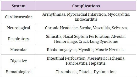
A prothrombotic effect is recognized by increasing platelet aggregation and adhesion and reducing the activity of prostacyclin E2 [4]. It raises body temperature due to an increase in muscle activity, vasoconstrictive heat loss, and hypothalamic temperature control loss due to the depletion of dopamine receptors. All the systemic effects described are responsible for the complications on the different systems with deleterious effects both in the short and longterm [5] (Table 1). Specifically, renal involvement is often considered, although less frequently, mediated by rhabdomyolysis; nonetheless, it was also described that renal involvement was directly associated with vasculitis, direct tubulointerstitial injury, and glomerular vasoconstriction [6]. In this document we describe two clinical cases of acute kidney injury associated with cocaine use, we present their clinical characteristics and we will briefly review the literature on this topic.

biomedres-openaccess-journal-bjstr

Table 1: Systemic complications of cocaine.

Case Report

Case 1: Clinical History

30-year-old male patient, with no previous medical records. He consulted the emergency department on intense right lumbar pain of 48 hours of evolution without radiation, associated with macroscopic hematuria without a fever, chills, or dysuria. When questioned, he highlighted the use of inhaled cocaine for 2 years. On admission physical examination, vital signs are normal, afebrile, saturating 98 (0.21). with compression pain in the right flank. No signs of peritoneal irritation. Frankly positive right fist percussion. The rest of the physical examination did not show alterations.

Laboratory Tests Show: Hto: 40% GB 11,800 (Ns 80% L 20%), normal urea and creatinine. Normal calcemia and uric acid, Total Bilirubin, Direct Bilirubin, FAL, TGP and TGO all of them at normal levels. Elevated LDH (x 5 times its normal value).

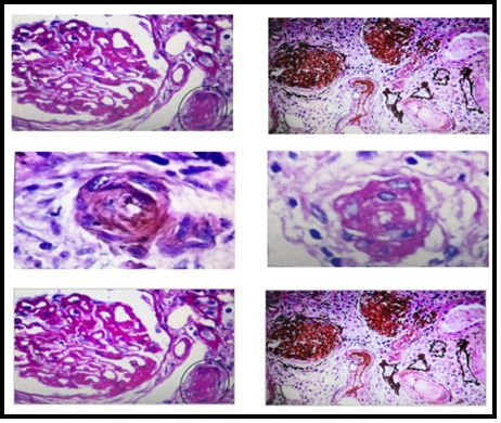
Urinary Sediment Examination: A field covered with isomorphic red blood cells is observed. Normal chest X-ray and electrocardiogram. The renal ultrasound did not show uronephrosis, urethral stones, or bladder abnormalities. A computed tomography (CT) scan showed hypodense and triangular external base images of the right kidney without post-contrast enhancement, characteristic of renal infarction (Figure 1). The case is interpreted as an acute renal infarction with preserved renal function associated with cocaine use. Treatment consisted of supportive measures with abundant hydration and opioid-based analgesia for pain management.

biomedres-openaccess-journal-bjstr

Figure 1: Renal biopsy images, performed by the Renal Pathology Service Hospital San Jose -Colombia, shows: thickening and wrinkling of capillary walls, arteries occluded by thrombus, concentric intimal hyperplasia, and onion-skinning.

Evolution: The evolution was favorable, and hematuria resolved at 72 hours.

Clinical Case 2: Clinical History

40-year-old male patient of African descent diagnosed with hypertension. The medical records show that the previous 6 months he was in antihypertensive treatment, had severe alcoholism, and was a heavy smoker. He consulted the [2] emergency department for mild acute infection by COVID 19 and decompensated heart failure.

Laboratories: Hematocrit 35%, hemoglobin: 12 g / dl, leukocytes: 9960, platelets: 219000, glucose: 122 mg / dl, BUN 86 mg / dl, creatinine: 2.77 mg / dl, sodium: 139 mEq / L, potassium:3.7 mEq / L, LDH: 305 IU / L, D dimer: negative, serologies, ANCA, anti- DNA, normal serum complement, negative viral serologies, normal thyroid profile. Toxicological tests are positive for the presence of cocaine in the blood.

Physical Examination: Arterial Tension: 200/110mmHg, heart rate. 87 beats per minute, respiratory rate 20 breaths per minute, temperature 36 degrees centigrade. Oriented in time, space and person; In the neck, it is observed jugular engorgement, crackles on bilateral pulmonary auscultation mainly in the bases, and lower extremity edema.

Electrocardiogram: In sinus rhythm with electrocardiographic data of left ventricular hypertrophy.

Renal Ultrasound: kidneys of preserved shape and size without alterations in the corticomedullary relationship.

Urinary Sediment: nondescript, without hematuria.

Evolution: He received supportive care, under monitoring in the intensive care unit, a negative fluid balance was induced with diuretics and later a coronary angiography was performed to evaluate possible coronary disease, which was reported normal. From the renal point of view, it was approached as an acute kidney injury AKIN II, with progressive renal function deterioration without anemia and without further data that suggests chronic kidney disease, consequently, a renal biopsy was performed.

Renal Biopsy Report: 25 glomeruli, 18 of them globally sclerosed, the remaining ones show ischemic signs of variable severity (capillary collapse, glomerular retraction, and pericapsular fibrosis), accompanied by podocyte alterations and in 2 of them, segmental sclerosis. The tubulointerstitial sector with pseudo thyroid classic atrophy (20%), fibrosis (20%), and focal mononuclear infiltrate. There are also groups of hypertrophied tubules with protein granules, cytoplasmic vacuoles, brush border loss, and isolated tubulitis. Arteries with moderate to severe intimal fibrosis, duplication of the elastic lamina, and presence of mucinous material in one of the branches. Arteries are mostly occluded by thrombi, concentric intimal hyperplasia, and/or sclerohyalinosis.

Immunofluorescence: IgG, IgA, and C1q: negative. IgM and C3 (2+) in accumulations, in areas of glomerular sclerosis, focal fibrinogen (+) in vascular walls, and Bowman’s capsule.

Histopathological Diagnosis: Thrombotic Microangiopathy.

Discussion

Cocaine is known to affect all components of kidney tissue and the pathophysiological mechanisms involved in toxicity are multiple [6]. Cocaine-induced rhabdomyolysis caused by of acute renal failure was the first to be described in the medical records, the suggested mechanisms are ischemia, direct cellular injury due to increased production of oxygen free radicals production, vasoconstriction, and the myoglobin crystals produced by cocaine-induced muscle necrosis [7]. Some researchers reported ANCA-associated which might be related to the consumption of substances used to adulterate cocaine such as levamisole or alcohol used for cooking it (toxic metabolites) or those that increase the psychostimulant effect, which is a regular practice in recreational consumers [8]. Acute renal infarction is another mechanism described [9]. In the clinical case, 1 we describe the usual form of presentation of acute renal infarction with lumbar pain and hematuria, in a patient with a clear record of cocaine use, it could be the reason. There was no alteration of renal function, in this case, due to unilateral renal involvement. Anuria and complete renal failure with the requirement for renal support have been described in cases of bilateral renal involvement, however, it is an even rarer form of presentation [10]. right renal involvement agrees with that some researchers described and it is estimated that it is due to a bigger size of the renal artery and a multiplied resistance to flow, which might be related to microthrombus formation.

Data from observational studies and multiple case reports show the association of cocaine consumption with cardiac and cerebral thrombosis, suggesting that cocaine itself has a marked prothrombotic effect [11]. At the renal level, the mechanisms of the genesis of the renal infarction described are based on observational studies and animal models that seem to involve a combination of marked vasoconstriction due to an increase in circulating catecholamines, elevated levels of endothelin 1, and micro thrombosis due to alteration of platelet aggregation and an increase in thromboxane synthesis as well as alteration of intrarenal hemodynamics by a mechanism that is not clear [10,11]. There is no established treatment for renal thrombosis associated with cocaine use and in the clinical case that we describe, since there was no alteration in renal function, its evolution was favorable only with established support measures. In Clinical case number 2, we describe a middle-aged man with a record of severe [2] polymedicated hypertension, acute kidney injury of unknown etiology, we performed a kidney biopsy that describes findings related to thrombotic microangiopathy.

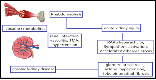
A positive result in the test for alkaloids together with a finding of elevated LDH and other demographic data of the subject points to the possibility of its relationship with the surreptitious use of cocaine after ruling out other potential causes. The Thrombotic microangiopathy was seldom considered a cause of acute [0] renal failure and accelerated hypertension in cocaine users, the mechanisms are not fully recognized, they could be related to the combination of platelet microthrombus formation and direct endothelial damage due to severe hypertension that characteristic of this condition [11,12]. No immunity alterations were described in cocaine use, in contrast with other [0] secondary causes of drugrelated thrombotic microangiopathy Although the prevalence of MIT is higher in women, the descriptions made in cocaine users place it predominantly in men, due to its higher consumption in this age group [13] (Diagram 1). Regarding the histological findings reported in our study, it can be verified that they belong to those usually described in thrombotic microangiopathies with renal involvement (notorious fibrinoid necrosis of the capillary tufts and glomerular arterioles) and that they reflect the renal tissue response to injury due to ischemia; [14] however, although the pathological findings mentioned in the recently described COVID 19 infection are multiple and affect all components of the renal tissue, the latter is associated with a predominantly tubulointerstitial and glomerular inflammatory involvement that is less common in patients with secondary thrombotic microangiopathies.

biomedres-openaccess-journal-bjstr

Diagram 1: Proposed pathophysiological mechanisms of renal injury induced by cocaine and/or its metabolites. MAT: Thrombotic Microangiopathy; RAAS: Renin-Angiotensin- Aldosterone System. Cocaine causes acute kidney injury by two mechanisms:
1) Indirectly After muscle injury, rhabdomyolysis occurs (the most frequent mechanism) and
2) Directly: Cocaine or its metabolites could induce direct injury by vasculitis, micro thrombosis formation, renal infarction, hypertensive nephropathy. Repeated episodes of acute kidney injury by mechanisms previously described would lead to the development of chronic kidney disease.

Some authors have reported viral inclusions in podocyte cytoplasm -not documented by the renal pathology group that evaluated the specimen in our description-, which reduces the prospects of a possible exclusively viral cause resulting in kidney damage without ruling it out completely [15]. Malignant hypertension as a mechanism of kidney injury associated with cocaine use has been supported by some authors in animal studies. Chronic cocaine use is [0] currently considered an independent risk factor for accelerated atherogenesis, which induces an increase in renin-angiotensin-aldosterone system activity, conditions hypertensive nephropathy [16] development. Although cocaine has not been directly established as a cause of chronic kidney disease, it is speculated that its chronic use is associated with repeated episodes of acute kidney injury together with malignant hypertension, they cause hemodynamic and structural changes due to oxidative stress, hypertension. Chronic disease mediated by vasoconstriction increased renin- angiotensin system activity, and accelerated atherogenesis causes increased mesangial matrix and tubulointerstitial fibrosis [17,18]. All of these conditions predispose to the development of chronic kidney disease.

Conclusion

Two cases are reported, the first with renal infarction, one of the rarest manifestations of cocaine-induced kidney damage without acute renal failure. The second case with cocaine- induced acute kidney injury without rhabdomyolysis or ischemic or toxininduced acute tubular necrosis. Renal failure with malignant hypertension was the main clinical manifestation in this patient. The histological characteristics were compatible with thrombotic microangiopathy. The mechanisms responsible for these pathological changes are unclear but are most likely multifactorial. Diffuse endothelial vascular injury due to direct toxicity or cocaineenhanced catecholamine release is likely a major contributor factor to the renal pathology observed in the 2 reported cases. Due to the diversity in the pathophysiological mechanisms involved in kidney damage, the final diagnosis usually requires a histopathological study; however, the importance of a comprehensive approach is highlighted, considering the possibilities in the differential diagnosis of acute kidney injury, particularly in young patients.


For more Articles on: https://biomedres01.blogspot.com/
Tuesday, February 27, 2024

Case Report: Elevated Hemidiaphragm Due to Liver Abscess of Biliary Origin


Case Report: Elevated Hemidiaphragm Due to Liver Abscess of Biliary Origin

Introduction

Elevated hemidiaphragm image in chest X-ray study is usually linked to thoracic problems including eventration, phrenic nerve paralysis and parenchymal lung volume loss [1]. Nevertheless, some upper abdominal abnormalities could lead to this phenomenon, which should be cautiously considered. In this article, we report a pyogenic liver abscess case due to rare complications of chronic gallbladder diseases.

Case Report

A 71-year-old man with a history of diabetes mellitus was admitted to the hospital complaining of right upper quadrant (RUQ) pain, fever and hiccups after meals lasting for one week. According to his description, there was no decreased appetite, nausea, vomiting, bowel habit change, dysuria, cough, chest pain or other respiratory symptoms. On admission, he was febrile with a temperature of 38.2°C and otherwise unremarkable vital signs. Abdominal physical examinations revealed RUQ tenderness without rebound tenderness. Laboratory studies demonstrated neutrophilic leukocytosis, an elevated high-sensitivity C-Reactive protein (hsCRP) and normal liver function test except a slightly increased γ-glutamyl transferase (γ-GT). Blood parameters included a white blood cell count of 20.0×10⁹/L (a normal range of 3.6-11.2), a neutrophilic segment of 82.9%, hsCRP of 19.37 mg/dL (a normal range of <1.0), a total bilirubin of 0.55 mg/dL (a normal range of 0.2-1.3), alanine aminotransferase (ALT) of 40 IU/L (a normal range of 5-40), γ-GT of 80 IU/L (a normal range of 8-50), and alkaline phosphatase of 106 IU/L (a normal range of 38-126).

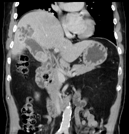
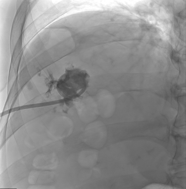
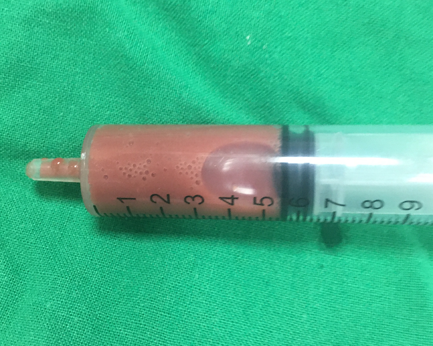
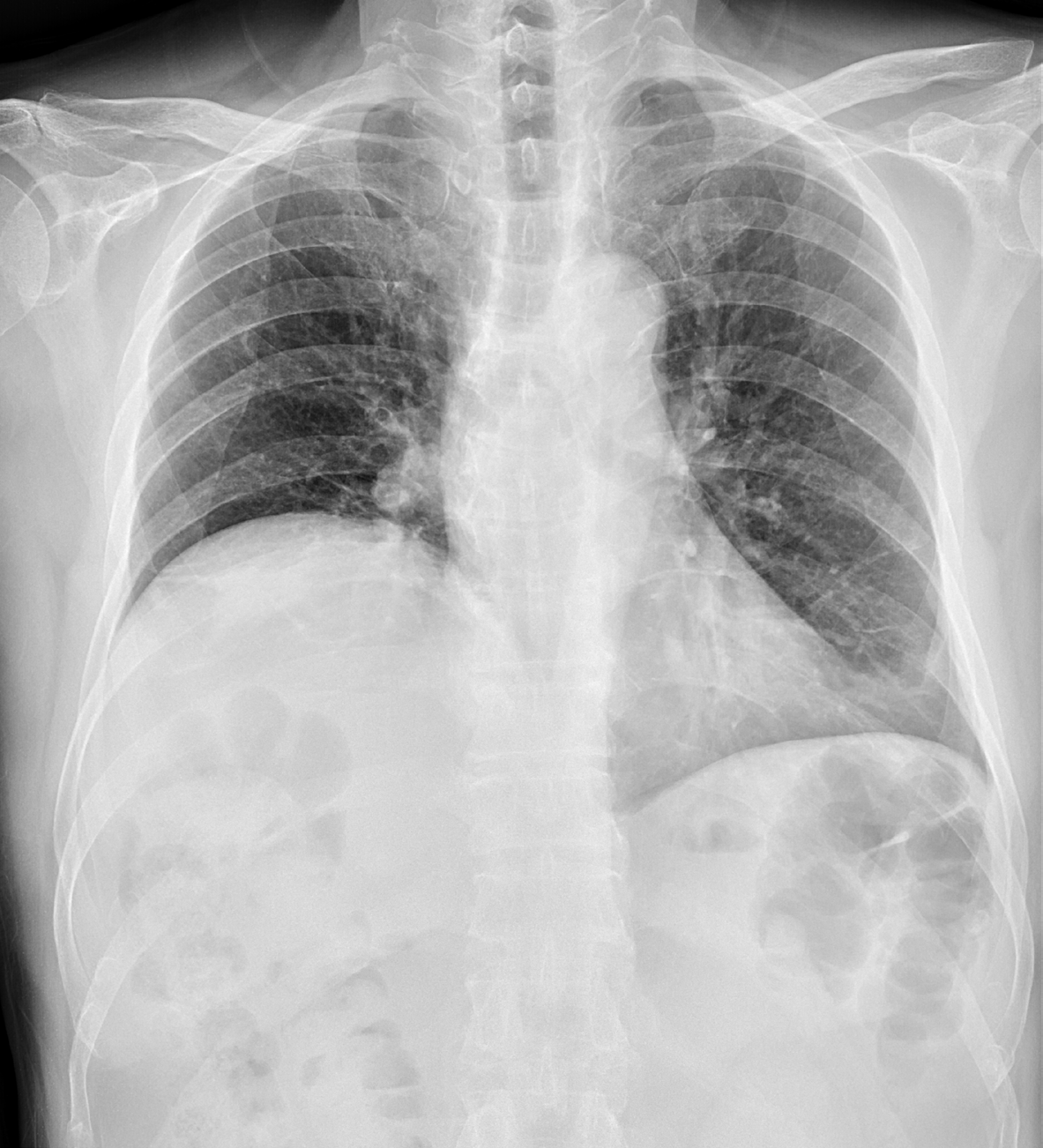
Chest X-Ray showed elevation of right hemidiaphragm (Figure 1). Sonography of the abdomen revealed a hypoechoic lesion in the right lobe of liver, and liver abscess was suspected. Urgent computed tomography (CT) of abdomen was then arranged to show one lobulated lesion (5.0×3.5 cm in size) with hypodensity and ring enhancement in right lobe of the liver, consistent with liver abscess (Figure 2). Furthermore, one gallbladder stone (2.5×1.5 cm in size) accompanied with gallbladder wall thickening and gallbladder fistula with the liver abscess were noticed. Therefore, antibiotic treatment with Ceftriaxone was administered. Percutaneous drainage of the abscess was performed on the next day (Figure 3), and brick red material was aspirated for pus culture (Figure 4). Consequently, culture result was positive for Escherichia coli. After antibiotic treatment and abscess drainage, the patient had no further symptoms and was discharged with antibiotics and cholecystectomy was advised at two weeks later.

biomedres-openaccess-journal-bjstr

Figure 1: Chest X-Ray of elevation of right hemidiaphragm.

biomedres-openaccess-journal-bjstr

Figure 2: Computed tomography shows one lobulated lesion with hypodensity and ring enhancement in right lobe of the liver, which is compatible with liver abscess.

biomedres-openaccess-journal-bjstr

Figure 3: Percutaneous drainage of the liver abscess.

biomedres-openaccess-journal-bjstr

Figure 4: Aspirated brick red material from the liver abscess.

Discussion

Development of an intrahepatic abscess on account of gallbladder perforation is a rare situation as reported [2,3]. Some predisposing factors may lead to the perforation including diabetes mellitus (DM), cholelithiasis, trauma or malignancy history, male gender and the elderly [3]. Moreover, DM played an important role in pyogenic liver abscess comorbidities along with malignant neoplasms [4]. Therefore, the senior man with DM history in our report suffered a higher risk of both gallbladder perforation and liver abscess. Clinical presentations of biliary origin liver abscess are not highly specific and sensitive. Common symptoms include higher body temperature, chilling, RUQ pain, weak and jaundice [2,4]. Chest X-ray usually serves as the primary image examination to screen an emergent febrile patient and abnormal findings, inclusive of elevation of right hemidiaphragm, right basilar lung atelectasis and pleural effusion, suggest the presence of liver abscess [1,5]. Sequentially, abdominal ultrasound is recognized as the first-line choice for identifying cholecystitis; however, CT is a more sensitive method to diagnose a perforation or visualize the hole sign between liver abscess and gallbladder, as compared with ultrasound [2,3].

Regarding the most common pathogen for liver abscess, Klebsiella pneumoniae should be the answer in most countries in Eastern Asia. However, the major pathogen resulting in liver abscess of biliary origin in a recent study performed in Eastern Asia was Escherichia coli, which is consistent with the culture finding in our case [4]. Moreover, the treatment suggestions included intravenous antibiotics and percutaneous drainage [2,3,5]. Cholecystectomy is usually advised to prevent the recurrence of cholecystitis [2].


For more Articles on: https://biomedres01.blogspot.com/
Monday, February 26, 2024

Comparison of Calcium, Chlorine, Iodine, Potassium, Magnesium, Manganese, and Sodium in Thyroid Tissue Adjacent to Thyroid Malignant and Benign Nodules


Comparison of Calcium, Chlorine, Iodine, Potassium, Magnesium, Manganese, and Sodium in Thyroid Tissue Adjacent to Thyroid Malignant and Benign Nodules

Introduction

Thyroid benign and malignant nodules (TBN and TMN, respectively) are the most common endocrine disorder worldwide. Moreover, in some parts of the world, especially those of current or former iodine deficiency, Thyroid Nodules (TN) are still an endemic disease [1]. Incidence of TBN and TMN has been growing steadily over the past four decades, despite the use of iodine prophylaxis in many countries [2]. Some factors causing this higher incidence of TN were described in literature [3] and analysis of these data shown intriguing links between the etiologies of TBN and TMN [2,3]. In other words, the factors contributing to increases in the incidence of TBN are the same as those contributing to increases in TMN. However, the current state of knowledge regarding TN demonstrates that the etiology and pathogenesis of TBN and TMN are still not enough understood, because there are many not adequately explored chemicals, which induced thyroid hormone perturbations leading to these diseases. For over 20th century, there was the dominant opinion that TN is the simple consequence of iodine deficiency [4].

However, it was found that TN is a frequent disease even in those countries and regions where the population is never exposed to iodine shortage. Moreover, it was shown that iodine excess has severe consequences on human health and associated with the presence of TN [5-8]. It was also demonstrated that besides the iodine deficiency and excess many other dietary, environmental, and occupational factors are associated with the TN incidence [3,9- 11]. Among these factors a disturbance of evolutionary stable input of many chemical elements (ChEs) in human body after industrial revolution plays a significant role in etiology of TN [12]. Besides iodine, many other ChEs have also essential physiological functions [13]. Essential or toxic (goitrogenic, mutagenic, carcinogenic) properties of ChEs depend on tissue-specific need or tolerance, respectively [13]. Excessive accumulation or an imbalance of the ChEs may disturb the cell functions and may result in cellular proliferation, degeneration, death, benign or malignant transformation [13-15]. In our previous studies the complex of in vivo and in vitro nuclear analytical and related methods was developed and used for the investigation of iodine and other ChEs contents in the normal and pathological thyroid [16-22]. Iodine level in the normal thyroid was investigated in relation to age, gender and some non-thyroidal diseases [23,24].

After that, variations of many ChEs content with age in the thyroid of males and females were studied and age- and genderdependence of some ChEs was observed [25-41]. Furthermore, a significant difference between some ChEs contents in colloid goiter, thyroiditis, thyroid adenoma and cancer in comparison with normal thyroid was demonstrated [42-47]. The present study was performed to clarify the role of some ChEs in the etiology of TBN and TMN. Having this in mind, the aim of this exploratory study was to examine differences in the content of Calcium (Ca), Chlorine (Cl), Iodine (I), Potassium (K), Magnesium (Mg), Manganese (Mn), and Sodium (Na) in thyroid tissue adjacent to TN using a nondestructive instrumental neutron activation analysis with high resolution spectrometry of short-lived radionuclides (INAA-SLR), and to compare the levels of these ChEs in two groups of samples (tissue adjacent to TBN and TMN, respectively). Moreover, for understanding a possible role of ChEs in etiology and pathogenesis of TN results of the study were compared with previously obtained data for the same ChEs in “normal” thyroid tissue [42-47].

Material and Methods

All patients suffered from TBN (n=79, mean age M±SD was 44±11 years, range 22-64) and from TMN (n=41, mean age M±SD was 46±15 years, range 16-75) were hospitalized in the Head and Neck Department of the Medical Radiological Research Centre (MRRC), Obninsk. Thick-needle puncture biopsy of suspicious nodules of the thyroid was performed for every patient, to permit morphological study of thyroid tissue at these sites and to estimate their trace element contents. In all cases the diagnosis has been confirmed by clinical and morphological results obtained during studies of biopsy and resected materials. Histological conclusions for benign nodules were: 46 colloid goiter, 19 thyroid adenoma, 8 Hashimoto’s thyroiditis, and 6 Riedel’s Struma, whereas for thyroid malignant tumors were: 25 papillary adenocarcinomas, 8 follicular adenocarcinomas, 7 solid carcinomas, and 1 reticulosarcoma. Samples of visually intact thyroid tissue adjacent to TBN and TMN were taken from resected materials.

“Normal” thyroids for the control group samples were removed at necropsy from 105 deceased (mean age 44±21 years, range 2-87), who had died suddenly. The majority of deaths were due to trauma. A histological examination in the control group was used to control the age norm conformity, as well as to confirm the absence of micro-nodules and latent cancer. All studies were approved by the Ethical Committees of MRRC. All the procedures performed in studies involving human participants were in accordance with the ethical standards of the institutional and/or national research committee and with the 1964 Helsinki declaration and its later amendments, or with comparable ethical standards. Informed consent was obtained from all individual participants included in the study. All tissue samples obtained from tumors and visually intact tissue adjacent to tumors were divided into two portions using a titanium scalpel to prevent contamination by ChEs of stainless steel [48]. One was used for morphological study while the other was intended for ChEs analysis. After the samples intended for ChEs analysis were weighed, they were freeze-dried and homogenized [49]. The pounded samples weighing about 10 mg (for biopsy) and 100 mg (for resected materials) were used for ChEs measurement by INAA-SLR.

To determine contents of the ChE by comparison with a known standard, Biological Synthetic Standards (BSS) prepared from phenol-formaldehyde resins were used [50]. In addition to BSS, aliquots of commercial, chemically pure compounds were also used as standards. Ten sub-samples of Certified Reference Material (CRM) of the International Atomic Energy Agency (IAEA) IAEA H-4 (animal muscle) weighing about 100 mg were treated and analyzed in the same conditions as thyroid samples to estimate the precision and accuracy of results. The content of Ca, Cl, I, K, Mg, Mn, and Na were determined by INAA-SLR using a horizontal channel equipped with the pneumatic rabbit system of the WWR-c research nuclear reactor (Branch of Karpov Institute, Obninsk). Details of used nuclear reactions, radionuclides, gamma-energies, spectrometric unit, sample preparation, and the quality control of results were presented in our earlier publications concerning the INAA-SLR of ChEs contents in human thyroid [27,28], scalp hair [51], and prostate [52,53].

A dedicated computer program for INAA-SLR mode optimization was used [54]. All samples for ChEs analysis were prepared in duplicate, and mean values of ChEs contents were used in final calculation. Using Microsoft Office Excel software, a summary of the statistics, including, arithmetic mean, standard deviation of mean, standard error of mean, minimum and maximum values, median, percentiles with 0.025 and 0.975 levels was calculated for ChEs contents in two groups of tissue adjacent to TBN and TMN. Data for “normal” thyroid were taken from our previous publications [42- 47]. The difference in the results between two groups of samples “adjacent to TBN” and “adjacent to TMN”, as well as between “normal” and “adjacent to TBN and TMN combined” was evaluated by the parametric Student’s t-test and non-parametric Wilcoxon- Mann-Whitney U-test.

Results

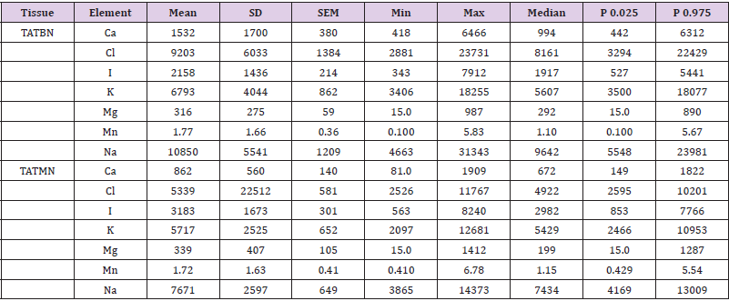
Table 1 presents certain statistical parameters (arithmetic mean, standard deviation, standard error of mean, minimal and maximal values, median, percentiles with 0.025 and 0.975 levels) of the Ca, Cl, I, K, Mg, Mn, and Na mass fraction in thyroid intact tissue samples of two groups “adjacent to TBN” and “adjacent to TMN”. The ratios of means and the comparison of mean values of Ca, Cl, I, K, Mg, Mn, and Na mass fractions in pair of sample groups such as “adjacent to TBN” and “adjacent to TMN” is presented in Table 2. Table 3 depicts certain statistical parameters (arithmetic mean, standard deviation, standard error of mean, minimal and maximal values, median, percentiles with 0.025 and 0.975 levels) of the Ca, Cl, I, K, Mg, Mn, and Na mass fraction in thyroid tissue adjacent “TTA” to TN (two groups “adjacent to TBN” and “adjacent to TMN” combined). The ratios of means and the comparison of mean values of Ca, Cl, I, K, Mg, Mn, and Na mass fractions in pair of sample groups such as normal thyroid tissue “NT” and “TTA” is presented in Table 4.

biomedres-openaccess-journal-bjstr

Table 1: Some statistical parameters of Ca, Cl, I, K, Mg, Mn, and Na mass fraction (mg/kg, dry mass basis) in thyroid tissue adjacent to thyroid benign (TATBN) and malignant (TATMN) nodules.

Note: M: Arithmetic Mean, SD: Standard Deviation, SEM: Standard Error of Mean, Min: Minimum Value, Max: Maximum Value, P 0.025: Percentile with 0.025 Level, P 0.975: Percentile with 0.975 Level.

biomedres-openaccess-journal-bjstr

Table 2: Some statistical parameters of Ca, Cl, I, K, Mg, Mn, and Na mass fraction (mg/kg, dry mass basis) in thyroid tissue adjacent to thyroid benign (TATBN) and malignant (TATMN) nodules.

Note: M: Arithmetic Mean, SEM: Standard Error of Mean, Statistically significant values are in bold.

biomedres-openaccess-journal-bjstr

Table 3: Some statistical parameters of Ca, Cl, I, K, Mg, Mn, and Na mass fraction (mg/kg, dry mass basis) in in Thyroid Tissue Adjacent (TTA) to thyroid benign and malignant nodules (combined).

Note: M: Arithmetic Mean, SD: Standard Deviation, SEM: Standard Error of Mean, Min: Minimum Value, Max: Maximum Value, P 0.025: Percentile with 0.025 level, P 0.975: Percentile with 0.975 level.

biomedres-openaccess-journal-bjstr

Table 4: Differences between mean values (MSEM) of Ca, Cl, I, K, Mg, Mn, and Na mass fraction (mg/kg, dry mass basis) in Normal Thyroid (NT) and Thyroid Tissue Adjacent to thyroid benign and malignant nodules (TTA).

Note: M: Arithmetic Mean, SEM: Standard Error of Mean, Statistically significant values are in bold.

Discussion

As was shown before [27,28,51-53] good agreement of the Ca, Cl, I, K, Mg, Mn, and Na contents in CRM IAEA H-4 samples analyzed by INAA-SLR with the certified data of this CRM indicates acceptable accuracy of the results obtained in the study of “adjacent to TBN”, “adjacent to TMN”, “NT”, and “TTA” groups of thyroid tissue samples presented in Tables 1-4. From Table 2, it is observed that in thyroid tissue adjacent to TMN the mass fraction of I is 1.47 time higher while mass fractions Cl and Na 42% and 29%, respectively, lower than in thyroid tissue adjacent to TBN. In a general sense Ca, K, Mg, and Mn contents found in the “adjacent to TBN” and “adjacent to TMN” groups of thyroid tissue samples were similar (Table 2). It allowed combine data obtained for two groups for the purposes of finding a common ChEs composition of TTA to TN and improving statistical characteristics of results for this group of samples (Table 3). From obtained results it was found that the common characteristics of thyroid tissue adjacent to TBN and TMN were elevated levels of Cl, I, and Na, which overdrew those in “normal” thyroid approximately in 2.2, 1.4, and 1.4 times, respectively (Table 4). Thus, if we accept the ChEs contents in “normal” thyroid glands as a norm, we have to conclude that with a nodular transformation the Cl, I, and Na contents in thyroid intact tissue adjacent to TN significantly changed.

Characteristically, elevated or reduced levels of ChEs observed in thyroid nodules are discussed in terms of their potential role in the initiation and promotion of these thyroid lesions. In other words, using the low or high levels of the ChEs in affected thyroid tissues researchers try to determine the role of the deficiency or excess of each ChEs in the etiology and pathogenesis of thyroid diseases. In our opinion, abnormal levels of some ChEs in TN could be and cause, and also effect of thyroid tissue transformation. From the results of such kind studies, it is not always possible to decide whether the measured decrease or increase in ChEs level in pathologically altered tissue is the reason for alterations or vice versa. According to our opinion, investigation of ChEs contents in thyroid tissue adjacent to TN and comparison obtained results with ChEs levels typical of “normal” thyroid gland may give additional useful information on the topic because these data show conditions of tissue in which TN were originated and developed.

Chlorine and Sodium

Cl and Na are ubiquitous, extracellular electrolytes essential to more than one metabolic pathway. In the body, Cl and Na mostly present as sodium chloride. Therefore, as usual, there is a correlation between Na and Cl contents in tissues and fluids of human body. Because Cl is halogen like I, in the thyroid gland the biological behavior of chloride has to be similar to the biological behavior of iodide. The main source of natural Cl for human body is salt in food and chlorinated drinking water. Environment (air, water and food) polluted by artificial nonorganic Cl-contained compounds, for example such as sodium chlorate (NaClO3), and organic Cl-contained compounds, for example such as polychlorinated biphenyls (PCBs) and dioxin, is other source. There is a clear association between using chlorinated drinking water, levels NaClO3, PCBs and dioxin in environment and thyroid disorders, including cancer [55-59].

Thus, on the one hand, the accumulated data suggest that Cl level in thyroid tissue might be responsible for TMNs development. However, on the other hand, it is well known that Cl and Na mass fractions in human tissue samples depend mainly on the extracellular water volume [60]. TN and thyroid tissues adjacent to nodules can be more vascularized than normal thyroid. Because blood is extracellular liquid, it is possible to speculate that more intensive vascularization could be the reason for elevated levels of Cl and Na in thyroid tissue adjacent to TB and TMN. If that is the only case, the equilibrium between Cl and Na increases has to be, however, in comparison with “normal” thyroid the change of Cl level in adjacent tissue is significantly higher than change of Na level. Thus, it is possible to assume that an excessive accumulation of Cl in thyroid tissue is involved in TBN and TMN etiology.

Iodine

To date, it was well established that iodine excess has severe consequences on human health and associated with the presence of TBN and TMN [4-8,61-64]. In present study elevated level of I in thyroid tissue adjacent to TBN and TMN was found in comparison with “normal” thyroid. Thus, on the one hand, it is likely that elevated level of I in thyroid tissue might be involved in the TN origination and development. On the other hand, however, elevated level of I in thyroid tissue adjacent to TN may explain by unusually intensive work of this tissue. Compared to other soft tissues, the human thyroid gland has higher levels of I, because this element plays an important role in its normal functions, through the production of thyroid hormones (thyroxin and triiodothyronine) which are essential for cellular oxidation, growth, reproduction, and the activity of the central and autonomic nervous system. As was shown in our previous study, TBN and, particularly, TMN transformation of thyroid gland is accompanied by a significant loss of tissue-specific functional features, which leads to a significant reduction in I content associated with functional characteristics of the human thyroid tissue [43-47]. Because the affected part of gland reduced productions of thyroid hormones, the rest “intact” part of thyroid tries to compensate thyroid hormones deficiency and work more intensive than usual.

Limitations

This study has several limitations. Firstly, analytical techniques employed in this study measure only seven ChEs (Ca, Cl, I, K, Mg, Mn, and Na) mass fractions. Future studies should be directed toward using other analytical methods which will extend the list of ChEs investigated in thyroid tissue adjacent to TN. Secondly, the sample size of TBN and TMN group was relatively small and prevented investigations of ChEs contents in this group using differentials like gender, functional activity of nodules, stage of disease, and dietary habits of patients with TN. Lastly, generalization of our results may be limited to Russian population. Despite these limitations, this study provides evidence on some ChEs level alteration in thyroid tissue adjacent to TN and shows the necessity to continue ChEs research of TN.

Conclusion

In this work, ChEs analysis was carried out in the thyroid tissue adjacent to TBN and TMN using INAA-SLR. It was shown that INAA-SLR is an adequate analytical tool for the non-destructive determination of Ca, Cl, I, K, Mg, Mn, and Na content in the tissue samples of human thyroid in norm and pathology. I was observed that in thyroid tissue adjacent to TMN the mass fraction of I is 1.47 time higher while mass fractions Cl and Na 42% and 29%, respectively, lower than in thyroid tissue adjacent to TBN. The common characteristics of thyroid tissue adjacent to TBN and TMN were elevated levels of Cl, I, and Na, which overdrew those in “normal” thyroid approximately in 2.2, 1.4, and 1.4 times, respectively and similar contents of Ca, K, Mg, and Mn. Thus, from results obtained, it was possible to conclude that the role of ChEs in etiology and pathogenesis of TBN and TMN is similar and exessive accumulation of Cl and I in thyroid tissue may be involved in the TN origination and development.


For more Articles on: https://biomedres01.blogspot.com/
Centrifugal Pneumatic-Sieve Fractionation of Barley and Wheat Flour for Maximum Recovery of Protein and Carbohydrate Fractions

  Centrifugal Pneumatic-Sieve Fractionation of Barley and Wheat Flour for Maximum Recovery of Protein and Carbohydrate Fractions Introductio...