Friday, June 28, 2024

Data Warehouses, Decision Support Systems, and Deep Technologies During the Global COVID-19 Pandemic


Data Warehouses, Decision Support Systems, and Deep Technologies During the Global COVID-19 Pandemic

Introduction

Data storage for businesses involves the storage of such information as stocks, raw materials, deposits, and other such information related to the daily operations of the business. The architecture of such a system needs to be aimed at data management. Data warehousing uses technologies that allow data from multiple sources to be compared and analyzed so that businesses can the consolidated data to make decisions (Simion, et al. [1]). The database is built to implement the volume and the requirements of the system and help project managers and organizational managers make decisions related to the development of the business structure or further daily operations (Simion, et al. [1]). Furthermore, database applications improve the reliability and efficiency of the user and the ability to make decisions, store, update, and get answers through reports (Simion, et al. [1]). Communication with the essential departments of the organization is facilitated by the efforts of the component dialog. The analysis highlights the structure between the data analysis and the simplest form of analysis is comparing the data with similar information. Other observations require techniques using analytical data based on mathematical theories which were developed to make correlations based on mathematical theories using products of a hypothetical nature compared with actual data (Simion, et al. [1]).

Deep Technologies

Over the past few decades, society has become overtly more data driven. With the takeover of social media, a need for learning how data and subjects relate to one another implies the need for a data processing method to consider emotion, sense, everyday practices, and the nexus between data and data technologies, Big Data, Internet of Things (IoT), and Artificial Intelligence may certainly be the future of all data transactions (Lee A [2]). Future data technologies must take all these factors into consideration and be able to process data at rates far exceeding current speeds and abilities. Parallel processing is beginning to play a key role in data interpretation. Thus, the benefit of using these technologies lies in capturing the scalar nature of data and focusing on the socio-technical processes behind data applications. It begins with a conceptualization of the Personhood of Data, which highlights exactly how distant the subject is from the data resulting in a conceptual language that then provides a method for analyzing the scalar nature of the data (Brynjolfsson Jin, et al. [3]).

Data, then, becomes a positivistic resource as the data highlights the humanity of the subject through each bit of datafied representation of humanity through the scientific representation of the subject’s world. So, with the use of deep technologies, data can be processed and analyzed and be used not only to humanize daily data activities, stresses, and emotions and sensemaking but to individualize our humanity in the surveillance of the data with separate and scientific representations of new data structures—the distant relationship of data and the embodied life represented by social media and activities only capable of being analyzed by deep technologies (Lee A [2]). The simplest form of analysis is comparing the data with similar data synthesized. In addition, information can acquire quality when using techniques of graphical representation that make these correlations, observation techniques analytical data based on mathematical theories, comparing actual data with the theoretical products of a hypothetical model, or observation techniques automatic based on data.

Big Data

The Internet of Things (IoT), Big Data, and Neural Networks are being considered as emerging networks that can comprehend complex automatic monitoring, identification, and management through a network of smart devices and parallel processing such as in a Neural network. Big Data has emerged as a viable and sustainable network that can analyze massive amounts of data from several sources including social media, sensors, attenuators, etc. to derive a sustainable decision by linking devices together and then developing consistent algorithms which can analyze, manipulate, and manage the connected systems so that a huge bulk of data can then be used for smarter decision-making and post-analysis for various reasons (Brynjolfsson Jin, et al. [3]).

IoT is a set of disparate devices connected via a common network such as Big Data analytics. The efficient use of the IoT in multiple areas has helped improve productivity and reduce errors (Brynjolfsson Jin, et al. [3]). Because smart devices are linked to the network, they can make smarter decisions and post-analysis via various purposes; in other words, the network is connected to these devices using Big Data, which improves the limited resources and management of data with those smart devices to improve power efficiency. Due to the inherent nature of Big Data, including the 7 Vs, improvements can be engaged in networking ability and various approaches in place for recovery, constrained energy, and the huge bulk of storage on the cloud. Therefore, data correlation and multiple characteristics of sensory data can be improved with the use of Big Data and deep technologies (Chu, et al. [4]).

Emerging Diseases and Deep Technologies

When using deep technologies, data scientists prefer R Studio or Python languages, however, they are limited in their speed and memory abilities. Scalability is one of the most important considerations when using machine learning models and parallel data summarizations (Al Amin, et al. [5]). Thus, a more successful computational data model can be presented using parallel data summarization because the model requires only a small amount of memory (i.e., RAM) and the algorithm works in three phases, producing a broader statistical and machine learning model which can handle datasets much bigger than the main memory. Designed with a vector-vector outer product with a C++ code to escape the bottlenecking that can happen in deep learning, this system is still faster and with a bigger memory than other parallel systems (e.g., Spark, Hive, Cassandra, etc.), which could become important in such cases as epidemiological disease tracing (Al Amin, et al. [5]).

Big Data plays a variety of important roles which critically support the world’s manufacturing, legal, financial, cybersecurity, and medical systems. Through open-source platforms like Hadoop, etc., information is shared among government and nongovernmental facilities regarding emerging diseases, predictions made through computational models, and cybersecurity underlaid for those who must shelter in place and work from home (Ahouz, et al. [6]). Johns Hopkins University Center for Systems Science and Engineering (JHU CCSE) has had the closest to actual live data as any other models with error rates at 4.71, 8.54, and 6.13%, respectively. Furthermore, these error rates were based on computational model error rates based on actual infection, death, and hospitalization rates based on data mining using Big Data analytics (Gupta, et al. [7,8]).

Conclusion

The strengths of Big Data analytics cannot be understated in this data-driven society where everything written on social media to the demographic makeup of the victims of the pandemic can be used as input into the computational model makeup for determining who may be future victims of the pandemic. According to the models predicted by JHUCCSE, a high rate of errors was unavoidable at the beginning of the pandemic. Contributing to the high error rates were the lack of knowledge about the disease, unknown diagnostics, and unknown patterns of susceptibility. However, as time has shown, knowledge of the factors surrounding the pandemic has contributed to a decrease in the error rates relative to the pandemic.


For more Articles on: https://biomedres01.blogspot.com/
Thursday, June 27, 2024

Cell Membrane Nano Vesicles as Drug Carriers: Biomedical Applications


Cell Membrane Nano Vesicles as Drug Carriers: Biomedical Applications

Introduction

Cell therapy replaces damaged tissue or cells with intact and living cells obtained from the patient (autologous cells) or a donor (allogeneic cells). Cell therapy may have its origins in 1931 when Swiss physician Paul Niehans attempted to cure a patient by injecting calf embryos. Various cell types have been engineered as novel therapeutics for multiple diseases and conditions due to evolving research and emerging innovative technologies [1-3]. Cells such as erythrocytes, platelets, cancer cells, leukocytes, stem cells, and bacteria may be used. Among these, erythrocytes have a high degree of biocompatibility when used as autologous cells or carriers of therapeutic agents [4,5]. Platelets have been extensively studied as novel drug delivery carriers for enhanced efficacy due to their critical roles in hemostasis and thrombosis and their newly discovered role in the development and metastasis of tumors [6,7]. Synthetic nanoparticles (NPs) have been used to diagnose and deliver drugs to patients suffering from various diseases [8-15]. NPS are synthesized from a variety of materials, including lipids [16,17], polymers [18], proteins [19-22], and metals [23-25]. NPS are frequently conjugated with targeting ligands or antibodies to deliver them to pathological sites in the body [26]. However, the biofunctionalization of NPs is insufficient to replicate the complex and multicellular interactions found in the human body, potentially limiting the efficacy of drug delivery via those nanotechnologies [27,28].

Intercellular communication is critical for survival and homeostasis maintenance in all multicellular systems. Communication occurs via two major pathways: direct cell-cell interactions and the secretion of soluble signaling molecules by cells. Recent research indicates that an additional mechanism, mediated by extracellular vesicles (EVs), plays a critical role in regulating intercellular interactions over short and long distances [29,30]. To effectively treat cancer, drug delivery systems targeting tumor cells have been developed via the design of targeting ligands conjugated to the surface of nanoparticles [31-34]. Arginineglycine- aspartic acid (RGD) is a peptide that binds to fibronectin [35] specifically. RGD [34,36] is also a specific ligand for integrin v3, which plays a critical role in tumor angiogenesis and metastasis [37], according to studies. Integrin v expression is significantly higher on endothelial cells lining tumor blood vessels than normal endothelial cells [38]. Integrin v3 is highly expressed on various tumor cells, promoting tumor metastasis [39] and facilitating the migration of immune cells into tumor tissues [40]. Thus, using RGD to deliver therapeutics to integrin v3 may be a novel strategy for tumor intervention [38,41,42]. Despite the remarkable progress described above, cell-based therapeutics face numerous obstacles before being widely adopted for clinical use with increased efficacy [43,44]. One of the most critical impediments is the inability to control the fate of injected cells in vivo, which is critical for increasing the percentage of cells reaching target tissues and decreasing off-target accumulation. Therapeutic cells transplanted into the recipient’s body may encounter undesirable host immune responses and frequently lose their therapeutic activity due to immune surveillance [45]. Another critical issue is that injecting drugs or drug-loaded nanoparticles into cells may cause damage to the cell membrane and alter the cell phenotype. Some cases may even be cytotoxic to the carrier cells [46].

Leveraging Cell Membranes to form Nano Vesicles as Efficient Drug Delivery Systems

Cell therapy has enormous potential for treating a variety of diseases. For example, genetically engineering isolated T cells from patients with chimeric antigen receptors (CARs) redirected T cell specificity and ushered in a new era of cancer treatment. The exciting news is that the US Food and Drug Administration recently approved CD19-specific CAR T cells to treat children and young adults with B-cell acute lymphoblast leukemia [47]. Additionally, clinical trials involving cell transplantation are being conducted to address cell function deficiencies [48,49]. Additionally, novel strategies for tissue engineering utilizing stem cells are currently being developed at various stages [50]. However, these approaches are frequently constrained by the difficulty of meeting clinicalscale demand for donor cells, as well as the lifelong requirement for immunosuppressive or other adjuvant drugs that enhance cell efficiency but have undesirable side effects [51]. To this end, cell engineering methods have been proposed to address concerns about rejection by host immune systems by immunoisolating the cells using microcapsules or three-dimensional scaffolds [52-54]. Although promising, complications arise due to the foreign-body response induced by the implant materials, which manifests as immune cell recruitment, fibrous deposits, restricted nutrient passage, and eventual cell death.

Additionally, additional work is necessary to evaluate the accuracy of transplantation and the extent of engrafted cells to optimize the therapeutic outcome [55]; as a result, other transformative cell engineering technologies are in high demand for biomedical applications. CCells’ ability to communicate with one another and their environment enables them to perform complex tasks and adapt sophisticated biological entities in the body system. As a classic example, the various proteins on the membrane of red blood cells (RBCs) facilitate information exchange with phagocytes, thereby alleviating complement-mediated attack and prolonging circulation [56]. Furthermore, it has already been illustrated that the recruitment of specific cells in response to chemokines contributes to immune responses, disease development, and tissue formation [57]. Using the knowledge gained from these processes, efforts have been made to incorporate therapeutic constructs into natural cells to develop the next generation of delivery platforms [57-59] As a further evolution,” top-dow” procedures have been proposed in which synthetic nanomaterials are disguised as cellular membranes to recapitulate the original cells’ critical functions and thus increase nanomaterials’ utility [60-62]. These approaches to cell engineering alleviate the host immune response and preserve the complexity and, most importantly, the biological functions of innate cells, conferring enhanced therapeutic efficacy. This Account will highlight recent research conducted in our laboratory that focuses on newly developed cell engineering technologies for cancer immunotherapy, targeted drug delivery, and diabetes treatment.

Cell Membrane Surface Repertoire / Different Sources/ Utility and Bio Functionality

It’s a hot scientific area right now, and exosomes play a vital part in transmitting information from cell to cell. Exosomes, as previously said, have the potential to be a next-generation biological instrument for the delivery of therapeutic compounds in the body. The ability to target the brain due to BBB permeability is one of the advantages of exosomes, which also have several advantages, including infinite secretion, artificial encapsulation of biofunctional molecules, controlled expression of synthetic proteins in exosomal membranes, (iv) low cytotoxicity, (v) regulated immunogenicity, (vi) efficient use of cell-to-cell communication routes, and (vii) low cytotoxicity. There are several problems to exosome-based delivery systems, including inadequate cell targeting and absorption effectiveness and insufficient cytosolic release of exosomal contents. Because of this, significant advances in the design of advanced exosome-based delivery systems are necessary shortly. Biofunctional peptide-modified exosomes were used to design and demonstrate a novel drug delivery system, completed and demonstrated. A pH-sensitive fusogenic peptide is used to promote the cytosolic release of exosomal contents in this approach, which is based on arginine-rich cell-penetrating peptide-modified exosomes for active macropinocytosis and intracellular delivery of therapeutic compounds [63]. Understanding the dynamic architecture of cell membranes is the most difficult component of membrane research to accomplish. However, it was not until the 1970s that it was discovered that lipids and proteins could migrate laterally inside the lipid bilayer, signaling that cell membranes function as fluids [64]. Asymmetrical lipid organization in the bilayer’s two leaflets was also demonstrated at the time [65], and this was confirmed afterward. Specific flippases are responsible for flipping lipids from one leaflet to the other to preserve the correct asymmetry under conditions of excessive energy consumption [66]. It was Vittorio Luzzati, in the 1960s, who found another remarkable property of lipid bilayers, demonstrating that they could fold into a variety of variants with distinct symmetries [67]. These are threedimensional structures with a high curvature, which periodically exhibit periodic cubic structures. This extraordinary property indicated that lipids have an unrivaled ability to construct a wide range of architectural structures in their environment. Additionally, the move from a planar bilayer structure to cubic membranes needs little energy, allowing cells to self-organize and self-organize dynamically in a large number of situations. Until recently, the only polymorphism that has found its way into biology has been cubic membranes [68,69].

Extraction of Cell Membrane & Formation of Nano Vesicles

Considering that they have a hollow core structure, cell membrane-derived vesicles are an excellent choice for coating material for various therapeutic cargo-loaded nanoparticle formulations. The coating of therapeutic nanoparticles made of various materials and forms, as well as the encapsulation of tiny molecules within their interior, have been accomplished using cell membrane-derived vesicles in recent years. Molecular core nanoparticles entrapped in cell membranes are designed to function either as drug transporters or as a therapeutic agent in and of themselves [70]. By coating a nanoparticle with the cell membrane, developers can take advantage of the synergistic properties afforded by the cell membrane and the core components to create new products. When loaded with drugs, nanoparticles with lipid bilayer structures, such as those found in cell membrane-derived vesicles, may operate as an additional physical barrier, inhibiting the release of the loaded drug(s) from the nanoparticle core [71]. Drugs encapsulated in polymeric core nanoparticles that have been successively coated with cell membranes have been shown to have a prolonged release after being injected. One investigation discovered that RBC vesicles released more than 50% of the encapsulated doxorubicin in the first 16 hours [72] after being implanted. Doxorubicin was encapsulated in a polylactic acid core and then coated with an RBC membrane, which was demonstrated to delay the release, with 50 percent release observed after 36 hours [73]. Due to the difference in release kinetics, it has been determined that the cell membrane can operate as a diffusion barrier. Encasing nanoparticles within vesicles formed from cell membranes makes it feasible to increase medication loading in the body. When PLGA nanoparticles were wrapped around cell membrane-derived vesicles, it was demonstrated that the doxorubicin loading content increased to 21 percent [74], as opposed to a maximum loading content of 10 percent when cell membrane-derived vesicles were not wrapped around a nanoparticle core [75]. A variety of small compounds, including doxorubicin [76], indocyanine green [77], and clarithromycin [78], as well as macromolecules, such as glucose oxidase [79] and growth factors [80], have been enclosed in core nanoparticles for delivery. One more benefit of using a cell membrane coating is that it improves the biocompatibility of the underlying material. Because cell membranes are composed of biodegradable and naturally occurring lipids, proteins, and carbohydrates, cell membrane coatings may potentially lower the cellular toxicity of the core material used in the coating. Metal [81,82], carbon [83], and gold [84] nanoparticles are among the nanoparticles that have been covered with cell membranes, according to the researchers. Immune cell opsonization and phagocytosis of plasma proteins have been demonstrated to be reduced when a cell membrane coating is applied [85]. This results in a longer circulation period for the core material. The ability of cell membrane-derived vesicles to adopt a variety of morphologies depends on the cell membrane’s fluidity. Core materials can be coated with cell membrane-derived vesicles in various shapes, including spherical [86], nanocube [87], and nanorod. In one application, iron oxide/manganese oxide nanocubes were coated with U-251MG cancer cell membranes to improve delivery to tumor tissues [87].

Characterization of the Nanovesicles

Vesicle size, polydispersity index, and zeta potential were determined at 25 1 °C using the Dynamic Light Scattering method and a Zetasizer Nano ZS instrument (Malvern Instruments, United Kingdom) [88,89]. Before being characterized at a scattering angle of 90°, samples were diluted with filtered phosphate buffer saline and then characterized again at the same angle. When determining the surface charge of drug-loaded vesicles, the Zetasizer Nano ZS was used (Malvern Instruments, Malvern, UK). After 60 seconds of investigation, the average zeta potential of the vesicles was calculated, which provided insight into the morphology of nano-transfersomes. When using an 80 kV voltage setting for transmission electron microscopy (JEM-1011, JEOL, Tokyo, Japan), pictures of nano-transfersomes can be obtained. An appropriate thin layer of the nano-transfersomes drop was allowed to develop and cure adequately on a copper grid. The samples were inspected with a transmission electron microscope [90]. The effectiveness of entrapment is determined (percent). This study used an Ultra Centrifuge (OptimaTM Max-E) to test the effectiveness of entrapment of EM-loaded nanotransfersomes vesicles for one hour at 40,000 rpm and four degrees Celsius [91]. It was necessary to properly separate the supernatant and quantify it using the HPLC method [92]. The entrapment efficiency was calculated with the help of the equation shown below.Regarding percent entrapment efficiency, the formula is (T - C)/T 100, where T signifies total electromagnetic field and C means electromagnetic field detected exclusively in the supernatant.

Future Prospects

We reviewed the present state of the art in cell membrane-covered nanoparticles and highlighted significant biological applications from a biomedical standpoint. Cell membrane-specific functions and qualities such as lengthy circulation time, immune evasion, antibioadhesion, and tissue-specific targeting are transferred to synthetic materials through biomimetic techniques. The goal of the biomimetic strategy is to produce cell-like nanoparticles utilizing a top-down assembly approach that bridges the gap between synthetic materials and biological entities, further motivating the design of theranostic systems in terms of their physical structure. A further advantage of the proposed technique is that it is free of chemicals and includes the bioactivity of membrane-related components into the nanoparticle surface-engineering process. Meanwhile, the amazing diversity of parent cells (RBCs, platelets, stem cells, cancer cells, immune cells, and microbes) provides a plethora of coating types that can be conjugated with nanoparticles for various biomedical applications due to the incredible diversity of parent cells. Cell membrane-covered nanoparticles will have many benefits over their uncoated counterparts in future applications such as in vivo drug delivery, bioimaging, and cancer therapy. More to the point, most synthetic nanoparticles can be covered by cell membranes, resulting in biomimetic platforms that are biocompatible and immunogenic regardless of their hydrophobicity, surface potential, size, or morphology. The use of a single kind or conventional types of cell membranes as coating materials, on the other hand, may restrict the range of functions available to the coating. As coating components in the future, it is recommended that mixed cell membranes or novel cell or bacterial membranes be utilized. Therapeutic nanoparticles will work better in vivo if they have a variety of integrated components that contain a wide range of biological moieties and functionalities. When considering the inclusion of ligands into cell membrane coatings to boost synergistic performance in biomedical applications, consider ligands made of antibodies, DNA/RNA, peptides, proteins, and enzymes, among other things. Furthermore, cell membranecovered synthetic nanoparticles are still hampered by complex preparation procedures, easy deactivation, large-scale synthesis, and challenging preservation; these difficulties should be explored and solved in the future, as well as by the limitations of current technology. Research into cell membrane-covered nanoparticles should also focus on translating findings from experimental studies to therapeutic applications.

Conclusion

This article discusses nanoparticles’ many designs and applications with various cell membranes in nanomedicine. Because these nano systems were initially designed to extend their circulation time. They were initially examined using red blood cell membranes, as these cells are known to circulate for extended periods in the blood. Due to this technology’s adaptability, it has been applied to the membranes of a range of different cell types, including white blood cells, platelets, cancer cells, mesenchymal stem cells, and beta cells. By leveraging this cellular diversity and unique features and the capacity to use a variety of nanoparticles and drug delivery systems, an entirely new sector of nanocarrier manufacture with multiple uses has opened up. Notably, cell membranes possess a variety of features, including the ability to regulate physical contact, immunological evasion, and homologous targeting. The remarkable technology demonstrated here is that the nanoparticles’ coating preserves the cell membrane’s characteristics. Membrane-coated particles may be utilized to prevent autoimmunity to a specific antigen in the future. As a result, the application possibilities for these nanocarriers are practically endless and undiscovered. Coating nanoparticles with cell membranes has generated substantial interest in the clinical area due to membranes’ immunomodulatory properties in treating diseases such as bacterial infections and cancer. Indeed, it has been demonstrated that injection of these coated nanoparticles enhances antigen-specific immunity. Additionally, this coating technology makes nano vaccines, which promote broad immunity to attack specific targets. We believe that nanocarriers will be created for use in medicine to promote human health in the future. To summarize, this technique possesses several advantages and cellular diversity, enabling it to be applied to a wide variety of medical conditions. As a result, it has not been deployed fully in a clinical context.


For more Articles on: https://biomedres01.blogspot.com/

Wednesday, June 26, 2024

Treatment of Severe COVID-19 Outpatients by Methylene Blue (First Report)


Treatment of Severe COVID-19 Outpatients by Methylene Blue (First Report)

Introduction

There are no evidence-based treatments that are appropriate for the management of COVID-19 outpatients. The incubation period is 2 to 14 days. Symptoms that typically appear within five days of exposure, are fever, dry cough, shortness of breath, fatigue, and feeding difficulty. To date, the only dexamethasone has shown efficacy in hospitalized patients with COVID-19 [1]. Early initiation of antiviral therapy for COVID-19 could improve clinical outcomes by halting clinical progression, and also might shorten the duration of viral shedding, potentially reducing onward transmission [2]. In the clinical setting for the management of severe COVID-19 patients, anti-viral agents, antibiotics, anticoagulants, immunomodulatory drugs, antioxidants, fluid therapy, and oxygen support are applied for treatment [3,4]. The results of our clinical trials (phase 1, 2, 3) for treatment of COVID-19 patients showed that the use of oral Methylene Blue (MB) leads to a significant decrease in-hospital stay and about a 10 % decrease in mortality in the treated group. We suggested that the addition of MB to the treatment protocols for severe COVID-19 patients was associated with significant clinical benefits [5-7]. MB has these properties such as anti-viral, antibiotic, anticoagulant, immunomodulatory, antioxidants, anti-hypoxemia, and anti-respiratory, which could be applied in the clinical management of COVID-19 outpatients as well [6-8]. Therefore, in this case series study, we used MB for the treatment of severe COVID-19 outpatients to evaluate its efficacy for these patients.

Material and Methods

This study was performed at Mashhad University of Medical Sciences, Mashhad, Iran, after ethics committee approval (IR. MUMS.REC.1399.122; ClinicalTrials.gov Identifier: NCT04370288; April 19, 2020) and taking written informed consent from patients. The clinical trial has been conducted according to the principles expressed in the Declaration of Helsinki. Methylene blue syrup (or powder) formulation: The compositions of the syrup were MB, vitamin C. The special formulation for MB (the reduced form) was patented (IR-139950140003002083) (on June 1, 2020, PCT). It should be noted that the current standard care protocols were applied according to WHO guidelines. In the current standard care protocols, outpatients receive supplemental oxygen, antibiotics, anticoagulants, corticosteroids, zinc, vitamin C, and vitamin D [3,4].

Results

Case 1

On December 10, 2020, a 60-year-old female, with a past medical history of Hypertension (HTN) and Ischemic Heart Disease (IHD), was admitted to the internal medicine department of the hospital due to headache, cough, myalgia, fever, reduced oxygen saturation (SpO2): 86%, and dyspnea which started 5 days before admission. Her RT- PCR was positive for SARS-CoV-2. Her blood work revealed White blood cell (WBC) counts: 6.4 ×10³/μL with 83% neutrophils and 11% lymphocytes, platelet count: 129×103/μL, lactate dehydrogenase (LDH): 870 IU/L, C-Reactive Protein (CRP): 120 mg/dL, total bilirubin: 1 mg/dL, aspartate aminotransferase (AST): 79 IU/L and Alanine Aminotransferase (ALT): 101 IU/L. Her lung High-Resolution Computed Tomography (HRCT) revealed diffuse bilateral Ground-Glass Opacities (GGOs) and consolidation in the peripheral lung regions. Both upper and lower lobes were involved. She was treated with Remdesivir (200 mg on the first day and 100 mg for 10 days), IFN-β (44 μg/sc daily for 4 doses), Meropenem 1 gr TDS, Ticoplanin (400mg/BID first day and then 200mg/BID), Azithromycin (500mg/day), and Dexamethasone (8 mg/day for 10 days).

After 8 days of treatment with her consent, the patient was discharged from the hospital with SpO2 (70%, on room air), and with the simple oxygen mask, SpO2 was 82-92%. She was treated as an outpatient in her home by oral MB powder as the last option (1mg/kg TID for 2-days, followed by 1mg/kg BID for the next days) along with standard care. There were no side effects or allergic reactions noted and just the color of urine became green. After 10 days of MB therapy, her SpO2 increased to 93-95% without oxygen therapy. The blood workup showed WBC counts: 7.8 ×10³/μL with 79% neutrophils and 20% lymphocytes, platelet count: 197×103/ μL, LDH: 501 IU/L, CRP: 9.1 mg/dL, total bilirubin: 0.9 mg/dL, AST: 45 IU/L, and ALT: 64 IU/L.

Case 2

On January 20, 2021, a 58-year-old male without any past medical history presented with symptoms (cough, myalgia, fever, shivering, respiratory distress, and SpO2: 90%) which started 8 days before treatment. His RT- PCR was positive for SARS-CoV-2. His initial workup showed WBC count: 7.6 ×10³/μL with 80% neutrophils and 17% lymphocytes, platelet count: 211×103/μL, LDH: 744 IU/L, CRP: 54 mg/dL, total bilirubin: 1.1 mg/dL, AST: 39 U/L and ALT: 47 IU/L. His lung HRCT revealed diffuse GGOs and consolidation in the peripheral lung regions. Both upper and lower lobes were involved. He was treated only with MB (1 mg/kg every 8 hours for two days, followed by 1mg/kg every 12 hours for the next days). After 5 days of MB therapy, his SpO2 increased by 98% and respiratory distress showed marked improvement. The blood workup exhibited WBC count: 6.3 ×10³/μL with 72% neutrophils and 23% lymphocytes, platelet count: 217×103/μL, LDH: 231 IU/L, CRP: 9 mg/dL, total bilirubin: 1.1 mg/dL, AST: 31 IU/L and ALT: 39 IU/L.

Case 3

On January 29, 2021, a 90-year-old female with a past medical history of HTN and IHD was treated by the administration of MB powder. She presented with fever, shivering, respiratory distress, and SpO2:60% which started 8 days before treatment. Her RTPCR was positive for SARS-CoV-2. Her initial workup showed WBC count: 6.9×10³/μL with 70% neutrophils and 25% lymphocytes, platelet count: 219×103/μL, LDH: 841 IU/L, CRP: 64 mg/dL, total bilirubin: 1.4 mg/dL, AST: 57 U/L and ALT: 81 IU/L. Her lung HRCT revealed diffuse bilateral GGOs and consolidation in the peripheral lung regions. Both upper and especially lower lobes were involved. Her cardiologist advised her just take her medicine and oxygen support. She was treated only with MB (1 mg/kg every 8 hours for two days, followed by 1mg/kg every 12 hours for the next days). After 2 days of MB therapy, her respiratory distress showed marked improvement. After 10 days SpO2 reached 85% (on room air) and after 15 days reached 94%. The blood workup exhibited WBC count: 9.4 ×10³/μL with 69% neutrophils and 31.2% lymphocytes, platelet count: 216×103/μL, LDH: 401 IU/L, CRP: 8 mg/dL, total bilirubin: 1.2 mg/dL, AST: 47 IU/L and ALT: 57 IU/L.

Case 4

On October 11, 2020, a 72-year-old male with a past medical history of severe diabetes mellitus was treated by the administration of Azithromycin (500mg/day), Favipiravir (200 mg, 8 pills daily for 5 days), and Dexamethasone (8 mg/day for 10 days). His symptoms include cough, myalgia, fever, shivering, respiratory distress, and SpO2: 78% started 6 days before treatment. His RT- PCR was positive for SARS-CoV-2. His initial workup showed WBC count: 10.9 ×10³/ μL with 75% neutrophils and 21% lymphocytes, platelet count: 312×103/μL, LDH: 841 IU/L, CRP: 81 mg/dL, total bilirubin:1.9 mg/ dL, AST: 67 U/L and ALT: 87 IU/L. His lung HRCT revealed diffuse bilateral GGOs and consolidation in the peripheral lung regions. There was no improvement after 10 days of treatment. He was advised to be hospitalized, but he refused it. On October 25, 2020, He was treated only with MB (1 mg/kg every 8 hours for two days, followed by 1mg/kg every 12 hours for the next days). After 7 days of MB therapy, his SpO2 increased by 93% and respiratory distress showed marked improvement. The blood workup exhibited WBC count: 8.7 ×10³/μL with 71% neutrophils and 25% lymphocytes, platelet count: 245×103/μL, LDH: 200 IU/L, CRP: 12 mg/dL, total bilirubin: 1.5 mg/dL, AST: 41 IU/L and ALT: 50 IU/L.

Case 5

On December 24, 2020, a 74-year-old female without past medical was treated by the administration of Azithromycin (500mg/ day), Remdesivir (200 mg on the first day and 100 mg for 4 days), Ceftriaxone 1 gr BID, Vancomycin 1 gr BID, and Dexamethasone (8 mg/day for 10 days). Her symptoms manifested as fever, shivering, respiratory distress, myalgia, and SpO2:86% started 2 days before treatment. Her RT- PCR was positive for SARS-CoV-2. Her lung HRCT revealed diffuse bilateral GGOs. Her initial workup showed WBC count: 14.2×10³/μL with 75% neutrophils and 21% lymphocytes, platelet count: 301×103/μL, LDH: 901 IU/L, CRP: 87 mg/dL, total bilirubin: 2.4 mg/dL, AST: 76 U/L and ALT: 87 IU/L. There was a drop of SpO2 to 75% (on room air) after 7 days of treatment and an increase in respiratory rate. On December 24, 2020, she was treated only with MB (1 mg/kg every 8 hours for two days, followed by 1mg/kg every 12 hours for the next days). After 2 days of MB therapy, her respiratory distress showed marked improvement and SpO2 reached 93%. The blood workup exhibited WBC count: 5.9 ×10³/μL with 72% neutrophils and 23.2% lymphocytes, platelet count: 219×103/μL, LDH: 207 IU/L, CRP: 9 mg/dL, total bilirubin: 1.7 mg/dL, AST: 51 IU/L and ALT: 59 IU/L.

Case 6

On December 1, 2020, a 67-year-old male without any past medical history was treated by the administration of Azithromycin (500mg/day), Remdesivir (200 mg on the first day and 100 mg for 4 days), Ceftriaxone 1 gr BID, Vancomycin 1 gr BID, and Dexamethasone (8 mg/day for 10 days), as he presented with fever, shivering, respiratory distress, myalgia, and SpO2:76% which started 5 days before treatment. His RT- PCR was positive for SARSCoV- 2. His initial workup showed WBC count: 11.1×10³/μL with 76% neutrophils and 20% lymphocytes, platelet count: 211×103/ μL, LDH: 821 IU/L, CRP: 77 mg/dL, total bilirubin: 1.6 mg/dL, AST: 57 U/L and ALT: 78 IU/L. His lung HRCT revealed diffuse bilateral GGOs. After 22 days of treatment with his consent, the patient was discharged from the hospital with SpO2 (82%, on room air), and with the simple oxygen mask, SpO2 was 91%. He was treated only with MB (1 mg/kg every 8 hours for two days, followed by 1mg/ kg every 12 hours for the next days). After 7 days of MB therapy, his respiratory distress showed marked improvement and SpO2 reached 93%. The blood workup exhibited WBC count: 5.7 ×10³/ μL with 71% neutrophils and 24.2% lymphocytes, platelet count: 251×103/μL, LDH: 289 IU/L, CRP: 8.9 mg/dL, total bilirubin: 1.7 mg/dL, AST: 47 IU/L and ALT: 57 IU/L.

Case 7

On December 1, 2020, a 60-year-old male without any past medical history was treated by the administration of Azithromycin (500mg/day), Remdesivir (200 mg on the first day and 100 mg for 4 days), Ceftriaxone 1 gr BID, Vancomycin 1 gr BID, and Dexamethasone (8 mg/day for 10 days). He was noted to have a fever, shivering, respiratory distress, myalgia, and SpO2: 78% started 8 days before treatment. His RT- PCR was positive for SARS-CoV-2. His lung HRCT was noted to have diffuse bilateral GGOs and consolidation in the peripheral lung regions. Both upper and especially lower lobes were involved. His initial workup showed WBC count: 15.4×10³/μL with 81% neutrophils and 15% lymphocytes, platelet count: 365×103/μL, LDH: 748 IU/L, CRP: 95 mg/dL, total bilirubin: 2.8 mg/dL, AST: 87 U/L and ALT: 97 IU/L. After 25 days of treatment with his consent, the patient was discharged from the hospital with SpO2 (84%) on room air, and with the simple oxygen mask, SpO2 was 93%. He was treated only with MB (1 mg/kg every 8 hours for two days, followed by 1mg/ kg every 12 hours for the next days). After 8 days of MB therapy, his respiratory distress showed marked improvement and SpO2 reached 93%. The blood workup exhibited WBC count: 8.7 ×10³/ μL with 74% neutrophils and 26.3% lymphocytes, platelet count: 314×103/μL, LDH: 215 IU/L, CRP: 2.5 mg/dL, total bilirubin: 1.5 mg/dL, AST: 51 IU/L and ALT: 59 IU/L.

Discussion

In this study, MB has been used as the last option for the treatment of severe COVID-19 outpatient who did not respond to Remdesivir, Interferon-β, and Favipiravir therapies. Seven patients recovered completely. Considering the properties of MB, the results of these case series study, and the results of the clinical trial phase 1, 2, 3 [5-7]; MB as an adjunct therapy, could be applied for the treatment of COVID-19 patients along with standard care protocol, which has a very high clinical benefit for recovery. Also in our another study, as the last option of treatment and rescue therapy, MB was administered to 83 patients who failed to respond to antiviral drugs; 72 patients recovered completely, and 11 patients died [9]. In a case-control study during the 2009 H1N1 influenza pandemic in Canada, early antiviral treatment is an established protocol to manage severe disease progression [10]. In general, it is recommended to start antiviral therapy as soon as possible for patients to prevent the viral disease progression [11,12]. This early intervention might be considered critically important to effectively reduce the SARS-CoV-2 viral load and clinical outcomes improvement by halting clinical progression [13]. This might be shortening the duration of viral shedding, which potentially reducing onward transmission. It is reported that approximately 35% of people with COVID-19 have not returned to their previous level of health 14 to 21 days after diagnosis [14]. These “long haulers” have a syndrome referred to as long COVID. Therefore, an early effective antiviral treatment by primary care physicians could reduce this event. It is reported that patients with a higher viral load on day 7 had a higher rate of hospitalization than those with a better clearance of viral RNA on day 7 [15]. On the other hand, there is currently no consensus on the specific antiviral drug for the treatment of COVID-19 patients. Considering the approved mechanism of antiviral effect of MB against the SARS-CoV-2 virus [16- 18], along with other important properties such as anti-hypoxemia activity, anti-respiratory distress activity, an inhibitor of nitrite production, antimicrobial agent, an inhibitor of reactive oxygen species, an inhibitor of xanthine oxidase, anti-platelet aggregation, antifungal agent, anti-inflammatory agent; MB could be considered as the drug of choice for early treatment in outpatients along with other supportive cares [5-7]. MB is in two forms: the oxidized form (oxidant: dark blue) and the reduced form (antioxidant, colorless). In plasma, MB is reduced to LMB which is excreted primarily in the urine. [19] MB has been widely used for more than 200 hundred years for the treatment of malaria (15 mg/kg), bipolar disorder (2-5 mg/Kg), vasoplegic syndrome (2 mg/kg), sepsis (1-2 mg/ Kg) and methemoglobinemia (1-2 mg/Kg) [20]. Reduced MB, as an anti-hypoxemia drug, converts Fe3+ in methemoglobin to Fe2+ so that the oxygen is bound to Fe2+ and can be transported. Also, MB is used as an antidote to paraquat poisoning, to treat ifosamide encephalopathy, to maintain blood pressure in patients with septic shock, and to support orthotopic liver transplantation [21]. In our study, we showed higher oxidative stress in COVID-19 patients [5]. The reduced form of MB on the other hand, as an antioxidant, quenches oxidative stress and decreases hypoxemia.

Conclusion

Since MB appears to encapsulate many of the required mechanisms for the treatment of COVID-19 patients and is an FDA-approved drug for methemoglobinemia, is inexpensive and ubiquitously accessible; these mark MB as an excellent treatment option for COVID-19 patients along with other standard cares. MB may help to avoid the overwhelming of healthcare systems. Larger, multi-centered studies are required to substantiate the efficacy of MB for treatment.


For more Articles on: https://biomedres01.blogspot.com/

Tuesday, June 25, 2024

Healthy Diseases Versus Unhealthy Diseases


Healthy Diseases Versus Unhealthy Diseases

Introduction

At first glance, the title seems curious: can there even be healthy diseases? After all, the effort of modern medicine is to eradicate diseases, with vaccinations being the method of choice. The goal of creating a disease-free human being is only too understandable. No one likes diseases, everyone wants to avoid them. On the other hand, it is curious that despite all these efforts, the goal is missed by far. The number of sick people is constantly increasing, with civilization diseases in particular showing a steady rise, if we consider only diabetes, cardiovascular diseases, cancer or dementia. This peculiarity is based on the fact that we lump the diseases together, even though they have significant differences. This article claims that there are healthy diseases and that there are unhealthy diseases. One could imagine that unhealthy diseases are those from which one predominantly dies and, conversely, healthy diseases are those from which one does not die. This is certainly not wrong, but it is too short-sighted. This peculiarity is based on the fact that we lump the diseases together, even though they have significant differences. This article claims that there are healthy diseases and that there are unhealthy diseases. One might imagine that unhealthy diseases are those from which one dies predominantly, and conversely, healthy diseases are those from which one does not usually die. This is certainly not wrong, but it is too short-sighted, because a patient with e.g. primary chronic polyarthritis rarely dies from it, but he suffers greatly throughout his life.

Definitions

The following definition would make more sense: a healthy disease prolongs life, an unhealthy disease shortens life. One could now ask what condition in the organism contributes to prolongation. The most important condition is undoubtedly detoxification, second is the reduction of aggressive substances such as free radicals, third is the supply of vital, essential substances such as vitamins, minerals, amino acids or antioxidants. According to this, healthy diseases could be defined as those that detoxify the body, and unhealthy diseases as those that do not detoxify the body or, conversely, poison it. Detoxification involves a simple, but rarely observed principle: what has left the body no longer harms it. This implies that the organism has an intelligence in such a way that it tries to eliminate harmful substances and, on the other hand, to retain useful substances. We can trust the organism with this intelligence, otherwise the species living on earth would not have survived for millions of years. The information necessary for this is probably stored epigenetically and executed as reflexes. For example, if we eat a food that is spoiled or harmful, we experience diarrhea as a highly sensible reflex.

Healthy Diseases

What would be the healthiest disease according to these criteria? It is the common cold. The sinuses and other toxin reservoirs can detoxify themselves with the help of the draining mucus, and the organs of the head are freed from waste products. The rhinitis will be more frequent the sooner the lymphatic drainage of the head is slowed down and/or reduced. A mucus-producing cough serves the same purpose, but by its nature it is primarily of the respiratory tract and lungs. These so-called diseases should not be suppressed or interrupted, one should rather increase the flow of mucus. Other detoxification and deacidification methods of the head and especially the brain are: Dandruff, rashes, tears, earwax, increased saliva production, shedding of hair, double chins, etc. They should be reduced and also removed, but their production should not be suppressed. In general, it should be noted that lymphatic flow as a detoxification pathway is not given enough attention nowadays. The lymphatic fluid has no drive of its own, but is activated by the vibration of the muscles. As a substitute for insufficient muscular vibration (e.g. due to cramping of the chewing and neck muscles), active vibration should be used such as humming “OM”. Vibration plates can also be considered. Technical electrosmog also plays a role. The lymphatic fluid is a second-order electrical conductor, making the lymphatic system as a whole a large antenna for 5G. The greater the electrosmog, the lower the lymph flow. Detoxification of metabolic toxins and waste products is a task for all of us. Acute inflammations caused by pathogens also serve us for this purpose. They should only be treated with antibiotics if they have been present for a longer period of time.

Unhealthy Diseases

This contrasts with primary chronic inflammations that are not (or no longer) caused by pathogens. They are typical of unhealthy diseases. They are called silent inflammations [1,2]. These are causally associated with most diseases of civilization. The five cardinal symptoms of inflammation - pain, redness, swelling, heat and loss of function - do not occur in silent inflammation. The crucial parameter is the highly sensitive hs-CRP, which indicates the NLRP3 inflammasome. It is a protein complex that increases the production of cytokines (including IL1-beta, IL18, IL6) and CRP. A CRP between 0.5 and 10 mg/L is indicative of silent inflammation. CRP is not only a marker, but a highly active substance. It should be reduced with gentle therapy. Instead of corticosteroids, natural remedies such as frankincense, turmeric, garlic, cinnamon, berberine etc. may be considered.

Importance

These silent inflammations are a prototype for unhealthy diseases. Furman, et al. [3] held them responsible for half of all deaths worldwide! Silent inflammations do not detoxify the organism, but poison it. Thus, the basis of a therapy is detoxification and deacidification. Unfortunately, these principles are not generally accepted until today. Conventional medicine does not know the principles, but treats with pharmaceutical agents such as antibody preparations. This will remain unsuccessful in the long run. Of course, welknown unhealthy lifestyles are associated with unhealthy diseases, such as lack of exercise and being overweight. Acids especially in the connective tissue are hardly considered. However, the consistency in the mesenchyme is hardened by acids and it can difficult yet perform its supply and detoxification function. We should consider deacidification as a life task. This can be achieved by reducing animal proteins and using root vegetables as the basis of the diet.

Conclusion

However, appeals to people to optimize their lifestyles have been shown to be overwhelmingly ineffective. Therefore, all that remains is a rethinking in the field of medicine, to distinguish between healthy and unhealthy diseases. The healthy diseases should be promoted instead of suppressed.


For more Articles on: https://biomedres01.blogspot.com/

Monday, June 24, 2024

Reducing the Barriers of Enjoying Dancing Performance for Deafblind People


Reducing the Barriers of Enjoying Dancing Performance for Deafblind People

Opinion

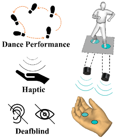
It was estimated that 0.2% of people around the world are living the server deafblindness [1]. For people with a combination of sight and hearing impairments, it is difficult to fully enjoy themselves in the art. Some researchers have dedicated themselves to helping deafblind people with artwork [2], but how to help people with deafblindness to “watch” the dance performance has not been studied adequately. Hereby, we offer an application proposal to the aforementioned open question and hope our idea could cause more social attention and academic interest. Figure 1 briefly illustrated the concept of the haptic application in dance performance for deafblind people. We could convert the dance performance (captured and analyzed from the human motion capture), for instance, the feet pace, to the haptic information (vibration, or ultrasound), thus the deafblind people can “feel” the dance performance from the foot space and dance rhythm. To realize the proposed object, following, but not limited to, professions and researchers are considered necessary to be involved: artist, psychologist, healthcare-givers, researchers from computer science, engineering, and robotics. Only with the cooperation from multidiscipline people, we could reduce the barriers for deafblind people and help them enjoy the art just like the rest of us. We strongly believe: the surprised face or the big smile of the deafblind people when they felt someone was just “dancing” on their palms, will make all tough research work worthy.

biomedres-openaccess-journal-bjstr

Figure 1: Concept of haptic dance performance for deafblind people.


For more Articles on: https://biomedres01.blogspot.com/

Saturday, June 22, 2024

Bilateral Erector Spinae Plane Block in the Management of Thoracic Spine Fracture: A Case Report Erector Spinae Plane Block for Spine Fractures


Bilateral Erector Spinae Plane Block in the Management of Thoracic Spine Fracture: A Case Report Erector Spinae Plane Block for Spine Fractures

Introduction

Spinal fractures are common and cause intense pain [1,2] due to the rupture of the vertebral body and local muscle contracture. Several analgesic techniques are usually proposed, including opioidbased analgesia, but the efficacy is often not optimal. Regarding ketamine, some studies with small numbers of participants showed significant effectiveness [3] but larger randomized clinical trials have not confirmed these results. Neuraxial techniques (epidural and intrathecal analgesia) have been shown to provide effective analgesia following elective spine surgery [4,5]hampering reconvalescense. We investigated the efficacy of patient-controlled epidural analgesia (PCEA, but are contraindicated in the context of vertebral trauma or vertebral fracture. The ultrasound-guided erector spinae plane block (ESPB), first described in 2016 [6], has been used in some cases of spinal surgery [7]but have various undesirable risks such as pneumothorax. The erector spinae plane block (ESPB including spinal decompression [8,9]but its effect on lumbar surgery is unclear. The aim of this study was to investigate the effect of the ESP block on postoperative opioid consumption and pain scores in patients undergoing spinal surgery.\nMethods\ nSixty patients undergoing open lumbar decompression surgery were randomly assigned to 2 groups. The ESP Group (n = 30. A case of EPSB in the emergency department was first reported in a patient with nonsurgical lumbar transverse process fracture [10]. The use of intraoperative ESPB was also recently reported to provide effective postoperative analgesia in a patient with traumatic rib and spine fractures [11]neuraxial techniques are often challenging or contraindicated due to spine fractures or coagulopathy. Erector spinae plane (ESP. We report here the use of ESPB as part of the analgesic perioperative treatment of multiple spinal fractures associated with refractory opioid-resistant pain. The patient provided written consent for the report of his case.

Case Description

A 50-year-old man presented with pathological spinal fractures secondary to bone metastases from a melanoma. Fractures were located at T11 and T12 vertebral bodies. Patient had severe pain (numerical rating scale [NRS] 7-9/10 at rest, 9-10/10 at mobilization) despite multimodal analgesia including paracetamol, a non-steroidal anti-inflammatory drug (NSAID) (intravenous ketoprofene, 100 mg every eight hours), a continuous clonidine infusion (600 μg per 24 hours), a continuous infusion of ketamine (0.1 mg.kg-1 per hour), and morphine (10 mg IV per 4 hours). Pain was refractory and the patient showed signs of morphine intolerance: nausea and vomiting, and bradypnea without pain relief. He also presented anxiety and depression secondary to pain with dark thoughts and no self-projection into future. Thirty six hours after the fractures, he was proposed an ESPB to relieve pain. The ultrasound guided ESPB was performed in the post anesthesia care unit (PACU) under surgical aseptic conditions.

The patient was placed in the left lateral decubitus position in his bed, and was monitored with pulse oximetry, non-invasive arterial pressure and electrocardiogram during and up to 30 minutes after the block. The ultra-sound-guided ESPB was performed by the first author (Figure 1), using a high frequency ultrasound probe (12-18 Hz) and ultrasound system HS 40® (Samsung Health Care, Korea). The skin was sterilized with an alcoholic povidone-iodine solution and the ultrasound probe was placed in a sterile bag. The probe was placed transversely at the T10 level to center the spinous process. Then, the probe was moved laterally in order to center the transverse process at the T10 level and was moved sagittally to obtain a parasagittal view. The same procedure was performed on each side. An 80 mm 22 gauge needle (UniPlex NanoLine®, Pajunk, Germany) was inserted in-plane in a cranio-caudal direction to guide the needle tip between the posterior fascia of the erector spinae and the tip of the transverses process of T11 and T12.

biomedres-openaccess-journal-bjstr

Figure 1: Sonographic anatomy for Erector Spinae Plane block (ESPB), Trapezius muscle (Tm), Erector Spinae muscle (Es), T9 and T10: Transverse process of the 9th and 10th thoracic vertebrae respectively. Ultrasound guided ESPB showing location of needle and local anesthetic (LA) deposited below the Es and spread of LA from the cephalad to the caudad direction.

Correct needle position was first confirmed by hydrodissection with 0.5 mL of 3.75 mg.mL-1 ropivacaine followed by the injection of 20 mL of the same solution to avoid exceeding the maximum recommended dose of 3 mg.kg-1 in total. Patient reported first pain relief 15 minutes after the injection. At 30 minutes post-injection he had mild pain at rest (NRS 3/10) and moderate pain at mobilization (NRS 5/10). He also showed positive mood change, looking more positively to the future and improving his morale. Patient was very well relieved for 16 hours following injection. Later, pain at rest became moderate (NRS 5/10) and remained similar to mobilization. Twenty-four hours after performing the ESPB, a spinal arthrodesis was performed to fix the spinal fracture. Another ESPB block was performed to insure intra and postoperative analgesia. The patient was pain-free (NRS 0/10) in the PACU. Pain was absent for 12 hours at rest (NRS 0/10) and of low intensity at mobilization (NRS 2-3/10). Thereafter, pain was mild at rest and moderate at mobilization.

Discussion

Bone tissue is a common target for metastatic cancers, with approximately 70% of patients with any metastatic cancer showing bones metastases [12]hypercalcemia, pathologic fracture, and spinal cord or nerve root compression. From randomized trials in advanced cancer, it can be seen that one of these major skeletal events occurs on average every 3 to 6 months. Additionally, metastatic disease may remain confined to the skeleton with the decline in quality of life and eventual death almost entirely due to skeletal complications and their treatment. The prognosis of metastatic bone disease is dependent on the primary site, with breast and prostate cancers associated with a survival measured in years compared with lung cancer, where the average survival is only a matter of months. Additionally, the presence of extraosseous disease and the extent and tempo of the bone disease are powerful predictors of outcome. The latter is best estimated by measurement of bone-specific markers, and recent studies have shown a strong correlation between the rate of bone resorption and clinical outcome, both in terms of skeletal morbidity and progression of the underlying disease or death. Our improved understanding of prognostic and predictive factors may enable delivery of a more personalized treatment for the individual patient and a more cost-effective use of health care resources.”,”container- title”:”Clinical Cancer Research”,”DOI”:”10.1158/1078-0432. CCR-06-0931”,”ISSN”:”1078-0432, 1557-3265”,”issue”:”20”,”- journalAbbreviation”:”Clin Cancer Res”,”language”:”en”,”note”:”- publisher: American Association for Cancer Research\nsection: Advances in Treating Metastatic Bone Cancer\nPMID: 17062708”,”page”:”6243s-6249s”,”source”:”clincancerres.aacrjournals. org”,”title”:”Clinical Features of Metastatic Bone Disease and Risk of Skeletal Morbidity”,”volume”:”12”,”author”:[{“family”:”- Coleman”,”given”:”Robert E.”}],”issued”:{“date-parts”:[[“2006”,10 ,15]]}}}],”schema”:”https://github.com/citation-style-language/ schema/raw/master/csl-citation.json”} . These bone metastases are very painful and alter quality of life. Some treatments are available to treat pain caused by bone metastases prior to their specific management (radiotherapy, surgery, minimally invasive surgery, kyphoplasty). Paracetamol, NSAIDs and opioids are typically used as first-line treatments, but with inconsistent efficiency. Ketamine does not seem to show significant effects on the management of these pains and has embarrassing side effects [3,13]including nociceptive, inflammatory, and neuropathic sources. Although opioids have long been a mainstay for perioperative analgesia, other non-opioid therapies have been increasingly used as part of a multimodal analgesic regimen to provide improved pain control while minimizing opioid-related side effects. Here we review the evidence supporting the use of novel analgesic approaches as an alternative to intravenous opioids for major spine surgery.”,”collection- title”:”Perioperative Management for Major Spine Surgery”,” container-title”:”Best Practice & Research Clinical Anaesthesiology”,” DOI”:”10.1016/j.bpa.2015.11.002”,”ISSN”:”1521-6896”, ”issue”:”1”,”journalAbbreviation”:”Best Practice & Research Clinical Anaesthesiology”,”language”:”en”,”page”:”79-89”,”source”:”Science- Direct”,”title”:”Non-opioid analgesics: Novel approaches to perioperative analgesia for major spine surgery”,”title-short”:”Non-opioid analgesics”,”volume”:”30”,”author”:[{“family”:”Dunn”,”given”:”Lauren K.”},{“family”:”Durieux”,”given”:”Marcel E.”},{“family”:”Nemergut”,” given”:”Edward C.”}],”issued”:{“date-parts”:[[“2016”,3,1]]} }},{“id”:1526,”uris”:[“http://zotero.org/users/2661041/items/ K3Q6Q6HK”],”uri”:[“http://zotero.org/users/2661041/ items/K3Q6Q6HK”],”itemData”:{“id”:1526,”type”:”article-journal”,” abstract”:”Background\nDelirium is a common and serious postoperative complication. Subanaesthetic ketamine is often administered intraoperatively for postoperative analgesia, and some evidence suggests that ketamine prevents delirium. The primary purpose of this trial was to assess the effectiveness of ketamine for prevention of postoperative delirium in older adults.\nMethods\ nThe Prevention of Delirium and Complications Associated with Surgical Treatments [PODCAST] study is a multicentre, international randomised trial that enrolled adults older than 60 years undergoing major cardiac and non-cardiac surgery under general anaesthesia. Using a computer-generated randomisation sequence we randomly assigned patients to one of three groups in blocks of 15 to receive placebo (normal saline. The gold standard management for analgesia is patient-controlled epidural analgesia (EA). In the case of spinal fracture, there is a contraindication to epidural analgesia. Moreover, EA has some risks and side effects, like epidural hematoma and low arterial blood pressure [4,14]. The paravertebral block is not described for spinal analgesia and has some risks, especially pneumothorax and low blood pressure.

Erector muscles of the spine consist of a group of three muscles (iliocostalis, longissimus and spinalis) located on the deep side of the back. Separated at the cranial part of the back, they join to form a common mass at the level of the sacrum. They allow the extension of spine in a symmetrical contraction. At the upper thoracic level, they are covered by the rhomboid muscle (T1-T5) and more superficially by the trapezius muscle (up to T12).

ESPB has been shown to be effective following spinal surgery [8,9]but its effect on lumbar surgery is unclear. The aim of this study was to investigate the effect of the ESP block on postoperative opioid consumption and pain scores in patients undergoing spinal surgery.\nMethods\nSixty patients undergoing open lumbar decompression surgery were randomly assigned to 2 groups. The ESP Group (n = 30, and very recently in cases of spinal trauma [11,12]. Cadaveric studies have confirmed the blockade of dorsal rami of multiple spinal nerves above and below the injection site when dye is injected below the fascia of the erector spinae muscle [6,15]. The ventral rami are blocked inconsistently and could be involved in the analgesic effects of ESPB without extension to the paravertebral zone. The local anesthetic spread, in both the cephalad and caudad directions is facilitated by the presence of the thoracolumbar fascia.

Thanks to the diffusion of the local anesthetic, we were able to perform the ESPB above the fractures. This allowed us to avoid injecting at the level of the fractured vertebra and to avoid possible anatomical modifications that could lead to difficulties in identifying the anatomical structures. ESPB is associated with a lower risk of side effects than the paravertebral block or epidural analgesia. This makes it faster to learn and probably easier to implement. We successfully tried to relieve this patient’s pain with the ESPB pending surgical management of this spinal fracture. This technique produced remarkable but temporary analgesia. An ESPB with continuous infusion via a catheter may be an interesting option for patients waiting spine surgery after a spinal fracture.


For more Articles on: https://biomedres01.blogspot.com/

Semaglutide in Obesity and Non-Alcoholic Fatty Liver Disease

  Semaglutide in Obesity and Non-Alcoholic Fatty Liver Disease Introduction Semaglutide injection was introduced in the US as an antidiabeti...