Gastrointestinal Tract Parasites of Camel in and Around Dire Dawa
Introduction
Camel (Camelus dromedarius) an important livestock uniquely adapted to arid and semi-arid environments [1] Camels in addition to serving as beast of burden and draft power produce milk, meat and wool, hair and hides [1]. Camel has a lot to do in pastoral social life in that it is the first among domestic animals to be considered as a social prestige in the world [2]. The dromedary has an estimated population of 18.5 million in the world [3]). In Africa, the dromedary camel population is about 15 million, which accounts 74% of the world’s camel population. Of these, 60% are found in Eastern parts of Africa i.e Somalia (6.2 million), Sudan (2.8 million), Kenya (0.9 million) and Ethiopia (1.7 million) [1,3]. The habitat on which camel lives generally is not suitable to development and transmission of parasites [4]. In spite of this, it harbors surprisingly diverse fauna of helminthes [5] and all classes of metazoan parasites were reported. Various groups of nematodes, trematodes, cestodes parasites of camel have been reported [5] and the major species include Moneizia, Strongyles, Haemonchus, Trichostrongyles, Fasciola, Tricuris and coccidian.
There has been little research done on disease of camel in Ethiopia. General field observations and a few surveys have indicated that camel diseases including parasitic diseases, low production low lifetime performance of female breeders and high calf mortality were the major constraints encountered in camels on Ethiopia [1]. There is some information on camel helminthes in Ethiopia. Some of these unpublished works on helminthes disease of camel in country include [2,5-11]. However, still there is a considerable gap in our knowledge of prevalence rate, the species of parasite involved, potential risk factors and economic impacts of gastrointestinal helminthes in camel population of Ethiopia in general and Dire Dawa area in particular.
Therefore, the objectives of this study were:
1. To estimate the prevalence rate of GIT helminthes in and around Dire Dawa.
2. To identify the species of GIT helminthes of camel in the study area.
3. To assess the potential risk factors of GIT parasites in camel of the study area.
Materials and Methods
Study Area
The study was undertaken in and around Dire Dawa administrative council in Eastern part of Ethiopia. Dire Dawa is located at 518km east of Addis Ababa at 9036’N and 40 52’E. The area has an altitude ranging between 226m and 950m.a.s.l. [12]. It has harsh climate with low unreliable and unevenly distributed rainfall with regular high temperature. The rainfall has bi-modal patterns with highest rainfall in July and August. The average annual rainfall varies700mm-900mm. the monthly mean maximum temperature ranges from 28.1oc I December and January and 34.6oc in May [12].
The administrative council is bordered to the South, South-east and South-west with Eastern Harrarghe zone of Oromiya Regional State and to the East, North-east, and North-west with Shinille zone of Somali Regional State. The total area of the administrative council is about 1,288.02 km2 [12]. Generally, livestock population of the study area recorded in [12]. exceeds 247,502of these goats make the highest proportion (51.26%) followed by cattle (22.09%); sheep (20.66%) and camel (2.24%).
Study Animal
A total of 200 camels were examined in and around Dire Dawa for GIT helminthes. This figure comprises randomly selected 123 adults and 77 young of which 100 were males and 100 were females. Sex selection was purposively done. The major area from where the samples came include Shinille, Melka-jebdu, Wursso, Issa village and Dire Dawa.
The Study Design and Methodology
The study was the cross-sectional survey in which the prevalence of the GIT helminthes had been estimated on the basis of routine fecal examination of individual samples. The fecal samples were collected from 200 camels directly from the rectum by using rectal gloves and place in universal bottle prepared with formalin solution until examined in the laboratory. Postmortem examination had been carried in the Dire Dawa municipal abattoir from 50 adult camels in addition to their fecal examination. During the survey, individual camel’s address, age and sex were recorded. Dental eruption and wear were used to consider age categorization [13]. Baesd on this bellow four years old considered as young and above four years as adult.
Laboratory Works
Coproscopic Examinations: A fecal sample of 10 to 15g was collected directly from rectum by using rectal glove and samples were placed in universal bottle half fill with formalin solution and tightly sealed. The samples were labeled and immediately dispatched to Dire Dawa regional veterinary diagnostic and investigational parasitological section laboratory. On some occasions samples were stored in +4oc frig for about 72 hours when immediate examination was not possible on the basis of wilson,[13] The laboratory technique employed were mainly qualitative fecal analysis i.e floatation, sedimentation, and Barman’s technique were practiced. Positive samples for helminthes egg examination were cultured at incubator temperature 37oc for aweek and then larvae identified by using Burmen’s technique [14]. Fecal culture was done by taking 19g of feces in a tray and then moistening with water if too dry or adding charcoal if too wet and incubating for a week.
Postmortem Examination: The survey was supported by postmortem examination on GIT helminthes of 50 camels (adult camels that were slaughtered at Dire Dawa municipal abattoir. Any parasite that was found in ruminal compartments of camel’s GIT examined grossly and microscopically from ruminal contents and identification was based on [13-15]. In addition, their location (sites within the lumen) and morphologies were used as reference frame in identification of adult parasites in their genus level.
Data Analysis: Data were entered into MS excel spread sheet and analyzed using SPSS 11.5 software (2002). Statistical analysis included comparison of GIT helminthes on the basis of age, sex, sampled areas. The prevalence in (%) was obtained by dividing the number of animals harboring a given helminthes to the total animal examined. P-value <0.05 was considered as statistically significant.
Results
Coproscopic Results
The present study revealed the existence of GIT helminthes in the study area with over all prevalence rates of 11.5% on the basis of coproscopical examinations. During this survey the GIT helminthes observed include Haemonchus spp, Tricostrongyles spp, Cooperia spp, Tricuris spp, Fasciola spp and Oesophagostomum spp which have significant importance both on health and economy. A different in prevalence rate of GIT helminthes between the young and adult camels was not statistically significant using x2 square analysis (p.>0.05) (Table 1). This shows that the prevalence rate of GIT helminthes was higher in young (18.18%) than adult camels (12.19%). In addition, analysis taken sex ways showed that the prevalence rate of GIT helminthes was higher in females than males. By x2 square analysis, the difference between prevalence of females (20%) and males (9%) was analyzed and found to statistically be not significant (p>0.05) (Table 2).
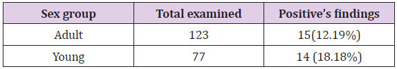
Table 1: Prevalence of GIT parasites in camel by age, irrespective of specie.
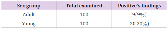
Table 2: Prevalence of GIT parasites in camel by sex, irrespective of species.
Postmortem Examination Result
A sample was collected from 50 adult camels and 5% of the samples were positive for different species of GIT helminthes. Based on sites on camels GIT and morphological appearance of the helminthes itself, the species of the helminthes was identified. As result, Haemonchus spp (4%), Trichostrongyles spp (2%), Tricuris spp (2%), and Fasciola spp (2%) were observed during postmortem examination. There was no mixed parasitic infestation in case of postmortem examination. On some occasions, Postmortem findings helped more in identifying exactly the species of parasites which was difficult during fecal eggs.
Occurrence of GIT Parasites in Different Study Areas
The frequency of occurrence of different parasites in different study areas irrespective of species shows presence of parasitic burden in all study areas. The frequencies of occurrences of different parasites in different study areas seemed almost the same (Table 3).
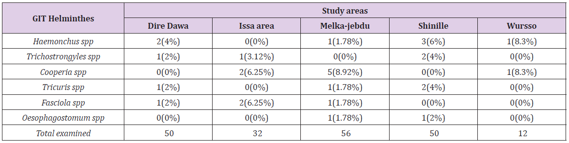
Table 3: Frequency of occurrence of different parasites in different study areas.
Discussion
This study revealed difference in the prevalence rate of GIT helminthes in coproscopic (29/200) and postmortem examination (5/50), which was 14.5% and 10% respectively. In addition, significant differences of GIT helminthes infestation between age groups, with higher infestation rate in young (18.18%) than adults (12.19%). this might be due to relative resistance of adult camels to GIT helminthes than younger. Similarities in frequencies of occurrences of GIT helminthes in the different study areas could be due to similarities in epidemiology and climatic conditions those study areas as well as their pastoral system of management. However, prevalence rates vary widely from region to region as well as from season to season within the same region [16]. Among those GIT helminthes examined, cooperia spp Oesophagostomum spp were absent in postmortem examination and Haemonchus spp (4%), Trichstrogyles spp (2%), Tricuris spp (2%), and Fasciola spp (2%) were prevalent. This could be due to resistance of adult camels for Cooperia spp and Oesophagostomum spp. this was agreed with survey done by [5,10,11].
Analysis that was taken in sex wise showed that the prevalence of GIT helminthes was higher in females (20%) than males (9%). This may be due to females being liable for many physiological stress-inducing factors like pregnancy and lactation [9]. There was no mixed infestation encountered during the study. This may be the reflection of pastures not being infected by different types of GIT helminthes. Camels acquire helminthes by grazing on infected pastures or ingesting larvae infected water [17]. During this survey the GIT helminthes found includes Haemonchus spp 7(3.5%), Trichostrongyles spp 4(2%), Cooperia spp 8(4%), Tricuris spp 4(2%), Fasciola spp 4 (2%) and Oesophagostonum spp 2(1%). Haemonchus spp (4%), Trichostrongyles spp (2%), Tricuris spp (2%) and Fasciola spp (2%).
Various works had documented; according to Rechards, [6] parasites encounter in Borena pastural area includes Trichostrongyles spp (85%), Strongloides (11%) and Tricuris spp (20%). In Harrargye pastural area, Birhanu, [7] reported Trichostrongyles spp (86%), Strongyloides (7.86%), Tricuris spp (43.6%) and cestodes (2.4%); Abebe, [8] reported Trichostrongyles spp (87.4%), Tricuris spp (49%) and cestodes (4.5%); Getachew, [9] reported Trichostrongyles spp (90.25%), Strongloids (10.7%), and Tricuris spp (28.9%) from Jigjiga and Lagahabur. Tenay, [10] reported Trichostrongyles spp (94.61%), Strongloids (8.09), Tricuris spp (45.6%) from Southern rangeland of Ethiopia (Borena). Theodros, [11] reported Trichostrongyles spp (94.61%), Strongloids (8.07%), Tricuris spp (72.43%) and cestodes (10%); Meles, [5] from Hararghe reported as Ttricostrongyles spp (86.94%), Strongloids (33.78%) and Tricuris spp (24.04%) and Ahmed, [2] in Eastern Ethiopia reported Trichostrongyles spp (71.3%), Haemonchus spp (64%), Nematodirus spp (34.8%), Tricuris spp (10.4%).In this study lower prevalence were reported as compared to those previous studies , this might be related to the wide use of antihelminthic drugs to the camels by owners that consequently reduce the overall prevalence rate in the study area.
Conclusion and Recommendations
The overall lower prevalence of GIT helminthes in camel of Dire Dawa and surrounding areas in this study suggests the natural resistance of the camel to the parasites due to the feeding habit and natural topography of the study areas as well as regular deworming habit of nomads. The study showed that Haemonchus spp and Cooperis spp, were found to be the most prevalent GIT helminthes in the study areas compared to other helminthes reported during the study. The result of present study has also revealed moderate spread of six GIT helminthes namely Haemonchus spp, Trichostrongyles spp, Cooperia spp, Tricuris spp and Oesophagostomum spp. Mixed infestation was not encountered both coproscopically and postmortem examinations. The prevalence rate of GIT helminthes was higher in young than adults and the prevalence rate was higher in females than males. Based on the above conclusive remarks the following recommendations are forwarded: 1. Public education about the importance of livestock management in parasite control.
2. Creating awareness on strategic parasite control methods.
3. Further epidemiological studies should be conducted in different agro-ecological zones and with different study designs.
For more Articles on: https://biomedres01.blogspot.com/


No comments:
Post a Comment
Note: Only a member of this blog may post a comment.