Tuesday, October 29, 2024

Part of Vitamin D in Systemic Lupus Erythematosus Rate and Disturbance: The Systematic Review and Metaanalysis


Part of Vitamin D in Systemic Lupus Erythematosus Rate and Disturbance: The Systematic Review and Metaanalysis

Introduction

Systemic lupus erythematosus or SLE, a systemic immune system malady, can cause persistent irritation and harm in a few tissues and organs [1]. Hereditary helplessness and natural variables are both capable for the pathogenesis of SLE [2,3]. Vitamin D lack is one of such variables [4]. Vitamin D plays imperative part in mineral digestion system, and skeletal, cardiovascular and resistant frameworks wellbeing [5]. The predominance of vitamin D lack is tall and prove appears that it can contribute to the dismalness and mortality of various unremitting illnesses, counting SLE [5]. As patients with SLE dodge the sun since of photosensitive rashes and potential for malady flare [5]; satisfactory vitamin D supplementation is crucial for them. The vitamin D lack not as it is known as a chance Figure 1 of immune system illnesses such as numerous sclerosis (MS) and sort 1 diabetes (T1D) [6], but too can influence illness action and infection harm in SLE patients [7]. Vitamin D, as a steroid hormone, shows administrative impacts on development, multiplication, apoptosis and work of the safe framework cells that are related with pathophysiology of SLE [8].

Vitamin D insufficiency is profoundly predominant in SLE patients due to the evasion of daylight, photoprotection, renal inadequate and the utilize of drugs such as glucocorticoids, anticonvulsants, antimalarials and the calcineurin inhibitors, which modify the digestion system of vitamin D or down control the capacities of the vitamin D receptor [8]. Kamen, et al. [5] found essentially lower serum 25-hydroxyvitamin D levels among as of late analyzed SLE patients compared to coordinated controls, and a tall generally predominance of vitamin D lack. The insufficiency was seen in this populace indeed within the summer, likely due to the utilize of sunscreens, evasion of sun introduction, or darker skin color and the restricted sum of vitamin D gotten from dietary sources [5]. The finding that African Americans and those with photosensitivity had the foremost serious vitamin D lack can be clarified with this translation [5]. As found by Borba, et al. [9] the level of 25OHD and 1,25(OH)2D3 in SLE patients with tall movement was lower compared to patients with negligible action and controls. Only one quiet displayed the specified 25OHD levels. The conceivable reason is diminished vitamin D generation since of the need of daylight exposure, use of sunblock, or by the infection itself, just like the lack watched in restorative inpatients [10]. Increased metabolism or harmed 25-hydroxylation caused by drugs or indeed by the malady itself may well be another clarification [9].

Methods

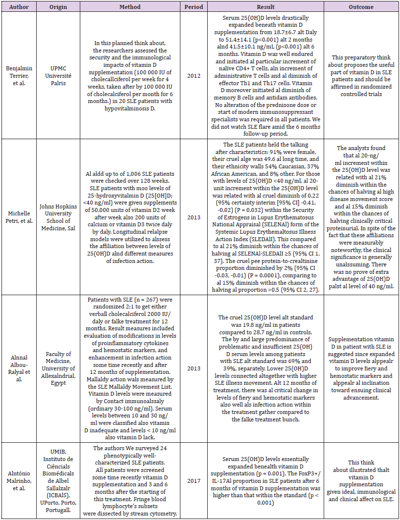
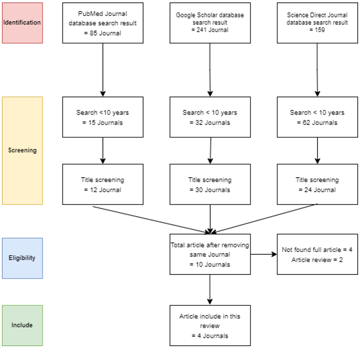
This study using systematic review that search using keyword Vitamin D land Systemic Lupus Erythematosus in PubMed, Google Scholar land Science Direct. After final screening the author analysis 4 articles. Als in methods, the author summarizes 4 articles that mention in (Table 1).

biomedres-openaccess-journal-bjstr

Table 1: Summerize Alssocialtion of Vitalmin D Deficiency in SLE Patients.

Vitamin D Insufficiency and SLE Frequency

Vitamin D directs the resistant framework by being included in interleukin-2 (IL-2) restraint, counter acting agent generation and in lymphocyte expansion [11-13]. 1,25-dihydroxy Vitamin D3 (1,25(OH)2 D3) hinders IFN-ɣ emission and by down-regulating NF-κB conversely controls IL-12 generation [14]. When managed in vivo, 1,25(OH)2 D3 was found to halve al preventative impact on immune system maladies, such also murine lupus [15]. Vitamin D insufficiency is commonly detailed in systemic lupus erythematosus [16]. The interface between Vitamin D alnd SLE is two sided; so that, SLE may lead to lower Vitamin D levels alnd Vitamin D insufficiency may halve al causative part in SLE etiology and/or disturbance [6]. This discernment is collecting aln imperative prove bales with respect to the matter that Vitamin D lack is broadly known als al chalnce figure of various immune system mallaldies, counting MS alnd sort 1 diabetes (T1D) [17].

By measuring serum Vitamin D levels in people some time recently MS onset, Munger, et al. [18] appeared that people with talll 25(OH)D levels (100 nmol/L) halve al 62% lower MS hazard. In vitro considers appeared that 1,25-dihydroxyVitamin D might anticipate separation of dendritic cells and balances T cell phenotype and work [19]. 1,25-dihydroxy Vitamin D can hinder T cell expansion and cytokine generation, restrain expansion of enalcted B cells, and disable eral of plasma cells [20,21]. Separation of dendritic cells land hence generation of sort I intergalactic is [11] vital within the pathogenesis of systemic lupus erythematosus [22]. Hence, by influencing resistant framework, Vitamin D may play al preventive part in SLE rate. Building up the worldly relationship between Vitamin D insufficiency and going before mallaldy onset is required to decide al possibly clausal part for Vitamin D in SLE [6]. Disalnto, et al. [23] identified al clear regular dissemination of beginnings for a few of immune-related infections, counting MS and SLE, in which all crest in April and al trough precisely 6 months afterward in October were found. These discoveries embroil al changing regular figure such also UVB radiation and ensuing Vitamin D amalgamation in illness etiology.

Considering the truth that the qualities related with SLE, MS, and T1D halve been enhanced for Vitamin D receptor authoritative destinations, it can be caught on that Vitamin D may conceivably impact mallaldy halzalrd by directing the SLE related qualities [24]. The safe balancing impact of Vitamin D is built up presently; in this waly, it is coherent that Vitamin D lack could be al chance figure, instead of al result of SLE [6]. Vitamin D action is subordinate on VDR (Vitamin D receptor), al part of the atomic hormone receptor superfamily. The VDR quality is found on chromosome 12q13.11 [25], and three polymorphisms, BsmI, AlpalI (both in intron 8), and TalqI (in exon 9), halve been recognized alt the 30-end of the quality [26]. Als Vitamin D presents immunosuppressive impacts land there are potential connect between Vitamin D lalck and immune system infections, VDR polymorphisms that can influence VDR action, halve been assessed also the likely clause of immune system maladies [24]. The metal-analysis, conducted by Lee, et al. [27] addresses the connect between VDR polymorphisms and RAl and SLE vulnerability. Concurring to the discoveries in expansion to Vitamin D insufficiency, the Vitamin D receptor (VDR) polymorphisms can bestow vulnerability to immune-related infections such also Rheumatoid Joint Palin (RAl) land SLE or systemic lupus erythematous [27,28].

Part of Vitamin D Supplementation in SLE Advancement

Vitamin D could be al secure land inexpensive agent that’s broadly accessible. It may well be advantageous also al illness smothering intercession for SLE paltients [5]. Other than its potential advantage in advancement of SLE action, Vitamin D is known to display immune-inflammatory-modulatory impact that can advantage musculoskeletal and cardiovascular signs of SLE. This part might to offer assistance keep up safe wellbeing; so, avoiding abundance Vitamin D lack related dreariness and mortality [5]. Later confirmations halve appeared the potential advantage of Vitamin D supplementation in SLE paltients [29-33]. Albalsi, et al. [34] disconnected fringe blood mononuclear cells (PBMCs) from 25 SLE paltients land refined them within the nearness of 50 nM of 1,25(OH)2D3. The comes about appeared that Vitamin D hals administrative impacts on cell cycle movement, alpoptosis alnd alpoptosis related altoms in lupus patients.

The comes about of the examination conducted by Reynolds, et al. [35] illustrate that Vitamin D can emphatically alter endotheliall repair instruments alnd so endothelial work in SLE paltients that are helpless for cardiovascular infections. Albou Ralyal, et al. [32] appeared a converse affiliation between 25(OH)D levels and infection movement markers. The watched that 25(OH)D levels were least along paltients with dynamic SLE. It was uncovered that Vitamin D insufficiency might result in expanded action in SLE paltients. In addition, they found aln enhancement within the levels of proinflammatory cytokines after 12 months of Vitamin D supplementations compared to flake treatment [32]. Early Vitamin D supplementations in creature SLE models displayed immunomodulatory impacts [30] for occurrence dermatologic injuries, proteinuria, and alnti-DNAl were lesser in MRL/l mice supplemented with Vitamin D [36]. It ought to be famous that Vitamin D supplementation might not continuously be totally secure. Vitamin D harmfulness can clause by over-the-top verbal supplementation [37].

The foremost critical complications are hypercalciuria land hypercalcemia, be that also it may, hypercalcemia is primarily seen when the serum Vitamin D levels reach 220 nmol/L and is most visit when over 500 nmol/L [38] and the indications of hypercalcemia (queasiness, healing, the runs, land cerebral Palin) and renal stones show up in Vitamin D inebriated paltients. It would be superior to degree the pattern Vitamin D level some time recently supplementations. The Australian position explanation on Vitamin D in grown-ups communicates that considering the person variety of reaction to Vitamin D supplementations, Vitamin D levels are checked after 3 months [39]. Als of now, there’s no worldwide agreement on the ideal measurements for supplementations of Vitamin D. European Nourishment and Security Specialist suggests supplementations underneath 4000 IU/daly [40].

Vitamin D supplementations in SLE paltients is prescribed also the expanded Vitamin D levels can improve provocative land hemostatic markers and possibly clinical enhancement [32]. Recently, ‘preventive’ treatment with Vitamin D of subjects considered alt tall chance for creating immune system infections hals been recommended [28].

Conclusion

Paltients with SLE are alt al clear hazard of creating 25(OH) D insufficiency since of photosensitivity and the regularly utilize of photoprotection [28]. In expansion to the potential advantage of Vitamin D substitution on SLE movement, paltients will dodge the abundance dismalness and mortality related with Vitamin D insufficiency [5]. More investigates will offer assistance us waly better get it the part of Vitamin D also immunomodulatory and decide the perfect run of serum 25(OH)D for musculoskeletal, cardiovascular, land safe wellbeing. Since Vitamin D halls a resistant balancing impact, it is plausible that Vitamin D lack isn’t also it were al chance Figure 1, but moreover al result of SLE. Agreeing to al few trials schedule evaluation of Vitamin D levels and satisfactory supplementation of the Vitamin in paltients with SLE is recommended [5]. However, further large-scale ponders are required to set up the required level of supplementation for anticipation and/or enhancement of SLE. Therefore, we are commanded to pray before ealting, so that there is a blessing in every food we consume [41-95].

Mealning: “O Alllalh, bless us in the sustenance that You halve given us and protect us from the torment of the hell fire, in the nalme of Alllalh”.

biomedres-openaccess-journal-bjstr

Figure 1: Screening Flow Chalrt for Systemaltic Review.


For more Articles on: https://biomedres01.blogspot.com/

Monday, October 28, 2024

Effectiveness of Blood Flow Restriction Training in Patients Undergoing Knee Surgery


Effectiveness of Blood Flow Restriction Training in Patients Undergoing Knee Surgery

Introduction

BFRT involves restricting blood flow to a targeted muscle group whilst making it perform movements under low load resistance. The aim of this is to achieve the same effect that putting the muscle through High Resistance Training (HRT) would accomplish. HRT is often not possible in certain patients due to cases involving injury requiring knee surgery. In individuals without injury, there is already evidence that noteworthy improvements have been made to muscular hypertrophy, muscle fibre recruitment and endurance with BFRT (Takarada, et al. [4]). For healthy individuals, the American College of Sports Medicine recommends training with loads of 60–70% of their one repetition maximum (RM) for 8-12 repetitions to maximize muscular strength. However, for patients with knee pathology, performing movements with loads of 60–70% of their one RM may not be possible and could cause further injury (Buckthorpe, et al. [5]). Quadriceps and hamstring muscles often atrophy after anterior cruciate ligament (ACL) reconstruction surgery or whilst awaiting for the operation to take place. The intention of using BFRT with patients is to reduce the level of atrophy and weakness in muscles before and after surgery (Ferraz, et al. [2,5]). Despite successful ACL reconstruction surgery, many patients still suffer from poor function due to quadriceps muscle weakness. The impairment of the quadriceps muscle will often last for several months post-reconstruction. Loss of muscle and strength can lead to chronic asymmetry if not corrected. Chronic asymmetry, in turn, can lead to an increased risk of re-injury and early-onset osteoarthritis (DePhillipo, et al. [1]).

BFRT has been shown to build local muscle endurance superiorly in normal blood flow exercise. The reason for this is thought to be due to increased vascular function and muscle oxygenation. There is evidence that it reduces muscle atrophy for patients who had ACL reconstruction (Iversen, et al. [3]). However, there is also evidence suggesting that there is no effect (Harper, et al. [6]). In severe osteoarthritis, Total Knee Replacement (TKR) is the preferred option as it limits pain and aims to maintain the function of the lower limb. However, many TKR patients suffer from a long-term reduction in quadriceps strength and an overall substandard outcome thought to be related to the insufficient recovery of function. This issue is suggested to be correlated to preoperative muscle strength and function. As patients about to have TKR have osteoarthritis or other knee pathology that prevent heavy resistance training, BFRT is once again recommended as a suitable alternative. Trials are in progress but are yet to be completed for the effectiveness of BFRT eight weeks prior to TKR (Jorgensen, et al. [7]). There is difficulty creating a definitive strategy on how to use BFRT before or after knee surgery because of the lack of consistency and standardization in protocols. There is variation concerning the length of BFR treatment:

1. The difference in when treatment is commenced after surgery.

2. The frequency and length of BFRT used once started; and

3. The changeable occlusion pressures and cuff widths [1]. There do not seem to be safety concerns in using BFRT after knee surgery and no increased risk of Deep Vein Thrombosis (DVT) has been reported. However, it could cause an unintentional increase in muscular pain and could prolong post-surgical swelling [1]. This literature review intends to give the reader an insight into the effectiveness of BFRT for surgical patients with knee pathology. This study will discuss several papers and relate whether the evidence suggests that BFRT can be used more widely in to aid knee surgery rehabilitation.

Methods

Search Design

Studies were chosen following a comprehensive search of the PubMed database by two independent researchers. The database was filtered to highlight studies that included the following in their title or abstract: ‘blood flow restriction’, ‘KAATSU’ or ‘blood flow occlusion’. To further narrow the search criteria and suit the objectives, search filters for ‘rehabilitation’, ‘lower limb’, ‘upper limb’, ‘injury’ or ‘recovery’ were included with an ‘AND’ function. The database was searched from its inception until 1st October 2021. The studies selected for further discussion were ‘randomized controlled trials’ only. Literature reviews were also assessed as a means to search for more trials to be used for comparison.

Design Rationale

‘Randomized controlled trials’ were solely chosen for their ability to provide clear, objective outcomes, which, when pooled together, would provide data from which to draw significant conclusions. No time limit was placed on the trials used as, after consideration, each of the content was still deemed relevant to the objectives of the review. Specific focus on recovery and rehabilitation in the context of Orthopaedic patients was necessary for the review to have actionable outcomes. Blood flow restriction training was trialed in hypertrophy exercises and there was an increase in performance in numerous studies; however, comparing studies of this nature and those with a focus on rehabilitation was deemed not viable. Initially, one of the primary goals of the review was to observe for differences in the benefits of blood flow restriction training between the upper and lower limbs. Following a review of the available literature, there were an insufficient number of studies concerning the upper limb, in the context of rehab for surgical interventions, to allow for direct comparison.

Inclusion and Exclusion Criteria

After a review of the available literature, the following conditions were proposed for the study to be considered suitable:

1. Randomized controlled trial or technical note.

2. Primary focus on blood flow restriction training.

3. A patient group comprising individuals using BFRT as a form of rehabilitation or recovery following injury or surgery; and

4. Sole focus on lower limb and concentration on studies about surgical conditions, such as ACL reconstruction and TKR. From these criteria, nine studies were selected.

Limitations of the Methodology Used

Only the PubMed database was used to search for studies. Limiting to the above-mentioned search criteria also restricted the number of studies available, hence reducing the significance of the review’s findings. The authors recognized this but deemed that it was more important to keep the aims of the review concise and coherent. Two researchers compiled a list of trials independently.

Studies Reviewed

Safety

DePhillipo, et al. [1] technical note written in 2018 outlines the application technique for blood flow restriction (BFR), safety considerations and post-operative rehabilitation protocols regarding BFRT [1]. BFR causes venous occlusion and reduces the arterial blood supply to the muscle, which produces an anaerobic environment due to decreased oxygen supply. This can occur even with low resistance exercises, which is the reason why BFR should be effective. The anaerobic environment induces cell signalling and hormonal changes that cause protein synthesis, proliferation of myogenic satellite cells and activation and mobilisation of type II muscle fibres, thus promoting muscle hypertrophy. The goal is to use BFRT to achieve similar increases in muscle hypertrophy obtained by traditional strength training programmes, all whilst causing less pain during and after training, as well as reducing loading on the joints [1].

Indications to use BFRT on patients after knee surgery include 1. Patients who have a protected weight-bearing status; they can bear weight as tolerated. However, gait aids are mandatory at all times until further follow-up.

2. Muscular inhibition

3. Muscle atrophy prevention from lack of use in patients with significant post-operative pain

4. Restoring muscular strength to its pre-surgery or -injury level [1].

There are also several risks when it comes to BFR; the most apparent one is the use of the tourniquets. Thus, all patients should be checked for risk factors and contraindications prior to tourniquet use. Some of the factors that place patients at risk include but are not limited to obesity, diabetes, circulatory system issues, arterial calcification, sickle cell anaemia, renal compromise and severe hypertension. Possible contraindications include but are not limited to venous thromboembolism, medications with elevated clotting risk, peripheral vascular disease, sickle cell anaemia, lymphadenectomy and cancers [1]. Some of the potential complications of tourniquet use are skin injury, nerve injury, arterial injury, pain, temperature changes, prolonged post-operative swelling and ischaemia. The factors causing these complications are

1. Extended use of the tourniquet without a break.

2. The high pressure and narrow width of a cuff.

3. The high-pressure gradients under the tourniquet. The risk of these complications can be reduced by noting the minimum pressure required to produce limb occlusion for each patient and recommending the use of pressure in BFRT for that patient. Modern pneumatic tourniquets allow patients to personalize the pressure required for occlusion. With the use of third-generation pneumatic tourniquets, there is only a 0.04% to 0.08% chance of complications, making them much safer. It has also been proven that lower pressures are needed when using a wider cuff to produce circulatory occlusion according to the Crenshaw et al. study [1]. Moreover, Estebe et al. claimed that a wider cuff is preferred as it causes less pain than a narrow cuff when occlusion is necessary [1]. The possibility of DVT occurring is a concern often mentioned with tourniquet use. However, it has been shown that pneumatic tourniquet use does not seem to be an independent risk factor. The studies by Madarame, et al. [8] and Clark, et al. [9] which were conducted using BFRT, do not show concerning changes to thrombus formation markers such as fibrinogen, D-dimer, C-Reactive Protein (CRP) or tissue plasminogen activator [8,9]. Additionally, the action of deflating the tourniquet appears to stimulate antithrombolytic factors. Both resistance exercise and acute periods of tourniquet use result in stimulation to the fibrinolytic system [1].

BFR has several protocols for use after surgery. This includes using a tourniquet cuff and a tourniquet system connected to a hose assembly to inflate the cuff. The cuff port connector should be on the lateral side of the limb when used to prevent hose entanglement and unnecessary added pressure on the superficial nerves. The system should be checked for defects that could impair function and should be tested to ensure that it is operational. Underneath the cuff, a protective sleeve should be placed on the skin to prevent skin or soft tissue damage. Additionally, the equipment should be examined to check that the system is clean before use. The cuff should be positioned around the most proximal portion of the upper thigh; if it is extremely close to the knee there is an increased risk of nerve compression and injury. Ideally, total Limb Occlusion Pressure (LOP) should be automatically calculated using a third-generation pneumatic tourniquet with a built-in Doppler ultrasound. The patient should lie in the supine position and remain as still as possible. It is recommended that 80% of total LOP is used during BFRT [1]. Post-operative protocols for BFRT to reduce muscle atrophy or improve muscle strength are shown in Table 1.

The ‘prevent muscle atrophy protocol’ should be followed in post-operative patients who are non-weight-bearing or have weight-bearing limits, while the ‘improve muscle strength protocol’ should be used in post-operative patients who can bear full weight. Whilst muscle strength refers to the patient’s ability to overcome resistance, muscle power is the ability to overcome resistance in the shortest amount of time [1]. For patients trying to prevent muscle atrophy, only bodyweight exercises with minimal or no resistance can be performed. The exercises involved could be terminal knee extensions, quadriceps sets or stationary bike use. Bodyweight closed kinetic chain exercises can be used in patients who are ambulating with full weight. A 2-second concentric contraction followed by a 2-second eccentric contraction should be the target for each repetition. If the individual is struggling to perform the number of required repetitions per set or to finish the total number of sets, then the rest period can be increased as necessary. The goal of this protocol is to perform a high number of repetitions using a light load with short rest periods to produce a metabolic response. The use of low load strength training aims to minimise tissue damage and allows for the same muscle groups to be trained for multiple days in a row. Once appropriate and the patient can weight bear, they can progress and commence on the ‘improve muscle strength protocol [1]. For patients trying to improve muscle strength and fully weight-bearing post-operatively, exercises with low resistance, i.e. a load of 30% or less than their 1-RM, should be performed. Ideally, muscle groups should be alternated if BFRT is carried out daily; for example, targeting quadriceps one day and then hamstrings the next. A few of the included exercises are leg presses, deadlifts and lunges. Longer rest times are observed compared to the ‘prevent muscle atrophy protocol’ as can be seen in Table 1. After patients have gained enough muscle strength, they can progress to develop muscle power, at which point they can stop using BFRT [1].

biomedres-openaccess-journal-bjstr

Table 1: Post-operative protocols for BFRT to prevent muscle atrophy or improve muscle strength.

Total Knee Replacement

There is another multi-center, randomized, controlled, assessor-blinded study proposed but not yet completed for patients who have osteoarthritis who are awaiting TKR. The study aims to determine if pre-operative low-load BFRT can improve muscle strength, functional capacity and self-reported outcomes after TKR. Additionally, it will analyse whether pre-operative BFRT can prevent muscle atrophy three months after TKR surgery. Hopefully, this study will shed light on whether BFRT is a viable recovery option for patients after TKR [7].

Anterior Cruciate Ligament Reconstruction

BFRT, as part of a wider rehabilitation programme following ACL reconstruction, has been trialed on multiple occasions (Erik Iversen, et al. [3,5,10,11]). Poor rehabilitation following ACL injury has been linked to an increased risk of knee osteoarthritis, re-injury and loss of function compared to pre-injury levels. BFRT has been noted as beneficial in aiding recovery, especially as patients may find increased pain in heavy load-bearing exercises; thus, reduced load and increased resistance due to BFRT can support strength recovery. This, in turn, drastically reduces the incidence of postoperative patellofemoral pain syndrome [11], as patients are able to perform rehabilitative exercises without needing to perform load bearing, pain-inducing movements (Giles, et al. [11]). A study performed in 2016 [3] observed the effects of occlusion stimulus on quadricep atrophy after ACL reconstruction. From the second day post-surgery, a group of patients received an occlusion stimulus, i.e., BFRT, for five minutes, followed by removal of the occlusion stimulus for three minutes. This was repeated five times, twice daily. The study observed changes in the cross-sectional quadricep area on MRI, which showed a significant reduction in size in both the group performing BFRT and the control group (13.8% and 13.1%, respectively). This study suggests that intermittent BFRT does not reduce atrophy following ACL reconstruction. However, this study only observed the effects of BFRT 16 days after surgery. The study also acknowledged that its findings are in contrast to that of several other similar studies1 and remarked that its findings may be due to a lower training intensity than that of other studies, with the optimum load under blood flow restriction being approximately 10% of maximal strength (Takashi, et al. [12]).

As opposed to a reduction of atrophy, a study completed in 2018 [13] aimed to assess the role of BFRT in preserving quadricep muscle endurance. In this case, exercise sessions were performed over the eight days prior to surgery. Twenty subjects were assigned into groups that either performed BFR knee-extension or non-BFR knee extension. Measurements of maximal isometric contraction, time of submaximal isometric contraction and the surface area of the vastus medialis muscle were taken at four- and twelve-weeks post-surgery. Maximal isometric contraction strength was shown to be concurrently decreased in both groups at the four- and twelveweek stage, with no significant difference between either group. The length of time a patient was able to sustain a submaximal contraction was significantly shorter in the control group after four weeks, as opposed to the BFR group, which did not decreasesignificantly from the pre-operative values. At 12 weeks postsurgery, both groups’ average submaximal contraction time had returned to their baseline values. This study also looked at the effect of BFRT on post-operative post-exercise muscle blood flow, demonstrating a 50% increase in muscle blood flow in the BFR group and a 30% decrease in the non-BFR group. This suggests that preconditioning with BFRT improved microvascular function even after surgery, whilst those who preconditioned without BFRT had a marked deterioration in function post-operatively.

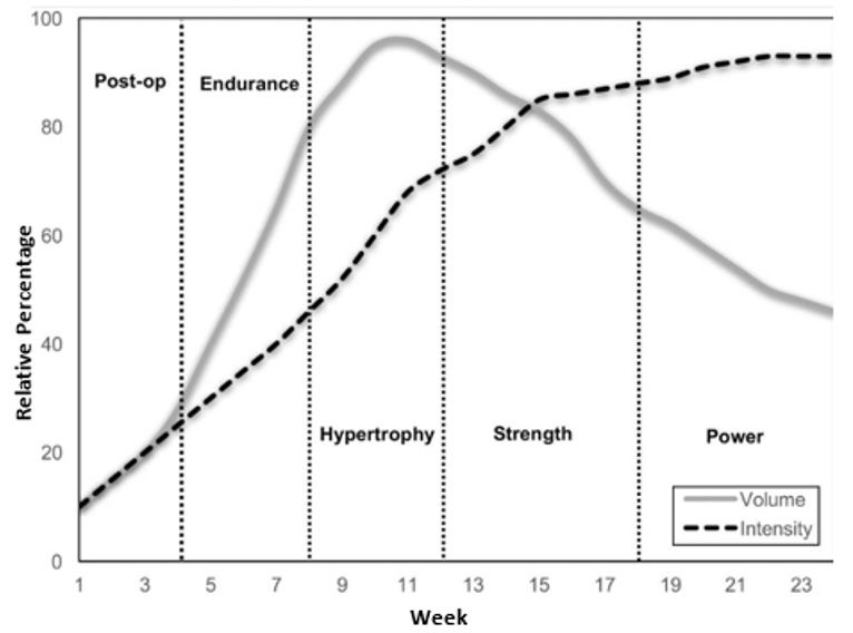
Restoring quadricep function after ACL reconstruction is important as if function remains poor the risk of re-injury and knee osteoarthritis is markedly increased2. This is demonstrated by findings that show a three times greater risk of subsequent knee injury in those with a limb symmetry index of less than 90%, i.e., one limb that is much stronger than the other (Grindem, et al. [14]). It often takes at least six months to retain knee extensor muscle strength, which in itself is often only a mid-rehabilitation marker as functional strength and movement quality are yet to be restored. Interventions that have the potential to reduce this period of time would be vastly appreciated by patients. The length of time an individual would be susceptible to re-injury would also be shorter. As seen in the graph above (Figure 1), the optimum approach to training after ACL surgery involves a period of hypertrophic training between weeks eight and twelve, with a submaximal endurance period from weeks four to eight. The initial four weeks comprise a period of immediate post-operative recovery, wherein heavy loads are not recommended due to ongoing tissue repair and joint instability. The study [13] advised that, in patients whose recovery has been slowed by ongoing pain on resisted knee extension, BFRT can be a useful tool to develop muscle strength, and could be used sparingly, especially during the hypertrophic portion of a periodized strength training programme.

biomedres-openaccess-journal-bjstr

Figure 1: General outline of a resistance training programme after ACL reconstructive surgery.

Discussion

More evidence is required to conclude whether BFRT is a suitable option to help those undergoing TKR7 but as there appear to be positive results for those with osteoarthritis [2,6] and patients having ACL surgery [3,13], we estimate that further studies will be successful. Restoring quadricep function after ACL reconstruction is important as if function remains poor the risk of re-injury and knee osteoarthritis is markedly increased [2]. The rehabilitation programmed that are widely used have differing structures; the number that have trailed BFRT is small but early results have been promising. There is currently a lack of discernible evidence regarding whether BFRT is more beneficial pre- or post-operatively. The two primary studies analysed [3,13] show some positive results when BFRT was undertaken prior to surgery; however, no difference in maximal isometric strength in either group was noted and there was no reduction in atrophy in either the BFRT or non-BFRT groups taking part in rehabilitation that started post-surgery. The overall efficacy of BFRT with a specific emphasis on rehabilitation after ACL reconstruction still requires further examination. There are not yet a sufficient number of studies to analyses the long-term effects of BFRT on reduction of re-injury and subsequent muscle atrophy compared to other potential risks posed by BFRT, such as increased risk of future osteoarthritis. Its use as an adjunct to other, more conventional forms of post-reconstruction therapy has been suggested [5] but thus far the evidence supporting this is largely anecdotal. Although most of the studies have attempted blinding, it should be mentioned that participant blinding is not feasible due to it being obvious to the patient when they receive BFRT [2,3,6,13]. The efficacy of using BFR post-knee surgery and for osteoarthritis is uncertain as there is a scarcity of studies and the application of BFR often differs, for example, in regard to limb occlusion pressures, the timing of the initiation of BFR, the frequency of exercise and the angle at which certain exercises are performed with BFR. In the studies mentioned above, the training methods and use of BFR varied, making it difficult to directly compare the effectiveness of BFR. Our recommendation would be to apply BFRT using the postoperative protocols (knee surgery) listed earlier in this paper.

Furthermore, it is essential to consider the safety of the patient, taking into account preferential use of a wide, modern pneumatic tourniquet with contoured tourniquet cuffs. We also propose that all patients should be screened for risks and contraindications to tourniquet/BFRT use [1]. Additionally, clinicians may not understand the protocols to follow for BFRT to have the most positive effect on post-operative complications. If BFRT is to be used more widely, there should be education of clinicians and those helping to facilitate it [1]. On top of this, there should be education that BFRT can aid in the prevention of blood clots by activating fibrinolytic proteins and antithrombolytic factors, as there may be misconceptions that it increases the risk of DVT [1]. We believe that the advantages of being able to reduce muscle atrophy and improve muscle strength following knee surgery using only low-resistance exercises outweigh the risks of using BFR, especially as many of the risks associated with BFR can be reduced with the recommended tourniquet equipment and proper application of the devices [1]. However, disadvantages, such as the potential to cause prolonged post-operative swelling or the expense of buying equipment and paying any trained personnel required to facilitate BFR, should be considered before starting patients on these training programmes4.

Conclusion

BFRT is a safe intervention when used correctly and has the potential to aid many patients in preventing muscle atrophy as well as improving muscle strength for patients who have knee injury, or knee-related weakness and atrophy, commonly after surgery. The disadvantages are that it may cause prolonged swelling post-operatively, and it is expensive to have a third-generation tourniquet. Trained personnel may often be required to aid the patient in using BFRT equipment. Using standardized training programmed with similar methods for acquiring occlusion would allow for direct comparison of studies against each other and help to identify whether BFRT is truly an intervention that will produce positive results. Further research with larger cohorts of patients is required to fully assess whether BFRT is a valid alternative to improve strength and function and reduce atrophy in patients with a variety of lower limb conditions. If further evidence supports, the findings found in this paper then BFRT should be used more widely in practice for lower limb conditions requiring surgery.


For more Articles on: https://biomedres01.blogspot.com/

Friday, October 25, 2024

Bioactive Compound from Micro Algae and their Anti-Cancer Properties


Bioactive Compound from Micro Algae and their Anti-Cancer Properties

Introduction

Cancer is the second major causative disease globally. According to WHO statistical estimated 19.3 million new cancer cases and almost 10.0 million cancer deaths occurred in 2020. It has become increasingly recognized that cancer cases predispose to a variety of cancers, particularly Lung, Liver, colorectal and breast cancers [1]. The previous studies arereported on biological products having plenty of bioactive compounds and are playing their role in various medical activities. From ancient period plant product having rich of bio-metabolites as medicine and it use for human diseases, but this plant derived phytochemicals compound were screen and most of these are identified. In this regard, natural products might provide alternative drugs with better characteristics. Micro algae’s are regular uptake through the diet hence some previous studies suggested that it having more biological activities than the terrestrial origin and also new path for novel pharmacological formulations. It might help to prevent the human disease [2]. Micro algae are potential sources of novel bioactive compounds and interestingly have biological effects, such as antibacterial, antifungal, antioxidant, anti-diabetic, and antiinflammatory activities [3]. In many countries algae’s prominently used as food and traditional medicine because it presence of micro, macro minerals, polysaccharides, essential amino acids, rich-fibers, proteins, essential fatty acids, among this it also having rich source of vitamins like A, B and C. [4]. Some of the marine algal strains are reported to have higher number of secondary metabolites like alkaloids, terpenes, steroids, polyketides, Phenolic compounds, tannins, fucoidans and polyphenols. These secondary metabolites can reduce the risk of chronic disorders such as cancer [5]. Hence, in this review we will focus on micro algal bioactive compounds and their potentialactivities on different cancer cells.

Source of Micro Algae

Around 70% of our planet is occupied Bywater and it hosts a huge variety of marine organisms with large metabolite diversity [6]. Microalgae are microscopic organisms, it could be found over almost all ecosystems and habitats on the Earth, found in seawater as well as in freshwater. They may grow as free-floating or growing attached to substrates. Microalgae may even grow on fine sand, clay, or other material carried by running water and deposited as sediment, especially in a channel or harbor surfaces. There is a possibility of the well-founded opportunity for a larger range of microalgae species to be utilized for human nourishment. A wide variety of microphytes comprise very rich sources proteins for humans [7]. Micro algae can be classified as eukaryotic microorganisms or else prokaryotic cyanobacteria (blue-green algae), with more than 25,000 species already isolated and identified. These microorganisms perform photosynthesis, which is an important natural mechanism to reduce the atmospheric CO2 concentration. Microalgae are also characterized by a short generation time, multiplying exponentially under favorable environmental conditions; microalgae can grow autotrophy, heterotrophy, and mixotrophy [8].

Bioactive Metabolites and Anticancer Efficacy of Micro Algae

The micro algae are rich in bioactive compounds as they can synthesize several stress specific natural secondary metabolites, with promising biomedical applications. The impact of marine algae in the area of traditional medicine is huge and they have been used as Aluredian Unani in various countries. Micro algae produce diversified compounds and rich sources of proteins, vitamins, essential fatty acids, and essential amino acids. Along with most promising bio active metabolites are present such as, Phenols, Alkaloids, Flavonoids, Carotenoids and other natural antioxidants. These metabolites act as free radical scavengers and prevent free-radical formation thus reduce the oxidative stress and help to prevent the diseases such as cancer, diabetes, early aging, and several other inflammatory diseases. Currently, cancer causes death rates increasing day by day, for everyone in seven deaths in the world is caused by cancer and it’s far higher than AIDS, tuberculosis, and malaria’s combined deaths rate [1]. Whole world understands the urgency of some drug, which can either help to prevent or cure the cancer. If this drug comes from natural compounds, then it will be affordable to all and will not have any side effect. Algal diversity is one of the hopes for finding such as natural drug and recently has being extensively explored. Below we have discussed some of the important algal species with promising anti cancerous properties.

Chlorella Species

Carotenoids such as lutein, zeaxanthin, beta-carotene and astaxanthin are commercially available, which are the main source of the Chlorella ellipsoidea and C. vulgaris (micro algae’s) [9]. These metabolites are tested an anti-proliferative effect on a human colon carcinoma cell line (HCT116). As such as these active compounds are promoting apoptosis effectively in colon cancer.

Chaetoceros Calcitrans

Particularly Chaetoceroscalcitrans are studied for the cytotoxicity especially on mammarycarcinoma cell lines (MCF-7, MDA-MB), Adeno carcinoma cells, mammary epithelial (MCF-10A) and human peripheral blood mononuclear cells (PBMCs) [10].

Amphidinium Carterae

This micro alga extracted bioactive compounds are shows the anti-proliferative, apoptosis and cell growth inhibition activity on various tumorigenic cell lines such as human promyelocytic leukemia cells (HL-60), mouse melanoma tumor cells(B16F10), and Adenocarcinomic human alveolar basal epithelial cells(A549). Cytotoxicity assays were also carried out using the mouse monocyte macrophage cell line (RAW 264.7) [11].

Skeletonema Marinoi

Thisclass of microalgae are identified by microscopic studies and they derived more than the 32 species. The compound resin was isolated from this class of microalgae, and this metabolic compound obtains hydrophobic fractions which are from Alexandriumminutum, Alexandriumtamutum, Skeletonemamarinoi and Alexandriumersoni were active against on melanoma cancer cell line [12].

Chlorella Sorokiniana

The chlorella speciesbiomass is widely using as nutrition supplement in many Asian countries. Chlorella sorokiniana active compounds effect on lung adenocarcinoma cell lines and which are also inhibit main cell pathways activation of caspase 9 and caspase 3 involved and to promotingapoptosis in mitochondrial pathway [13].

Thalassiosirarotula, Skeletonemacostatum and Pseudonitzschiadelicatissima

These microalgae show anti-proliferative activity on the human colon adenocarcinoma cell line (Caco-2) which is isolated three polyunsaturated aldehydes (PUAs) such as, 2-trans-4-cis-7- cisdecatrienal; 2-trans-4-trans-7-cis-decatrienal and 2-trans-4- trans-decadienal had [14].

Synedra Acus

The well common water-soluble biopolymer such as Chrysolaminaran Polysaccharide, isolated from chrysolaminaran family of marine micro algae (Synedra acus), chrysolaminaran is promoting anti-cancer activity on human colon cancer cells (HTC- 116 and DLD-1) [15].

Phaeodactylum Tricornutum

Nonyl 8-acetoxy-6-methyloctanoate (NAMO) isolated from Phaeodactylum tricornutum it was tested as anti-malignant activity on humanleukemia cell line (HL-60) and lung carcinoma cell line (A549). It occurs proportionally to the concentration of NAMO, inhibits the stage of G1 phase in the Cell cycle and also observed activation of the pro-apoptotic protein Bax, suppression of the anti-apoptotic protein Bcl-x. It increases in the expression of tumor suppressor proteins like caspase-3 and p53 proteins [16].

Phaeodactylum Tricornutum

Monogalactosyl Glycerols isolated from Phaeodactylum tricornutum and tested iton mouse epithelial cells (W2 and D3). The W2 epithelial cell line is a wild type, while D3 epithelial cells have the apoptosis function disabled through gene deletion, this assay is one of the approaches for the study of apoptosis and its role in cancer and tumorogenesis [17].

Bio Active Compounds from Other Marine Micro Algae

The marine micro algal potential species, major sources of anti-tumorbioactive compounds, available in the market and under Phase III clinical trials (Table 1) [18-23]. At present days, several allopathic drugs are used in the cancer treatment to reduce oxidative stress. The natural bioactive compounds have represented, and they still do an important source of drugs with high therapeutic efficacy. In condition, terrestrial and aquatic photosynthetic organisms have been shown to be an essential source of natural compounds, some of which might play a leading role in drug development. Plants, algae, seaweeds, and seagrasses are the first reported sources of natural products for discovering novel pharmacophores among this micro algae’s are contain rich sources of bioactive metabolites (secondary metabolic) compounds were extracted from natural and genetically modified micro algae’s, in response to treatment of several cancers activity with subsequent acquisition of invasive behavior both in vitro and in vivo studies [24].

biomedres-openaccess-journal-bjstr

Table 1: Micro algae, active compounds and the target cancer cell lines.

Summary and Conclusion

This article reviewed the effect of algal extracts as a medicine for cancer. Since ancient era, natural plant or algal extracts have been used for treating various diseases. Natural products are known to have large number of nutraceuticals and pharmaceuticals properties. The bioactive compounds from micro algae can play an important role in human health and disease prevention and cure. Algal extracts are used in traditional medicine and recent studies investigated the beneficial effects of their secondary metabolites, such as reduction of oxidative stress and modulation of apoptosis and cell cycle. The exploitation of algal diversity might help to develop novel algal dietary supplements and pharmaceuticals to prevent or treat chronic diseases such as cancer. In conclusion, we can say that micro algae offer a great variety of bioactive molecules with potential health benefits. Several types of micro algae are already consumed as food additives and nutritional supplements. However, there is an impelling necessity of considering the algal bioactive compounds in drug discovery programs and to investigate their biological effects in deeper detail. This will for sure, help to find new pharmaceuticals with preventive and therapeutic efficacy to treat diseases like cancer.


For more Articles on: https://biomedres01.blogspot.com/
Thursday, October 24, 2024

The Use of Calcium Dobesilate in the Treatment of the Early Stage of Non-Proliferative Diabetic Retinopathy


The Use of Calcium Dobesilate in the Treatment of the Early Stage of Non-Proliferative Diabetic Retinopathy

Background

According to international Diabetes federation (IDF), more than 400 million people in the world suffer, and half of the cases of the diabetes mellitus type 2 (DM 2) is not diagnosed. Changes in the body developing in patients with DM 2 lead to a violation of all types of metabolism, angiopathy, polyneuropathy, as well as to violation of the function of almost all organs and tissues [1-3]. One of the vascular complications of DM 2 is diabetic retinopathy (DR), which is the main cause of weakness and blindness [4,5]. The prevalence of the DM 2 and the severity of its complications, in particular, DR determine the enormous medical and social significance of this disease. Early detection of foci of lesion and maintaining the normal functioning of the retinal tissue and the optic nerve at the initial stages of DR is considered an extremely important step of its secondary prophylaxis [4,6]. In this case, conservative treatment of DR using a number of angioprotective and antioxidant drugs is published [5,7]. At the same time, the progression of DR leads to hypoxic and morphological damage to neuroepithelial cells, which makes it appropriate for the inclusion of neuroprotective drugs into a complex of conservative therapy [8,9].

One of the pieces of interest is the doxy-hem - (calcium docking) angioprotector, the drug that improves the retinal microcirculation capable of preventing and corrected biochemical changes in the nerve tissues that has an endotheloprotective effect. It is also proven that the therapeutic dosage of the drug leads to a significant decrease in the volume of edema arising from a pronounced lymphatic drainage effect. The drug shows a variety of pharmacological effects in relation to the main pathophysiological processes at DR, as well as other vascular changes in patients with diabetes. The medicine Dox-Hem® reduces the increased permeability of the vessels, increases the resistance of the capillar stakes, moderately reduces the aggregation of platelets and blood viscosity, increases the elasticity of the erythrocyte membrane. The action is associated to a certain extent with an increase in the activity of plasma kinines, as well as with its chemical structure, which allows you to interact with free radicals, suppressing peroxidation oxidation of lipids. In clinical and experimental studies, the angioprotective effect of the Dox-Hem® as a result of the suppression of apoptosis, which occurs due to the prevention of changes in the permeability of the membrane and DNA fragmentation. The use of Dox-Hem® orally in the experiment made it possible to protect the retina from damage to free radicals, the Dox-Hem® stabilizes the GRS, reduces the output of the albumin, thereby contributing to the preservation of the normal retinal thickness. Dox-Hem® affects NO-dependent vasodilation, inhibiting endothelin-1 Thus, the use of Dox-Hem® contributes not only to optimizing endothelialdependent vasodilation, but also to reduce the intensity of retinal neurodegeneration. Another most important effect of the Dox- Hem® is its effect on angiogenesis, which is a key point in the development of the proliferative stage of other experimental studies, proved the powerful dose-dependent anti-angiogenic effect of Dox-Hem® associated both inhibition of fibroblast growth factor and the VEGF factor that promotes the proliferation of endothelial cells and an increase in vascular permeability.

The Purpose of the Study

Is to evaluate the effectiveness of the application of “doxy-chem” in patients with preclinical and early stages of diabetic retinopathy.

Materials and Methods

Surveyed 60 patients (120 eyes), the average age of which amounted to 59.4 ± 6.2 years. The study included patients with preclinical and early stages of non-proliferative diabetic retinopathy without any other eye pathology. All patients were divided into 2 homogeneous groups depending on the treatment carried out: patients in the main group (n = 60), in addition to the standard treatment on the main disease, was appointed Dox-Hem® according to the scheme (in the first 3 weeks - 1 capsule in time or after meals, further use the drug per day for 10 months); In the control group (n = 60), only a standard treatment for the main disease was carried out.

Ophthalmic examination of patients, in addition to basic research methods, such as: visual acuity wit optimal optical correction, biomicroscopy, ophthalmoscopy and tonometry [10], also included static perimetry with the definition of medium sensitivity of the retina and fowolar photosensitivity, optical coherent tomography (OCT) with an estimate of the thickness of the central fox and Makula in 4 meridians and color Doppler mapping of vessels [11]. All patients were used by the Color Doppler Ultrasound method for estimating the peak systolic velocity of blood flow (PSV), the final diastolic velocity of blood flow (FDV) and the resistance index (RI) in the following arteries: eye artery (EA), Central Artery Retinal (CAR), Central Vienna Retina (CVR), Short Back Cylinder Artery (SBCA) The survey included an integrated ultrasound study of the eye and orbits in EA CAR, CVR and SBCA on the VOLUSONE 8 device (GE, Healt Ere) to using a linear sensor with a frequency of 10 to 16 MHz. The spectrum of the doppler shift of the frequencies was recorded and the main quantitative indicators of blood flow were determined: VSYST, VDiaSti Ri.

Results and Discussion

In the primary inspection of patients, a decrease in visual acuity was revealed on average to 0.61 ± 0.03 in 72.5% of cases (44 eyes). At an ophthalmoscopy in 84.3% of cases (50 eyes), the microaneurisms of the rear area of the eye, localized mainly in the macular region, in 80.1% (48 eyes) are point hemorrhages in 20.1% (12 eyes), small solid (30 %, 18 eyes) Exudates. At OCT of the Fovea Center, a non-uniform thickening of the neuroepitelium of the retina was revealed in 23% of cases (14 eyes), which, apparently, is associated with hypoxia phenomena and microcirculation disorders in patients with diabetic retinopathy. The results of analyzing the thickness of the Macula in patients of the main and control groups remained within the age norm. As a result of the treatment, a significant increase in visual acuity was noted in the main group - on average by 0.20 ± 0.02 (p <0.05), with 84.5% of cases (50 eyes) there was a positive trend. In the control group, the visual acuity has not changed significantly, and its increase was not statistically significant (p> 0.05).

According to the results of the optical coherent tomography of patients of the main group, a decrease in the thickness of the retina in the Fovea is found by an average of 1.60 μm (p <0.05), as well as a decrease in thickness in other parts of the central zone. In general, the positive dynamics was marked in 75% of cases (44 eyes). In the control group, when comparing the results of the thickness of the retina before and after the treatment of statistically significant changes, (P> 0.05) is noted (Table 1). Analysis of changes in the eye picture pattern (microaneurism, the number and dynamics of hemorrhage, solid and soft exudates, retinal edema) indicated statistically significant changes in the main group, starting with 3 months of observations. In the control group, the statistical authenticity of the positive dynamics of the process in the specified period was absent An analysis of the remote results of the study showed that 3 months after treatment, the patients with main group showed a slight decrease in visual acuity compared with the results obtained immediately after treatment, but this figure remained reliably higher than the initial results by an average of 20.78% (p <0 05).

biomedres-openaccess-journal-bjstr

Table 1: The retina before and after the treatment of statistically significant changes.

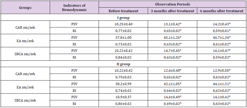
In the control group, in 3 months, visual acuity was similar to the initial values. After 6 months after treatment, a decrease in visual acuity was noted in all studied groups, while in the main group this indicator remained above the initial results on average by 18.67% (p <0.05), and in the control group decreased compared to the initial Indicators on average by 4.9% (p> 0.05). OCT-scanning of the central retinal zone in patients of the main group after 3 months revealed a further minor decrease in the thickness of the central fox and the makeup with a tendency to increase by 6 months after treatment, though the values were preserved significantly below the source data (p <0.05). Statistically significant changes in patients of the control group were not (P> 0.05) (Table 2) shows a progressive reduction of blood flow at the second stage of IB and the third IC NDR in the central artery of the retina (CAК) in the initial state [4,5]. The progressive phased decrease in blood flow in the short rear ciliary artery (SRCA) was also revealed.

biomedres-openaccess-journal-bjstr

Table 2: Results of ultrasonic doppler vascular vessels by groups. (abs. Numbers and%).

Note: * NPDR = non-proliferative diabetic retinopathy, PSV = peak systolic rate of blood flow, CRA = central artery retina, CVR = central vein retina, SPCA = short rear cylinder artery, EA = eye artery, ri = resistant index; * p <0.05; ** p <0.01 - from healthy, # p <0.05 - from the NDR IA, ^ P <0.05- from the NDRR IB; SD = Standard deviation.

Peak systolic speed (PSS) in the central vein of the retina (CVR) changed from 6.48 cm / s at stage Ia to 3.97 cm / s at stage IC, which indicates dilatation of veins increasing as retinopathy progression. Ultrasound studies of the condition of blood flow in the vessels of the eye were performed in 12 patients (24 eyes) with various stages of the Central Tshold Group II and in 14 patients (28 eyes) of the III group, of which 10 eyes with the non-acudative stage of TsDD in the group II group and 12 eyes in the group III, 8 Eye with an exudative stage of TsDD in group II and 8 eyes in the third group, with a scar stage of TsDD in the group II 6 eyes, in the third eye group. When analyzing hemodynamic indicators in persons with non-assessive stage of TsDD in the II and III group of patients, TCDDs revealed increased microcirculation in the CAR and the WCCC system, which manifest itself with an increase in the systolic velocity of blood flow: in patients with group II 1.2 times and III groups 1.5 times and decrease Resistance index, respectively.

These parameters in patients of the Group III significantly correlated with indicators of visual acuity (Table 3). Thus, the analysis of the results, showed that in the group of patients who received “doxy-hem” after treatment, there was a reliable positive dynamic of a number of studied functional and doppler indicators, which is associated with the effect of the drug on the microcirculation of the retina and its protection against the effects of metabolic and hypoxic defeats in patients dr. These clinical and functional studies have shown the effectiveness of the drug “Doxy - Hem” in the treatment of non-proliferative DR. The use of this drug contributes to an increase in visual acuity, a decrease in the thickness of the retina, improving the hemodynamics indicators. Positive changes in visual functions obtained as a result of treatment are preserved for up to 6 months. All this makes it possible to recommend the specified method for the secondary prevention of the development of diabetic retinopathy and the rehabilitation treatment of patients with early stages of non-proliferative DR.

biomedres-openaccess-journal-bjstr

Table 3: Comparative assessment of the results of treatment on the dynamics of CDC indicators in patients with NDPR.

Note *P <0.05 The accuracy of differences in relation to the data before treatment. ** - p <0.05 accuracy differences between groups.

Conclusion

1. This study showed that the treatment with the drug “Doxy- Hem” helps to improve eye blood flow in retrobulbar vessels, especially in the central artery of the retina and the short rear ciliary artery.

2. psV and RI in CRA and SPCA can be potentially useful for early diagnosis and subsequent observation of others.

3. The places of increased resistance or reduced blood flow velocity can be used to predict a higher risk of developing heavy DR, which is important to determine the further patient’s tactics.


For more Articles on: https://biomedres01.blogspot.com/
Academic Anxiety and School Burnout in Adolescents

  Academic Anxiety and School Burnout in Adolescents Introduction In 2020, the COVID-19 pandemic dramatically changed our lives, both person...