Thursday, January 30, 2025

Appraisal of Target Determination for Radiotherapeutic Management of Hepatic Tumors


Appraisal of Target Determination for Radiotherapeutic Management of Hepatic Tumors

Introduction

Although not included within the group of most common cancers, hepatic tumors comprise a leading cause of cancerrelated mortality around the globe [1-4]. Unfortunately, majority of affected patients present with advanced disease at diagnosis with high tumor burden and impaired hepatobiliary function which typically limits utilization of curative-intent management by surgery, Orthotopic Liver Transplantation (OLT) or Radiofrequency Ablation (RFA). Prognosis is typically poor particularly in the setting of unresectable tumors and extensive venous involvement. Within this context, patients with unresectable hepatic tumors may receive systemic therapies, arterially directed therapies, and Radiation Therapy (RT) [1-4]. Use of RT has been historically limited to palliative management [5]. Nevertheless, revolutionary advances in the field of radiation oncology and related disciplines have paved the way for contemporary radiotherapeutic approaches aiming at cure as well as symptomatic palliation [1-4]. Introduction of stereotactic irradiation has improved radiotherapeutic outcomes with improved accuary and precision under stereotactic immobilization and image guidance. Delivery of higher doses of radiation with stereotactic RT strategies have been possible with improved normal tissue sparing by steep dose gradients around the target volume. However, stereotactic irradiation is typically focused on relatively smaller and well-defined targets. Using ablative doses of radiation to treat relatively smaller targets has underscored the importance of accurate treatment volume definitions for optimal radiotherapeutic management. Herein, we assess incorporation of multimodality imaging for precise target definition of hepatic tumors.

Materials and Methods

In this study, we aimed to investigate whether multimodality imaging-based RT target volume definition improves interobserver and intraobserver variations to achieve optimal target definition for precise radiotherapeutic management of hepatic tumors. Within this context, RT target volume determination by incorporation of Magnetic Resonance Imaging (MRI) or by Computed Tomography (CT)-simulation images only has been comparatively evaluated. Ground truth target volume has been used for actual treatment and for comparative analysis and has been collaboratively determined by a group of experts after following meticulous evaluation, colleague peer review, and consensus. All patients were referred for radiotherapeutic management after detailed evaluation by a multidisciplinary team of experts to elucidate the indication with consideration of alternative therapeutic approaches, patient, tumor, and treatment characteristics. In the context of irradiation, lesion size, localization and association with surrounding critical structures, symptomatology, and contemplated results of radiotherapeutic management have been thoroughly assessed at the outset.

After collaborative decision making for irradiation, all patients underwent RT simulation at the CT-simulator (GE Lightspeed RT, GE Healthcare, Chalfont St. Giles, UK) for acquisition of treatment planning images. Planning images were acquired and sent to the delineation workstation (SimMD, GE, UK) via the network for contouring of treatment volumes and normal tissues. Either CTsimulation images only or registered CT and MR images have been used for target definition. Target definition by CT only and with incorporation of CT-MR registration was comparatively evaluated. Treatment dose calculation was performed individually for each patient in the Treatment Planning System (TPS) unit with consideration of electron density, CT number and HU values in CT images by also considering tissue heterogeneity. Synergy (Elekta, UK) linear accelerator (LINAC) has been utilized for precise RT with routine incorporation of IGRT techniques such as electronic digital portal imaging and kilovoltage cone beam CT for treatment verification.

Results

Reports of American Association of Physicists in Medicine (AAPM) and International Commission on Radiation Units and Measurements were considered in RT planning by expert radiation physicists by taking into account relevant normal tissue dose constraints. Calculation of treatment dose was performed with consideration of electron density, CT number and HU values in CT images by taking into account tissue heterogeneity. Optimal target coverage was prioritized in treatment planning while normal tissue protection was considered within the preset dose volume constraints. Definition of ground truth target volume was accomplished by a group of experts after thorough collaborative assessment, colleague peer review, and consensus to be used for actual treatment and comparative analysis. Synergy (Elekta, UK) LINAC was used for irradiation with routine utilization of IGRT techniques including kilovoltage cone beam CT and electronic digital portal imaging. Target definition by CT-only imaging and by CT-MR registration-based imaging was evaluated with comparative analysis. This study revealed that ground truth target volume was identical with target determination by CT-MR registration-based imaging for radiotherapeutic management of hepatic tumors.

Discussion

Worldwide, a significant proportion of cancer related mortality is caused by hepatic tumors [1-4]. Unfortunately, majority of affected patients succumb to their disease due to advanced disease at presentation and unresectability with reduced hepatobiliary function. Curative-intent management by surgical resection, Orthotopic Liver Transplantation (OLT) or Radiofrequency Ablation (RFA) may be limited by high tumor burden and extensive venous involvement at the outset. Within this context, prognosis is typically poor for patients with hepatic tumors. Radiotherapeutic management with external beam RT and stereotactic irradiation has been used for treatment. While utility of RT was historically limited for palliation, contemporary radiotherapeutic strategies have been developed to combat with hepatic tumors to achieve rigorous management aiming at cure in selected patients. Oligometastatic disease has been recently more aggressively managed by ablative therapies to achieve optimal treatment outcomes. Stereotactic irradiation in the form of Stereotactic Radiosurgery (SRS) and Stereotactic Ablative Body Radiotherapy (SABR) has been introduced as a viable radiotherapeutic modality providing high ablative doses to hepatic tumors with optimal normal tissue sparing through steep dose gradients around the target volume. Robust stereotactic immobilization and image guidance are prerequsites of ablative stereotactic RT approaches.

In addition, optimal target definition is an indispensable component of successful stereotactic irradiation strategies. Treatment simulation for RT is typically performed by CT simulation in overwhelming majority of cancer centers as part of current radiation oncology practice. However, optimal visualization of hepatic tumors may be accomplished by multiphase imaging studies to differantiate sources of blood supply and kinetics of contrast enhancement [6]. In the era of multimodality imaging for hepatic tumors, Magnetic Resonance Imaging (MRI) may provide additional information for optimal target definition. The superiority of MRI in offering improved soft tissue contrast resolution may have important clinical implications for radiotherapeutic management of hepatic tumors. Image resolution and contrast of CT and MRI may be different throughout the human body. While bone-air density differences may be more successfully differentiated in CT, soft tissue differences may be more successfully distinguished in MRI [7-9]. In the millenium era, there is a trend towards incorporation of multimodality imaging to improve outcomes of radiotherapeutic management for hepatic tumors as well as several other cancers throughout the human body [10-44]. At this standpoint, our study may have implications and add to the accumulating data about incorporation of multimodality imaging-based target definition for radiotherapeutic management for hepatic tumors.

There have been many critical advances in the discipline of radiation oncology with introduction of sophisticated treatment equipment and adaptive RT approaches, Intensity Modulated RT (IMRT), Image Guided RT (IGRT), Adaptive Radiation Therapy (ART), Breathing Adapted Radiation Therapy (BART), molecular imaging methods, automatic segmentation techniques, and stereotactic RT [45-82]. Management of cancer with contemporary radiotherapeutic strategies is an evolving field of active investigation and there is still room for further improvements. We conclude that visualization of data from multiple imaging modalities and incorporation of contemporary image registration and fusion techniques may dramatically improve target definition for more precise radiotherapeutic management of hepatic tumors. Admittedly, further studies are needed to shed light on this critical issue.


For more Articles on: https://biomedres01.blogspot.com/
Wednesday, January 29, 2025

5HT1A Receptor Transcripts are Found in Retzius Cells; Interneurons Inhibit Neuritic Regeneration by Serotonin


5HT1A Receptor Transcripts are Found in Retzius Cells; Interneurons Inhibit Neuritic Regeneration by Serotonin

Introduction

Following CNS injury, the organism loses its movement abilities to various degrees depending on the injury´s extension. This problem´s complexity has led to a dearth of information on the processes and elements that participate in CNS regeneration and recovery from injury. Clinically, this is reflected in the lack of totally effective treatments that could induce neuritic regeneration in injured neurons and hence, to the functional recovery of patients. Many factors play a role in neuritic regeneration such as neuronal identity, the extracellular matrix, microglia, and soluble factors among which, neurotransmitters are pivotal. The neurotransmitter serotonin has multiple functions [1], including a role in the neuritogenesis of hippocampal, cortical, spinal, and invertebrate neurons [2-4]. It has been reported to promote neuritogenesis [5,6], and to possess inhibitory properties [7,8]. Likewise, its receptors are determinants of the neurotransmitter´s functions. There are 7 types, and all have various sub-types [9]. As the neurotransmitter, its receptors have been associated with neuritogenesis [10-12], and various types of these receptors regulate the process [12-15]; there are also reports referring that R5HT1A inhibits neuritogenesis [16].

These observations indicate that the regenerative properties of various neuronal types may depend on the receptor they express, and in particular, they suggest that the inhibition of neuritogenesis by serotonin in RZ neurons is due to the presence of the 5HT1A receptor. The leech CNS has been used as a model to approach and attempt to answer various neurobiology questions and support this type of research, since its CNS transcriptome is present systemically [17], and in single, identified neurons [18]. The CNS is formed by 32 ganglia, the 21 intermediate ganglia with approximately 400 neurons, that regulate and control the organism´s functions. In every ganglion, Rz interneurons harbor serotonin and control many features of the animal´s behavior; AE motor neurons control the circular muscle annuli of the body, and the AL1 neurons possess great neuritic regeneration capacity. These 3 neuronal types respond differently in culture to serotonin by extensively growing or inhibiting their growth according to their identity [19]. In this study, we cloned and designed probes for the 1A and 2 serotonin receptors, to hybridize their genes in the CNS ganglia; we demonstrated that RZ neurons possess transcripts for the serotonin 1A receptor. These results suggest that Rz neurons decrease their neuritic growth in the presence of serotonin by activating the 5HT1A receptor.

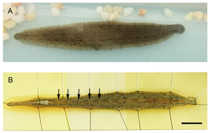
The Phylum Annelida was used for this study: Haementeria officinalis is a Mexican leech from our Neuronal Regeneration Laboratory animal colony (Figure 1A). The procurement and maintenance of Haementeria officinalis samples have been previously described [19]. These organisms were originally collected in the lakes and dams of the central Mexican plateau by a researcher authorized by the local “Direction of Agricultural Development”. All procedures were approved by the Institutional Committee for the Care and Use of Laboratory Animals (INRCICUAL No. 18/18). The organisms weighed between 0.5 and 0.8g. The animals were anesthetized with 9% ethanol, for 20 minutes. They were fixated with pins on a wax dish, and the ganglion chain was exposed (Figure 1B), extracted, and fixated in a dish with silicone to conduct the hybridization procedures. The probes were designed based on the Hirudo medicinalis leech transcriptome data, to detect the neuronal transcripts [17].

biomedres-openaccess-journal-bjstr

Figure 1:

A) Image of a Mexican leech of the species Haementeria officinalis. Dorsal image.

B) Diagram of ventrally opened CNS. Its ganglion chain formed by 21 intermediate ganglia can be observed, as well as the ganglia fused in the head and tail, in the extreme ends of the ganglion chain. The arrows point to the ganglia from the 7th to the 11th. Scale bar=1cm.

For leech Haementeria officinalis R5HT1A cDNA probes were used. Fragment of R5HT1A (from Hirudo medicionalis transcriptome [17]), leech genes were obtained by RT-PCR. First-strand cDNA was synthesized with a First-strand cDNA Kit (Roche Applied Science) and 1μg of RNA of the ganglion chain from leech weighed between 0.5 and 0.8g. The following primers (5′to 3′) were used: R5HT1A 5′primer, 5 ́-TCGTCCAACTGCACTCTCTC-3′ and 3′primer 5′-AGGGGTTGAAGAGGCTGTTG-3′ (corresponding to region 166- 368). PCRs were performed in a total volume of 25 μl using Taq DNA polymerase (Invitrogen). The cycling conditions were 15 seconds at 94°C for denaturation, 30 seconds at 55°C for annealing, 1 minute at 72°C for elongation, and then 30 minutes at 72°C after the last cycle (35 cycles). The PCR products were cloned into pCR®-Blunt IITopo ® vector (Invitrogen 45-0245). The long of the fragments was confirmed by restriction assay.

Digoxigenin 11 UTP-labeled single-stranded RNA probes were prepared using a DIG RNA labeling kit (Roche Applied Science) according to the manufacturer’s instructions. Tissues from ganglion chain from leech were treated with 1μg/ml proteinase K (preincubated at 37°C) for 5 minutes at room temperature, then washed with PBT and incubated with hybridization buffer at 65°C for 15 minutes, after corresponding Digoxigenin-labeled probe was added and incubated overnight at 65°C. Next morning, samples were washed and incubated with AP-labeled anti-digoxigenin antibodies at 4°C overnight. Next day, sections were washed and revelated with BM Purple and observed under a Zeiss Discovery Microscope, and photographed with a Zeiss digital camera and processed with Zen blue software (Zeiss).

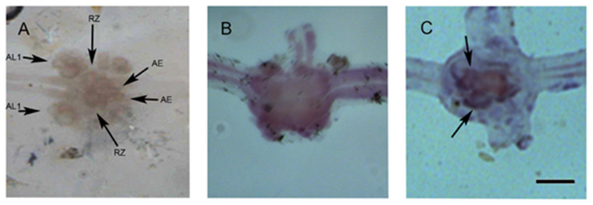
We made a probe that recognizing the 5HT1A receptor transcript for in situ hybridization. The probe that was designed and synthesized based on the transcriptome of the leech consortium [17] was functional, and binded to the Rz neuron. It was developed with anti-digoxigenin and detected with X-Purpura. The results were obtained from nine intermediate ganglia between the 7th and 11th ganglions from 3 different leeches. On figure 3, three ganglia are showed. The RZ, AL1 and AE neurons are in the ganglion without label (Figure 2A). On Figure 2B, the ganglion labeled with sense probe. The pair of RZ neurons that express the 5HT1A receptor had the label of antisense probe (Figure 2C. Arrows).

biomedres-openaccess-journal-bjstr

Figure 2:

A) Ganglion showing various identified neurons such as Retzius (Rz) interneurons, anterolateral 1 (AL1) neurons, and body annuli erector motor neurons (AE). The attached image shows a ganglion. In each ganglion, 2 sectioned nerves known as connective, are observed: the left one is anterior and to the side of the animal´s mouth while the right one is posterior, to the side of the tail. Laterally from each ganglion, 2 pairs of sectioned nerves are observed; they innervate the animal´s body. B) Hybridization with the sense probe.

C) Hybridization of the antisense probe of the 5HT1A receptor in an intermediate ganglion, developed with X-purpura. The ganglion in image A was hybridized with an “antisense” probe that recognizes the transcript of the 1A serotonin receptor. It is recognized by the arrows on the RZ neurons. Scale bar=100μm.

Conclusion

The probe that we obtained was functional and recognizes by hybridization some neuronal types, including Rz neurons; this suggests that these cells contain the transcript for the serotonin 1A receptor. This probe was designed on the basis of the leech transcriptome [17] and was inserted in the pCRR – Blunt II – topo R vector. The probe measures 202 bp that correspond to the 166-368 region of the 5htr1a gene of Hirudo medicinalis. Although the probe was designed based on the transcriptome of the Hirudo medicinalis leech, it also recognized neuronal transcripts of a different species: Haementeria officinalis, revealing the similitude in the sequences of the 5HT1A receptor in both species. Retzius neurons contain the R5HT1A transcripts, suggesting that this receptor´s expression leads to neuritogenesis inhibition mediated by serotonin. Thus, this receptor may play a role in the regulation of neuritogenesis in these neurons, as previously reported in other preparations that describe an inhibitory effect of the 1A serotonin receptor on neuritogenesis [16].

The inhibition of neuritic regeneration may be mediated by the 5HT1A receptor´s induction of a current promoting potassium entry into the cell, leading in turn, to a state of hyperpolarization that induces the inhibition of neuronal electric activity [20,21]. Further, electric activity is a mechanism regulating neuritogenesis via intracellular calcium regulation [22], and neuritic regeneration is inversely proportional to the calcium concentration in the neurite growth cones [23]. In the future, specific blockage of this receptor could be able to modify the regeneration of Rz neurons. However, we must test for the presence of other serotonin receptors, also in the Rz neurons. In other neurons, the final response may be mediated by an equilibrium in the activation of the serotonin receptors [14,15]. It is very relevant to understand the fundamentals of neuritic inhibition in Retzius neurons to evaluate its performance in organisms in vivo and in controlled culture conditions.


For more Articles on: https://biomedres01.blogspot.com/
Monday, January 27, 2025

Erectile Dysfunction and Metabolic Syndrome. An Entity Little Addressed


Erectile Dysfunction and Metabolic Syndrome. An Entity Little Addressed

Introduction

Metabolic syndrome (MetS) is characterized by: insulin resistance, excess weight, glucose intolerance, elevated triglycerides, decreased HDL and alterations in blood pressure. This also generates low-grade inflammation and prothrombotic state [1]. The National Cholesterol Education Program Adult Treatment Panel III (NCEP-ATP III) for MetS are:

1) Waist circumference ≥ 90 cm in men or ≥ 80 cm in women;

2) TG ≥ 150 mg/dl;

3) HDL-C < 40 mg/dl in men and < 50 mg/dl in women;

4) Systolic blood pressure ≥ 130 mmHg and diastolic ≥ 85 mmHg, or antihypertensive drug treatment; and

5) Fasting plasma glucose ≥ 126 mg/dl, or prior diagnosis or treatment of Diabetes Mellitus [2].

All of these are considered risk factors for the development of erectile dysfunction. Either as part of the complications due to endothelial damage, neuropathy or secondary to the consumption of drugs to control these pathologies. Erectile dysfunction (ED) is defined as the constant or recurrent inability to achieve and/ or maintain a sufficient penile erection for sexual satisfaction [3]. It is important to consider it as an early clinical manifestation of generalized vascular disease and as a risk factor for cardiovascular outcomes [4].

Etiology

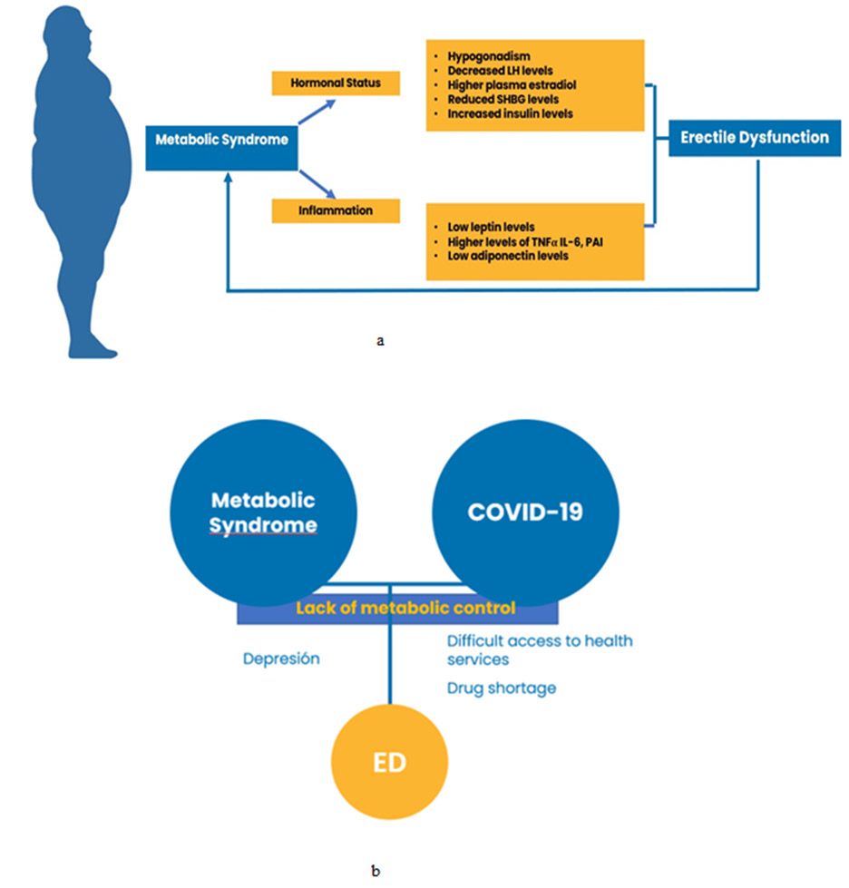
ED can have various etiologies and it is essential to identify the underlying diseases, such as: pathologies of the endocrine, central nervous and cardiovascular system. The prevalence of ED increases with age, however, those patients who present with diabetes mellitus have an increase in the prevalence of this compared to those who do not diabetic: 23% vs 11% respectively worldwide [5]. In Mexico, 45% of diabetic patients will have ED. This depends on the severity and evolution of diabetes however diabetes mellitus increases up to twice as much the risk of developing erectile dysfunction [6]. There are other etiologies associated with ED, such as: low levels of vitamin D and COVID-19 [7]. The latter becomes relevant since we continue to be on alert for this pandemic. Recently, the presence of viral particles in seroconverted COVID-19 patients in the endothelial cells of the penis has been studied, even after recovery [8]. What generates an association between ED and Covid-19 without forgetting the inflammatory state and endothelial damage that characterize this virus and its pathophysiology through the union with ACE-2 receptors, the affectation to mental health, the metabolic lack of control and the shortage of medicines generated as collateral damage of the COVID-19 pandemic are also factors that favor the appearance of ED [9-11] Figure 1.

biomedres-openaccess-journal-bjstr

Figure 1:

a. Increased adiposity leading to interactions metabolic syndrome and Erectile Dysfunction.

b. Metabolic Syndrome and COVID-19 as risk factors for ED.

Overweight and obesity, as well as the number of factors associated with MetS is directly proportional to the frequency of occurrence of ED, that is, the greater the components of the SMet, the greater the risk of developing it by up to 53.3% [5].

Likewise, there are drugs associated with ED used mostly for the control of SMet and for major depressive disorder mainly, such as: Alpha blockers, benzodiazepines, beta blockers, clonidine, digoxin, histamine H2 receptor blockers, ketoconazole, alpha-methyldopa, monoamine oxidase inhibitors, phenobarbital, phenytoin, selective serotonin reuptake inhibitors, spironolactone, thiazide diuretics and tricyclic antidepressants [3]. In addition to these, the use of statins to improve endothelial function for their pleiotropic effects is controversial since it has been reported in various clinical trials and preclinical trials to offer no benefit at the level of erectile function. Mostly reported with the use of simvastatin, compared to the rest of the statins. There are several theories that support the involvement of O2 and nitric oxide (NO) in statin-induced changes in endothelial function and suggest lower availability of NO for signaling, possibly through reaction with O2 to form peroxynitrite. On the other hand, erectile function has also been evaluated before and after the start of statin therapy, where the IIEF decreased in score and improves after one week of stopping treatment with these, as well as the increase in the percentage of the prevalence of patients with ED after the start of therapy [12,13].

Pathophysiology

Vasodilation is the main physiological point of erection that is mediated by NO in the penis. Phosphodiesterase-5 (PDE- 5) has a key role in erectile physiology, as it contributes to the conversion of cGMP to GMP which contributes to the relaxation of smooth muscle with calcium- dependent channels to generate vasodilation [14]. The synthesis of NO from arginine, through nitric oxide synthase (NOS), which requires a series of cofactors and cosubstrates, as they are not sufficient or are oxidized, the NOS produces superoxide anion, which interacts with the NO to produce peroxynitrites, generating vasoconstriction and this accelerates endothelial damage and culminates in atherosclerosis, creating a proplatelet state and finally ED [15]. MetS is a risk factor for the development of ED due to impaired endothelial function due to chronic low-grade inflammation managed by these patients through various biochemical and immunological pathway. The accumulation of adipose tissue is a triggering factor for MetS which generates changes in the body at the hormonal and inflammatory levels. Generating hypogonadism due to decreased LH levels, increased plasma estradiol, and hyperinsulinemia. At the same time, there is a decrease in the levels of leptin and adiponectin with an increase in proinflammatory markers, increasing the risk for ED by contributing to the decrease in testosterone [16] Figure 1a. There are also other risk factors that decrease testosterone levels, mentioned below [3].

Risk Factors

• Smoking

• Obesity

• Sedentary lifestyle

• Metabolic syndrome

• Chronic alcohol use

• Hypothyroidism/hyperthyroidism

Diagnostic Approach

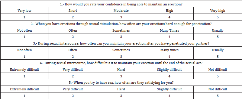
Perform clinical history to evaluate concomitant diseases, chronic use of medications and finally evaluate the presence or not along with the severity of ED using the IIEF test. The consultation of the patient suffering from erectile dysfunction should be carried out in a comfortable environment respecting and safeguarding the patient’s privacy. In order to diagnose and know the degree of ED, Rosen et al. formulated the International Index of Erectile Function (IIFE). Which is a validated tool and very practical and easy to use in everyday medical consultation. Its elaboration included the selection of 5 questions that evaluate different parameters of the ED in the last six months. The IIFE consists of 5 questions covering: erectile function, orgasmic function, sexual desire, satisfaction with sexual intercourse and overall satisfaction. Each with 5 possible answers with a score of 5-25. In addition, it demonstrates high sensitivity and specificity to detect changes in erectile function in response to treatment. The degree of ED is rated from 0 to 25 points with lower score greater severity of SD [17] (Charts 1a & 1b).

biomedres-openaccess-journal-bjstr

Chart 1a: IIEF test.

biomedres-openaccess-journal-bjstr

Chart 1b: ED score and severity.

IIEF Test

Another of the US scales that exist to evaluate the degree of ED is the SEAR (Self-Esteem And Relationship Questionnaire), which was designed to specifically evaluate psychosocial aspects associated with ED. It consists of 14 items, with 5 possible response options, through which the following 2 dimensions are evaluated with respect to the last 4 weeks: Sexual relations (items 1-8) and Selfconfidence (9-14), which in turn is divided into 2 subdimensions: Self-esteem (items 9-12) and Relationships in General (items 13 and 14). Finally, the questionnaire evaluates the degree of patient satisfaction with erectile function in general with respect to the last 4 weeks (final item) [18] (Charts 2 & 3).

biomedres-openaccess-journal-bjstr

Chart 2: SEAR quiz.

biomedres-openaccess-journal-bjstr

Chart 3: SEAR quiz answers.

Treatment

biomedres-openaccess-journal-bjstr

Figure 2: Multidisciplinary management of ED.

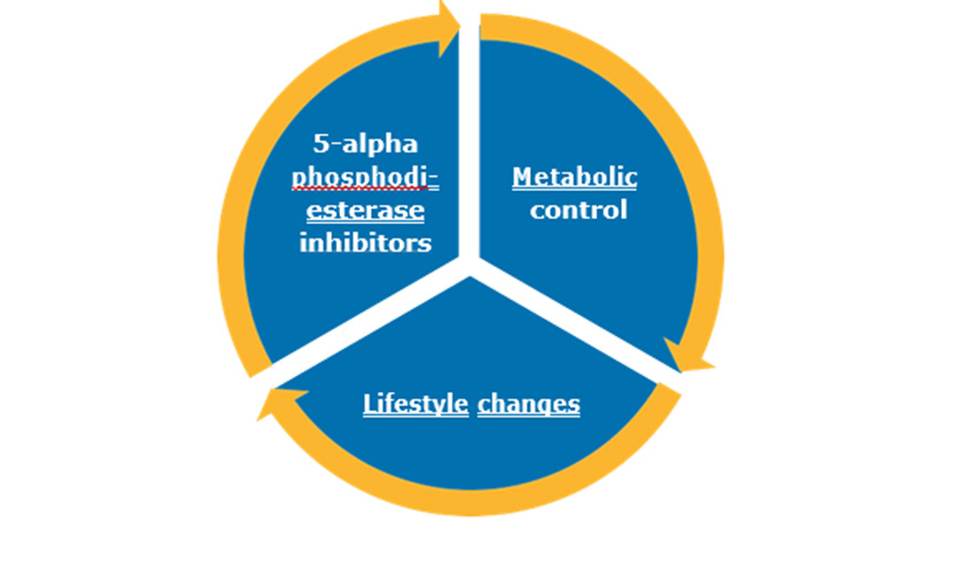
The treatment of a patient who presents ED and also SMet factors will require a multidisciplinary management that involves the metabolic control of concomitant diseases, offer changes in lifestyle with hygienic-dietary measures and finally pharmacological and non-pharmacological treatment as an alternative to these measures. Figure 2. In the case of those patients who present use of drugs that affect erectile function, it will be important to offer some replacement therapy.

Lifestyle Modifications

These interventions are necessary to reduce the risk of the appearance of ED and also improve this condition, such as: hygienicdietary measures and exercise. Hygienic-dietary measures include changes in diet and include those foods that have shown usefulness in ED, such as: the use of the Mediterranean diet and the consumption of red fruits, such as: blueberries, cherries, raspberries, blackberries, eggplant. etc. The use of the Mediterranean diet regains importance because the favorable response in endothelial function is proven, reducing the risk for major cardiovascular events, as well as the incidence of them [19]. The consumption of flavonoids in the diet, such as red fruits stimulate the synthesis of nitric oxide since they inhibit RhoA/Rho kinase pathway and stimulate [20]. Also 60 to 90 g of walnut improve markers of inflammation and endothelial damage. It is also recommended to include almonds, chestnuts, walnuts, hazelnuts and pine nuts [21]. It is also part of it as a preventive treatment to reduce the risk of cardiovascular diseases and therefore complications such as ED.

Pharmacological Treatment

biomedres-openaccess-journal-bjstr

Figure 3: Characteristics of 5 phosphodiesterase inhibitors.

The inhibitors of alpha 5-phosphodiesterase. Phosphodiesterases are a superfamily of enzymes whose main action is to inactivate cyclic adenosine monophosphate (cAMP) and cyclic guanosine monophosphate (cGMP) which are second messengers of prostacyclin and NO respectively, which promote vasodilation. In the case of the enzyme 5 phosphodiesterase, its main function is to accelerate the degradation process of cGMP and limit the vasodilator effects of NO [22]. Because of this it is the pharmacological target of sildenafil, tadalafil and vardanafil. These three with pharmacokinetic parameters distinct from each other, however, have all demonstrated adequate efficacy and tolerability for the treatment of ED. It is administered every 24h and each offers a different half-life of 10 to 36 h on which the duration of the pharmacological effect depends. Figure 3. The most common adverse reactions are: headache, flushing, myalgia, rhinitis, among others [1]. Several studies have reported the consumption preferences of these three, where both men and women have preferred Tadalafil by up to 70% [3,23,24]. This can be due to multiple reasons and one of them is the ease in the dosage that it presents and the duration of its half-life of up to 36 h that is greater than the rest of the drugs which can generate a mostly prolonged effect on erectile function [22]. Likewise, it was observed that with the use of Tadalafil significantly improved the severity of ED evaluated with the IIEF with a change of 10 to 18 points in this score p<0,013 3,24. Mentioning in an important way that these investigations have been based on consumption preference on the part of patients and not on pharmacological superiority.

Non-Pharmacological Treatment

For the purpose of this article, we refer to the nonpharmacological treatment of shock wave therapy as one of the alternatives that exists for this pathology. It is considered an innovative and painless treatment and its main mechanism of action is to generate neovascularization and thus favors the synthesis of NO. Generating various shots through a transducer that generates waves of different intensities directed towards the corpora cavernosa, with an approximate duration of 20 min. These sessions can be practiced 1-2 times a week [25]. An 80% success rate in erectile function has been reported in patients using this therapy with a response at 6 months with respect to baseline evaluating them with IIEF in Mexico [26]. However, it is important to mention that no treatment today is curative, it helps to improve erectile function while employing them.

Other Treatments for ED

Sex therapy, local treatments with intracavernous injections of prostaglandin analogues, hormonal therapy in the presence of clinical signs suggestive of an androgenic deficit confirmed by biological tests by the determination of a testosteronemia lower than normal and finally the surgical implant consisting of the surgical placement of two implants, one in each cavernous body, which produces a mechanical erection. They are indicated when the other treatments fail [4].

Conclusion

ED is an age-dependent pathology whose prevalence increases derived from the factors associated with SMet, so it is imperative the adequate control of the underlying diseases to reduce the risk of the appearance of cardiovascular diseases and therefore of ED. The approach of the patient with ED should always be multidisciplinary, starting by generating an adequate metabolic control and individualizing the pharmacological therapy to inquire about adverse reactions of each patient and assess concomitant therapy existing in SMet to reduce the risk of ED associated with medications. Today there are multiple therapeutic approaches aimed at improving erectile function, however, it is important to consider the preference of the patient diagnosed with ED. Mentioning that nothing is healing to date and only transiently improves erectile function. The use of statins for their already known pleiotropic effects on endothelial function remains uncertain within the improvement in erectile function of these patients. The prevention of this pathology lies primarily in reducing the risk of the appearance of fators associated with SMet. the Mediterranean diet, red fruits and seeds will always be part of the first step to cardiovascular prevention. Not forgetting that the metabolic syndrome and the current COVID-19 pandemic contribute to the increase in the prevalence of ED.


For more Articles on: https://biomedres01.blogspot.com/
Friday, January 24, 2025

Centrifugal Pneumatic-Sieve Fractionation of Barley and Wheat Flour for Maximum Recovery of Protein and Carbohydrate Fractions


Centrifugal Pneumatic-Sieve Fractionation of Barley and Wheat Flour for Maximum Recovery of Protein and Carbohydrate Fractions

Introduction

A feature of the grain farm in Russia is a wide variety of cultivated and processed crops used as raw materials for the production of food products of the population. These crops include both wheat and rye, traditional for the flour milling industry, and cereal crops - buckwheat, rice, barley, oats, millet, etc. The grain of each grain crop is a natural storehouse of the most valuable components necessary for human nutrition: proteins, fats, carbohydrates, vitamins, macroand microelements, minerals, etc. [1-4]. The main goal of mankind during the entire period of its development of grain plant foods was to increase the efficiency of grain use through a more complete use of its natural power, achieved through a deeper separation of cereal grains into anatomical parts. The level of modern development of flour milling technologies makes it possible to extract the endosperm from the grain of the main crops with an efficiency of 90%, the embryo with an efficiency of 25-70% of such crops as wheat and corn, for other crops this process has not been developed. There is no effective process for extracting the aleurone layer from the grains of the crops in question. Although the high value of the protein of this layer is known, it remains inaccessible to human nutrition due to its dense packaging in the fiber layer. Developments have not been completed and therefore there are practically no domestic technologies for extracting protein and starch by the “dry” method from the endosperm of grains of all crops [5-7].

The creation of modern technologies that allow the isolation of individual anatomical parts (endosperm, embryo, aleurone layer, shells) from the grain with the further production of components with a concentrated content of chemical elements (protein, fat, starch, minerals, etc.) from them is the basis of the progressive concept of flour milling production. At the same time, it is necessary to develop technologies for obtaining flour from various crops of a wide range of targeted purposes at the request of consumers, healthy food products. The relevance of the production of such types of flour was due to the high nutritional value of these cereals, their taste and nutritional advantages [8-9]. As a result of solving the problem of effective separation of cereal grains into anatomical parts and their subsequent separation into protein and carbohydrate fractions - flour concentrates, the raw material base of bakery, confectionery, pasta, food concentrate, meat, dairy, canning production will be significantly expanded. For example, barley flour is characterized by a low content of fat, resistant to storage, amino acid composition, not balanced in lysine and threonine, contains mucus that improve the functioning of the gastrointestinal tract. A feature of barley flour is a large amount of polysaccharide (-glucan, which has a cholesterol-lowering effect. Barley flour contains a lot of calcium, phosphorus and iodine [10-12]. The problem is relevant because one of the ways to reduce protein deficiency, improve the quality of nutrition and, as a result, improve human health is to create food products enriched with vegetable protein. Due to their functional features, proteins not only increase the biological value, but also significantly improve the quality of food products, give them dietary and medicinal properties [10-12]. The aim of our research is to develop a technology for separating wheat and barley flour into anatomical parts using pneumatic classification for maximum extraction of protein and carbohydrate fractions.

Objects and Methods of Research

For research, commercial batches of barley and wheat grains were used. Peeling of the original barley grain was carried out on a laboratory peeler of the ZSHN type with a capacity of up to 100 kg / h with a speed of 1500 per minute. The peeled grain was crushed in a laboratory rotary shredder with a passage deck with a hole size of 0.5 mm The circumferential velocity of the grinding rotor was 90 m / s. The crushed product was sifted in a laboratory sieve with an oscillation radius of R = 50 mm and an oscillation frequency of n = 200 rpm on sieves with a hole size of 100, 63, 40 and 30 μm. Centrifugal pneumatic-sieve classification was carried out on a laboratory rotary classifier with a speed of up to 4000 revolutions per minute. The rotary pneumatic classifier consists of the following main parts: a hopper, a vibrating feeder, an apparatus, boxes for collecting products, a filter with a box, a fan and material pipelines. All components of the laboratory unit are combined into three units: a power supply unit consisting of a hopper and a vibrating feeder; apparatus unit, including apparatus and boxes for collecting products; pneumatic transport unit, which includes a fan, filter and material ducts. The frequency of rotation of the rotor was regulated using a frequency regulator. In the process of centrifugal pneumosite vibration separation, the starting product is low-ash light barley and wheat flour, and the components are starch and protein particles, mainly extracted into high-protein or starchy fractions.

Results of the Study

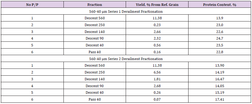
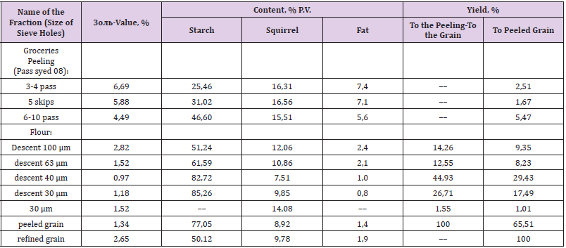
At the first stage of research, modeling of barley grain grinding was carried out according to the developed technological scheme. The scheme includes 5 consecutive stages of peeling barley grain and sorting of peeler sediments. For five passes through the peeler, flower films, fruit and seed membranes, the aleurone layer and the embryo were removed. The yield of peeled barley grain was 65.5%. The braned peeling products were sieved on a sieve with a cell size of 0.8 mm to remove the abrasive particles of the flower films. In the future, it is advisable to grind them to 200-150 μm for use as a food additive in the form of a source of macro- and microelements and components of dietary fiber. The moisture content of barley grain after peeling was 13.2%. In the second stage of research, the peeled barley grain was crushed in a laboratory rotary grinder. The descent from the sieve with a hole size of 100 μm was re-crushed, and then re-sieved on sieves of 100, 63, 40 and 30 μm. As a result of sieving, five fractions of barley flour were obtained. In these fractions, as well as in peeling products, in the grain after purification and in the peeled grain, the protein, starch and fat content was determined. Table 1 shows the fractional composition and protein content in the peeling products of barley grain. As a result of grinding barley grain according to the developed technological scheme, products with the following characteristics were obtained. The grain grinding balance is shown in Table 2.

biomedres-openaccess-journal-bjstr

Table 1: Fractional composition and protein content in peeling products of barley grain.

biomedres-openaccess-journal-bjstr

Table 2: Yield of fractions of barley flour and its chemical composition.

Table 2 shows that the fractions of barley flour of different sizes differ in protein, starch and fat content. The highest starch content is distinguished by fractions obtained with a sieve with holes of 40 and 30 μm (82.72% and 85.26%, respectively). In larger fractions, the starch content drops sharply (51.24% and 61.59%), which is explained by the presence of shell and protein particles in them. The presence of shells and protein is also reflected in the increase in the ash content of these fractions. The protein content correlates with the ash content of the fractions, with the minimum ash content (0.97%) and the protein content (7.51%) differing in the fraction obtained by passing a sieve with holes of 63 μm and descending from a sieve with holes of 40 μm. The maximum protein content (14.08%) is characterized by the smallest fraction obtained by the passage of a sieve with a hole size of 30 μm. At the same time, the protein content in it is 1.9 times higher than in the fraction with the lowest protein content (7.51%). The fat content decreases with a decrease in the size of the fractions. The largest fraction contains 3 times more fat (2.4%) than the fraction obtained by the descent of a sieve with holes of 30 μm (0.8%). This can be explained by the uneven distribution in the fractions of particles of the embryo rich in fat. The embryo is more plastic than the endosperm and therefore is less susceptible to grinding. As a result, the particles of the embryo mainly fall into larger fractions. This, in part, explains the relatively high protein content (12.06%) in the fraction obtained from the sieve with holes of 100 μm. This fraction also includes particles of the aleurone layer, the cells of which are characterized by a high protein content.

At the third stage of research, centrifugal pneumosite fractionation of barley flour was carried out to isolate starchy, protein and lipid fractions. As a result of modeling the production of carbohydrate components of barley grain by shock grinding and sieve classification, a starchy fraction of barley flour was obtained (sieve convergence with a hole size of 40 μm and 30 μm) with a starch content of 83.7%, which is 6.7% higher than in peeled grain, and 1.67 times higher than in grain after purification. The yield of this fraction was 71.64% to the peeled grain and 46.92% to the grain after purification. At the fourth stage of research, centrifugal pneumosite fractionation of barley flour was carried out to isolate starchy, protein and lipid fractions the basis of the developed method of extracting protein from wheat flour by the “dry” method is the destruction of the protein matrix and the production of its starch-free particles. These particles have dimensions in the range of 0-18 μm. Small (2-9 μm) and medium (10-18 μm) starch grains have the same sizes. Large starch grains have a size of more than 18 μm [8]. As a result of this destruction, it becomes possible to isolate barley protein particles in the process of pneumatic classification, as lighter and smaller.

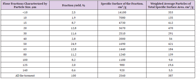
In standard wheat flour, small particles of protein are relatively small Table 3. This flour is obtained by the roll method of grinding on flour rolling rolls with a diameter of 250 mm, with an intervalian clearance of 10-20 μm and a ratio of 1: 1.25 speeds of fastrotating and slow-rotating rollers. The roughness of the grinding rollers is 2.5-4.0 μm. It is possible to increase the concentration of free small protein particles by grinding standard flour by the impact-abrasion method in disintegrators, by the impact method in the entoleitors or by the roll method in roller machines. The effectiveness of the impact-abrasion method. Table 3 presents data on the granulometric composition of the initial (control) sample of wheat flour of the highest grade and samples of flour crushed on an experimental disintegration machine to varying degrees of dispersion. This degree of dispersion was characterized by the specific surface area of the sample, determined on the PSKh-4 device. Table 4 presents the data of experimental studies on the specific surface of 10 fractions of wheat flour of different sizes.

biomedres-openaccess-journal-bjstr

Table 3: Granulometric composition and specific surface area of samples of starting and crushed wheat flour.

biomedres-openaccess-journal-bjstr

Table 4: Specific surface area of wheat flour fractions.

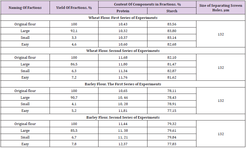
At the final stage, the study of the process of centrifugal pneumosite separation of wheat flour was carried out in two versions. In the developed technology for the production of wheat protein concentrates, this process can be used in one of these variants. In the first version, in this process, it is proposed to obtain such a fraction of free protein particles in which its content in accordance with the task should be 1.5-2.0 times greater than in the original wheat flour. In the second embodiment, it is first intended to obtain a fraction into which all free protein particles are extracted, together with small starch grains, and then divide them into fractions with the greatest possible efficiency of protein extraction into one of these fractions on a centrifugal-axial rotary pneumatic classifier. They were carried out at the pilot plant of centrifugal pneumosite separation of flour, dunst and cereal mixtures. In the experiments, the following parameters of the pilot installation were adopted: the diameter of the body is 200 mm, the diameters of the screen and the mesh cylinder are 200 and 150 mm, respectively. The first series of experiments was conducted to determine the effect of the size of the holes of the separating screen on the output and quality of the fractions. The sizes of the holes of the separating screen varied in the range of 118-315 μm. Metal-woven sieves were used as a separating screen. According to the results of the experiment, the size of the holes of 132 μm was adopted, which provides the yield of the passing fraction of 15% of the initial amount of native baking flour received for processing.

biomedres-openaccess-journal-bjstr

Table 5: Results of the study of centrifugal pneumosite separation of wheat and barley flour.

The second series of experiments was carried out to determine the effect of air flow and the speed of the air mixture at the entrance to the sieve cylinder of the apparatus on the efficiency of the fractionation process. Based on the results of this series of experiments, an air mode was chosen, in which the amount of the light fraction does not exceed 8%, and the small fraction 7%. The summarized results of the two series of experiments are presented in Table 5. As can be seen from Table 5, regardless of the size of the flour fractions in wheat flour, the starch content is higher by 3-4% than in the fractions of barley flour, and the protein content of the fractions of wheat and barley flour are practically comparable.

Findings

a) To obtain a concentrated protein fraction, it is advisable to carry out centrifugal pneumosite fractionation of crushed wheat flour, extracting small (up to 18 μm) light particles of protein and starch into one fraction, the yield of which should not exceed 15%.

b) The final separation of protein and starch particles should be carried out on the basis of the difference in their density in the centrifugal-axial rotary pneumatic classifier.

c) The highest starch content is distinguished by fractions obtained with a sieve with holes of 40 and 30 μm (82.72% and 85.26%, respectively). In larger fractions, the starch content drops sharply (51.24% and 61.59%), which is explained by the presence of shell and protein particles in them. The presence of shells and protein is also reflected in the increase in the ash content of these fractions.

d) The protein content correlates with the ash content of the fractions, with the minimum ash content (0.97%) and the protein content (7.51%) differing in the fraction obtained by passing a sieve with holes of 63 μm and descending from a sieve with holes of 40 μm. The maximum protein content (14.08%) is characterized by the smallest fraction obtained by the passage of a sieve with a hole size of 30 μm. At the same time, the protein content in it is 1.9 times higher than in the fraction with the lowest protein content (7.51%).


For more Articles on: https://biomedres01.blogspot.com/
Recent Methods in Cancer Treatment and Diagnosis

  Recent Methods in Cancer Treatment and Diagnosis Introduction The large number of cancer patients and the resulting deaths made cancer the...