Complex PCL Avulsion and Hoffa Fractures with a Unique Posteromedial Approach: A Case Report
Introduction
Posterior Cruciate Ligament (PCL) avulsions are rare injuries of the knee joint. What makes this injury even more complex is its association with Hoffa fracture, which is an intra-articular distal femur fracture affecting one or both of the femoral condyles in the coronal plane Hoffa [1] and constitutes around 0.65% of all femur fractures [2]. The posteromedial knee approach has been suggested as an appropriate approach to posterior tibia plateau, and femoral condyle fragments fixation with a safe complication profile and good clinical outcomes [3,4] Yet, not many surgeons are familiar with this approach. A thorough literature review was conducted on PubMed, Web of Science and Google Scholar till July 2021. It is to the best of our knowledge that no such cases with this combination of injuries and this surgical approach have been reported in the literature before. This case report presents an unusual PCL avulsion injury associated with an ipsilateral lateral femoral condyle Hoffa fracture. Also, we highlight our surgical treatment and patient’s clinical, functional, and radiological outcomes 18 months following management of this unique injury pattern.
Case Report
A 35-year-old gentleman sustained a road traffic accident after developing an epileptic generalized tonic-clonic seizure while driving his car. He complained of severe left knee pain and swelling. On physical examination, the patient was hemodynamically stable, conscious, and oriented. The left injured knee was moderately tender, swollen, with superficial abrasions anteriorly. The range of motion was limited to 0-30 degrees of extension-flexion. Also, he had tenderness over the lateral femoral condyle area. Varus stress test was suggestive of grade 2 injury of lateral collateral ligament of the left knee. The posterior drawer test could not be assessed at that time due to the severity of pain but tested positive intraoperatively. All compartments were soft with intact lower limbs neurovascular exam. Preoperative radiological evaluation with plain radiographs & CT scan of the left knee revealed a comminuted tibial plateau fracture extending posteriorly with multiple intra-articular fragments, comminuted Hoffa fracture of lateral condyle of the left femur (Figures 1 & 2).
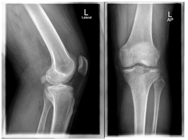
Figure 1: Preoperative AP & Lateral X-rays.
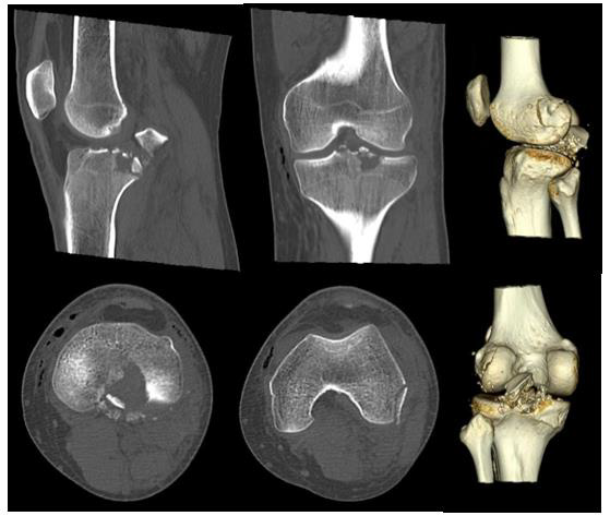
Figure 2: Preoperative knee CT scan.
Open reduction and internal fixation of left PCL avulsion and lateral femoral condyle fractures was performed by a senior knee orthopaedic surgeon at a trauma 1 center. In prone position and after scrubbing, prepping and draping the left lower limb, a proximal thigh tourniquet was inflated. Then a posteromedial knee approach was taken as described by Burks and Schaffer [5]. First, the PCL avulsed fragment was reduced and fixed with one 3.5 millimeters lag screw and a washer. Next, another posterior lateral 1 x 3 cm fragment was fixed using a 3.5 mm lag screw. Finally, a backup fixation was done with No.5 non-absorbable braided suture in the PCL and tied around the screw and washer. Furthermore, a small posterolateral incision, as an extension of the posteromedial approach in a lazy “S” fashion, was made on the left lateral femoral condyle, and a shell of bone was fixed with two headless 3.5 mm screws. Special knee tests were negative at the end of the procedure. Fluoroscopy confirmed excellent reduction and hardware placement.
Outcome & Follow-up
The patient was observed for a total follow-up period of 18 months following surgery and had frequent clinic visits at 1, 6, 12 and 18 months with the following findings (Table 1). Thorough clinical and radiological assessment was done in each visit, validated knee functional outcome scores including Tegner activity, Lysholm score and Oxford knee score (OKS) were utilized. Radiographic and bed side images at final follow up are shown in Figures 3 & 4, respectively.
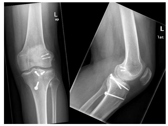
Figure 3: Final AP & Lateral X-ray at 18 months follow-up.
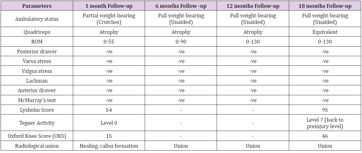
Table 1: A summary of the postoperative follow up parameters. PWB: Partial weight bearing. FWB: Full weight bearing.
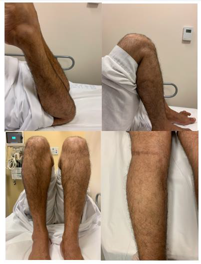
Figure 4: Clinical images at the final follow-up period of 18 months postoperatively.
Discussion
PCL avulsion fractures are rare and challenging injuries. While the best treatment options remain controversial, poorly treated PCL avulsions might lead to debilitating long term consequences of an unstable knee, malunion and nonunion [6,7]. These fractures can also be associated with other bony and ligamentous injuries around the knee joint, increasing surgical complexity. PCL avulsion fractures often have a similar mechanism of injury to PCL intrasubstance tears, such as dashboard injury, where an anteroposterior force applied to a flexed knee and sports traumatic injuries with a knee in hyperextension position [8]. Also, Hoffa and tibial plateau fractures are associated with high-energy trauma such as motor vehicle accidents, particularly in the younger population. While non-operative management could be considered in some PCL avulsion cases with minimal displacement of less than 5mm [9], most surgeons advocate for surgical fixation, taking into account various factors including fracture characteristics, displacement, comminution, and associated injuries [10,11]. The use of posteromedial knee approach has been reported in the literature with some technical variations, however, a recent paper describe the method used in our case and showed excellent radiological and functional outcomes [12] (Figure 5).

Figure 5: Intraoperative clinical image of the posteromedial knee approach used (from a previous case).
Patient Perspective
“After the horrific accident and how badly injured I was, I never thought I will be back on my legs walking. I’m really thankful to have reached this level of activity and getting back to my normal life”
For more Articles on: https://biomedres01.blogspot.com/


No comments:
Post a Comment
Note: Only a member of this blog may post a comment.