Saturday, July 25, 2020

In Vitro Tensile Properties, Hydrolytic and Oxidative Permeability of the Biomedical Thermoplastic Polyurethane (TPU) / Organohectorite Nanocomposites

In Vitro Tensile Properties, Hydrolytic and Oxidative Permeability of the Biomedical Thermoplastic Polyurethane (TPU) / Organohectorite Nanocomposites


Introduction

Polyurethanes are potential materials for biomedical applications due to their exceptional mechanical properties and biocompatibility Xiao & Chen [1]. However, when TPUs were used alone without nanofiller addition, they commonly failed to achieve the required mechanical and physical performance as biomaterial Osman & Osman et al. [2,3]. The addition of nanoscale inorganic particles into the TPU will improve the mechanical, physical and properties and biostability of the host material, further improving its quality as biomaterial. The poly (dimethylsiloxane) (PDMS) / poly (hexamethylene oxide) (PHMO)-based TPUs represent potential candidates for biomedical application due to their improved mechanical properties and biostability over polyether based TPUs Andriani et al. [4]. However, to develop the PDMS/PHMO-based TPU for biomedical applications, it is crucial to ensure that its mechanical properties and biostability meet the requirements of a biomedical device. Previous study showed that the ambient mechanical properties of the PDMS/PHMO based TPU were successfully improved by incorporating the organically modified organohectorite Osman et al. [3] Thus, this project intends to further investigate the potential of PDMS/PHMO based TPU nanocomposites for biomedical applications by assessing their in vitro mechanical performance and biostability.

Experimental Method

The polymer matrix used is commercially known as ElastEon™ E5-325 TPU consists of a 1000g/mol poly(dimethylsiloxane) (PDMS) and 700g/mol poly (hexamethylene oxide) (PHMO) mixed soft segment in a 98:2 (w/w) ratio, and a hard segment composed of alternating 4,4’-methylene diphenyl diisocyanate (MDI) and 1,4 butanediol (BDO) sequences. The hard segment concentrations are 32.5 wt%. The synthetic hectorite that used as nanofiller in the TPU was supplied by Kobo Products, Inc. Its average platelet length is approximately 30 to 50nm. Four types of surface modifier were used to surface modified the hectorite: Ethoquad 0/12, choline chloride, octadecyl trimethyl ammonium (bromide) (ODTMA) and dimethyldioctadecylammonium chloride (DODMAC). The PDMS/PHMO based TPU nanocomposites containing 2 wt% organohectorite were prepared by using solvent casting method. The procedures were explained elsewhere Osman & Osman et al. [2,3]. (Table 1) summarizes the formulation of all the nanocomposite samples.

In Vitro Tensile Test


The preliminary studies on the in vitro mechanical properties was carried out as a first step towards predicting the potential improvements in long-term in vivo performance of the TPU nanocomposites for long term implant applications. The test samples were conditioned in the environmental chamber containing H2O2 solution for 4 weeks before proceeding to the tensile test. The tensile test was performed according to ASTM D638 method with ten replicates of dumbbell sample from each material. Mean values of tensile strength, Young’s modulus and elongation at break were taken for comparison between materials.

Hydrolytic Permeability Test (in PBS Solution)

Hydrolytic permeability test using PBS solution was done to study the hydration characteristic of the studied materials and relate it with their barrier properties. Rectangular test samples with a dimension of L= W=3cm was cut, oven dried for 1 hour at 50 °C to remove the moisture and immediately weighed to determine the initial mass. Next, the samples were immersed in the beaker containing phosphate buffered saline PBS solution and conditioned at 37 °C, which approximates human body temperature. The sample mass was recorded after every 7 days of immersion (until 4 weeks’ time). The hydration characteristic of each material was determined from the percent increase in mass after each weeks of immersion, using the following calculation:
Increase in mass (%) = (Hydrated mass − Initial mass) x 100 / Initial mass

Oxidative Permeability Test (in H2O2 Solution)

Oxidative permeability test using H2O2 solution was done to study the barrier characteristic of the studied materials and relate it with their in vitro tensile properties. Rectangular test samples with a dimension of L= W=3cm was cut, oven dried for 1 hour at 50 °C to remove the moisture and immediately weighed to determine the initial mass. Next, the samples were immersed in the beaker containing hydrogen peroxide (H2O2) solution and conditioned at 37 °C, which approximates human body temperature. The sample mass was recorded after every 7 days of immersion (until 4 weeks’ time). The oxidative permeability of each material was determined from the percent increase in mass after four weeks of immersion, using the following calculation:
Increase in mass (%) = (Final mass − Initial mass) x 100 / Initial mass

Results and Discussion

In vitro Tensile Properties of Host TPU and TPU Nanocomposites

Tensile testing was carried on the host TPU and nanocomposites upon 4 weeks of H2O2 exposure to investigate the effect of oxidative exposure on the mechanical properties of these materials. Figures 1-3 compare the average values of modulus of elasticity, tensile strength, and break elongation of samples after four weeks of this oxidative treatment. Among all the samples, 2HeD-C nanocomposite shows the highest value of modulus of elasticity. The neat TPU showed the lower value for modulus of elasticity but higher value for elongation at break as compared to the TPU nanocomposites. This shows that the incorporation of the nanofillers impart in increasing the stiffness and rigidity of the TPU matrix. Good nanofiller-TPU interactions would bring stiffening effect to the host TPU molecular chains Rao [5]. Furthermore, the incorporation of the modified hectorite into the TPU structure resulted in the lowering of its tensile strength, therefore it is suggested that the presence of organic surfactant brings catalytic effect for degradation of the TPU upon oxidative exposure.
Figure 1: The modulus of elasticity of host TPU, 2He, 2HeE, 2HeE-C, 2HeO-C and 2HeD-C, 2HeD-C nanocomposites.
biomedres-openaccess-journal-bjstr
Figure 2: The tensile strength of host TPU, 2He, 2HeE, 2HeE-C, 2HeO-C and 2HeD-C, 2HeD-C nanocomposites.
biomedres-openaccess-journal-bjstr
Figure 3:The elongation at break of host, 2He, 2HeE, 2HeE-C, 2HeO-C and 2HeD-C, 2HeD-C nanocomposites.
biomedres-openaccess-journal-bjstr
However, with the presence of the hydrophobic dual modifier (HeD-C) on the hectorite surface, the modulus of the TPU increased to a larger extent. This could be probably due to enhanced compatibility between the most hydrophobic organohectorite and the TPU matrix system that contains hydrophobic PDMS soft segment. As a result, the molecular motions of soft segments were greatly restricted. According to the previous work (Andriani), the hectorite with dual surface modification (HeE-C) is more hydrophobic than its single modification counterpart (HeE). Enhanced hydrophobicity of the hectorite through dual surface modification allows more preferential interactions between the hydrophobic soft segments and the organohectorite nanofiller, thereby it induces more restricted soft segment mobility Osman & Osman et al. [2,6,7].

Hydrolytic Permeability of the TPU/Organohectorite Nanocomposites

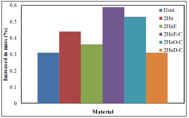
Changes in the weight of the neat TPU and TPU nanocomposites before and after exposed in PBS solution at 37 °C for four weeks were observed to determine the permeability of the materials. The increase of mass is assumed as a result of water permeability inside the TPU structure. From the data obtained as shown in (Figure 4), the water permeability of host TPU sample was the same level as the 2HeD-C nanocomposite but lower compared to other samples. This means that the water permeability ability of TPU was retained after the incorporation of 2wt% HeD-C nanofiller but increased with the addition of other types of nanofiller. This might be due to 2HeD-C organohectorite with dual modifications (DODMAC/CC) possesses the most hydrophobic characteristic, since the DODMAC has two long alkyl chains Osman, Andriani et al. [3,8]. On the other hand, 2HeE showed the highest water uptake in PBS solutions. This might be due to Ethoquad has the hydroxyl (OH) group in the structure, therefore it possesses the least hydrophobic characteristic Osman, Andriani et al. [3,8]. From the result, 2HeE-C shows the second highest increased in mass percentage.
Figure 4: Increase in mass percentage after 4 weeks of PBS (37°C) exposure.
biomedres-openaccess-journal-bjstr
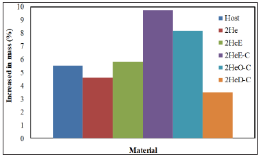
Figure 5: Increase in mass percentage after 4 weeks of H2O2 (37 °C) exposure.
biomedres-openaccess-journal-bjstr
This might also due to the present of OH group in the Choline chloride structure. Oxidative Permeability of the TPU/ Organohectorite Nanocomposites (Figure 5) shows the effect of oxidative agent on the TPU nanocomposites. As stated by Heath, Cooper and Lawrie [9], most of the TPU materials are prone to oxidation especially the one contains polyether in the soft segments. The impermeability towards oxidative agent may hinder the oxidative degradation process in the TPU. In this experiment, the increase of mass is assumed as a result of oxidative permeability inside the TPU structure. The TPU incorporated with HeD-C shows the lowest increased in mass upon H2O2 exposure. This might due to the presence of more compatible organosilicate as nanofiller in the TPU matrix, which contribute to greater bonding and interface interactions between the nanofiller and the TPU matrix, thereby resulting in more tortuous path for the entering of the oxidants.

Conclusion


The PDMS/PHMO-based TPU nanocomposite incorporating organohectorite with dual surface modification system (most hydrophobic characteristic) has better in vitro mechanical properties than the TPU nanocomposite incorporating organohectorite with single modification. This could be associated with enhanced compatibility between the nanofiller and the host TPU as the TPU contains the hydrophobic siloxane chains in its soft segment structure. This led to enhanced matrix barrier towards oxidative and hydrolytic permeability.

Rapid Progress in Diagnosis of ANCA-associated Vasculitis-https://biomedres01.blogspot.com/2020/07/rapid-progress-in-diagnosis-of-anca.html

Thursday, July 23, 2020

Rapid Progress in Diagnosis of ANCA-associated Vasculitis

Rapid Progress in Diagnosis of ANCA-associated Vasculitis


Introduction

Anti-neutrophilic cytoplasmic antibodies (ANCA) associated vasculitis (AAVs) is a part of chronic autoimmune inflammatory disorders affected blood vessels which usually affect small-sized vessels. AAVs include three types, granulomatosis with polyangiitis (GPA), eosinophilic granulomatosis with polyangiitis (EGPA), and microscopic polyangiitis (PMA). Despite it is common to misdiagnose them due to their overlapping with other disorders; the advancement in diagnostic approaches may contribute somehow to solve the problem of false diagnosis. In this minireview, we tried to report and summarize the most important tools in diagnosis of AAVs three types.

GPA Diagnosis Update

GPA diagnosis needs proper examination and writing down of clinical features to exclude the confusing clinical presentation. In addition, the laboratory analysis of serum ANCA autoantibodies and histopathology report of tissue specimen is not usually adequate for final diagnosis. Even though the early diagnosis of GPA is hard because of symptoms diversity and confusing histological reports, but these still important to proper management for this confused and aggressive disease. The symptoms in ear, nose, and larynx may be the first clinical features of GPA vasculitis. Thus, an attention should pay to think about GPA from otolaryngologist, particularly in otitis media cases which received treatment by antibiotics for a long period without positive respond. In orbital or sinonasal biopsies from GPA patients, increased IgG4+ plasma cells can be seen; this may cause misdiagnosis with IgG4-related disease [1]. Build on the above, the ideal path for GPA diagnosis aimed to identify, characterize and evaluate ENT signs and symptoms in GPA patients to reach earlier identification for the disease. Meanwhile, we are in need to do comparative studies that explain the diagnosis and prognosis of GPA by sinonasal imaging even if the sinus imaging in GPA gives nonspecific findings. Unfortunately, the available works of literatures were not outstanding to introduce a conclusion lead us to exact diagnostic tools. In 2011, the American College of Rheumatology (ACR) proposed classification criteria for GPA as a following:
a) Nasal or oral inflammation: Painful or painless oral ulcers or purulent or bloody nasal discharge.
b) Abnormal chest radiograph: Pulmonary nodules, fixed pulmonary infiltrates or pulmonary cavities.
c) Abnormal urinary sediment: Microscopic haematuria with or without red cell casts.
d) Granulomatous inflammation: Biopsy of an artery or perivascular area shows granulomatous inflammation.
At least two or more of these criteria should present to provide a sensitivity of 88% and a specificity of 92% [2]. On the other hand, a professional radiologist who has the skill to find the relationship between imaging findings and vasculitis knowledge is a very important issue. Indeed, radiologists should have the ability to interpret the laboratory test findings and connect them with imaging features together with the clinical presentation. The accumulation of the above-mentioned parameters related to radiologist may lead to a meaningful differential diagnosis. Recently, a novel procedure in clinical imaging, fluorine-18- fluorodeoxyglucose (FFDG) uptake into GPA lesions in positron emission tomography (PET) had a strategic potential importance in the diagnosis of GPA. But, differentiation between malignant and benign lesion still a challenge [3,4].

EGPA Diagnosis Update

Due to its overlapping with bronchial asthma, diagnosis of EPGA includes many issues. In cases of eosinophilic airways allergy with family history of EGPA, we should think seriously about EGPA. Meanwhile, clinical reports suggest that sensitization to fungi resulting in allergic bronchopulmonary mycosis (ABPM) especially aspergillosis may contribute to EGPA pathogenesis. So, the probability of EGPA coexist is high during the course of disease. Also, EGPA may coexist with GPA in a case referred as polyangiitis overlap syndrome. Additionally, cardiac involvement e.g. Löeffler ndocarditis in EGPA may diagnosed by using a sensitive procedure, cardiac magnetic resonance imaging (CMRI) to detect cardiac lesions. It helps to detect patients who need combining therapy and helps evaluate the therapeutic effect. Furthermore, in small intestines involvement in EPGA, the capsule endoscopy, the smallbowel radiography is convenient procedure for the diagnosis of EPGA lesions in the intestine and the evaluation of their clinical course [4,5].
Lanham’s criteria, ACR classification criteria and Chapel Hill definition are classification criteria and definitions commonly used for EPGA diagnosis: Lanham; asthma, eosinophilia > 1.5 ×10⁹ /l, and clinical or pathological evidence of vasculitis involving at least two organs. ACR 1990; asthma, eosinophilia > 10%, neuropathy (monoor polyneuropathy), non-fixed pulmonary infiltrates, paranasal sinus abnormalities, and extravascular eosinophil infiltration on biopsy [6]. Chapel Hill Consensus Conference 1994; eosinophil-rich and granulomatous inflammation involving the respiratory tract, necrotizing vasculitis affecting small to medium-sized vessels, and associated with asthma and eosinophilia. More important, the eosinophilia and edema of submucosa in EGPA is greater than in eosinophilic pneumonia [4]. Though it is difficult to point specific biomarkers in EGPA especially during times of glucocorticoid use, C-reactive protein to serum albumin ratio may provide a way in the management of AAVs including EGPA [7].

PMA Diagnosis Update

MPA diagnosis is basically performed using clinical presentations, computed tomography imaging, pulmonary or renal tissue sample, and laboratory investigations especially, serum ANCA autoantibodies analysis using immunofluorescence microscopy and ELISA. Actually, an important role can be achieved by chest computed tomography in patients with MPA to identify the abnormalities in a wide range of anatomic areas, including the whole airway. Meanwhile, fascial vasculitis in a patient with MPA can be diagnosed by magnetic resonance imaging. In addition, prostate screening should be performed while screening for malignancies in individuals diagnosed with vasculitis, including MPA, especially in the case of elderly adults with risk factors and alerting symptoms. Moreover, during vasculitis assessment cytomegaloviruses (CMV) should kept in mind because CD28- and CD57+T cells were associated with latent CMV infection but not with GPA or MPA diagnosis [3,4,8].

Conclusion Remarks


The idiopathic etiology of AAVs and uncommon occurrence contributed to increasing diagnosis difficulties. In the last few years, there were a plenty of scientific reports related to AAVs diagnosis and management especially those related to new clinical presentation. However, they couldn’t reach the goal of proper diagnostic approaches and treatment to secure AAVs patients. Actually, the need for a new protocol which considers the current researches outputs is an urgent necessity. Finally, we always recommend activating the communication between health workers, especially radiologist, histopathologist, otolaryngologist, laboratorians, rheumatologist, and all internal medicine branches physicians to reach the optimum diagnosis for AAVs.
The Use of Ionic Implantation for The Medical Materials Modifying-https://biomedres01.blogspot.com/2020/07/the-use-of-ionic-implantation-for.html

More BJSTR Articles : https://biomedres01.blogspot.com

The Use of Ionic Implantation for The Medical Materials Modifying

The Use of Ionic Implantation for The Medical Materials Modifying


Introduction

Modern medicine is not possible without the use of steel tools and implants. Usually, in order to meet the needs of physicians, manufacturers of tools use chrome and chromium-nickel corrosion-proof alloy steels 30Cr13, Cr13 40, 95Cr18 [1,2], AISI 301 (similar to 07Cr16Ni6), AISI 316L (analogue 03Cr17Ni14Mo3) [3,4] for multiple uses and special alloys [5] and steel ShKh15 (analogue 52100) [2] for disposable products. To produce materials that are in contact with biological media also often stainless steels [6] are used. But in this case steel is required much corrosion resistance and wear resistance [7]. While the key issue is the biocompatibility of the material of the implant and the environment in which it works [8]. One of the ways to succeed in creating such a modern medical material is surface modifying already existing metals and alloys [9]. A promising direction in the synthesis of new materials using the modifying surface is ion-plasma technologies, in particular, the method of ionic implantation [10-14]. The essence of ionic implantation, as technology of impact on the properties of materials, is the introduction of high-energy target ions in the surface layer and their accumulation with the subsequent creation of a surface film. Thanks to this the new distribution of surface energy and change of mechanical, physical-chemical, optical, and other characteristics of the processed sample are achieved. Therefore, the purpose of the work was to investigate the properties of stainless steel and steel ShKh15 as the materials that are widely used in medicine, after the ionic implantation.

Synthesis of Samples and Methods of Investigation

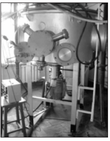
Treatment of the samples was carried out by titanium ions in nitrogen plasma using installation of ionic implantation (Figure 1). Mode of implantation corresponded to fluence near 5·1017 cm-2. The energy of the ions was about 20 keV. It allows to introduce them to a depth of up to 1 micron. The pressure in the vacuum chamber, which provided the ion jet did not exceed 10-4 Pa. Research of the surface hardness were carried out using the device “NOVOTEST T UD”. Sclerometric analysis of adhesive properties was performed with the help of a microhardness meter “PMT-3”. The state of the surface was shown wits using optical microscopy (microscope “MIM- 7”, additionally equipped with digital camera “Kodak Easy Share C1013”). Corrosion resistance was tested for long-term exposure to solutions of sulfuric acid and with subsequent gravimetric analysis.
Figure 1: General view of the installation of ionic implantation
biomedres-openaccess-journal-bjstr

Results and Discussion

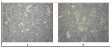
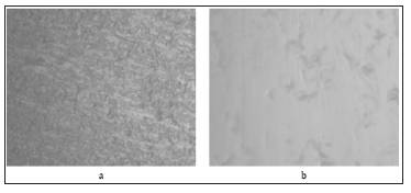
As a result of the studies a substantial increase in the hardness and strength of the surface for steel 12Cr18Ni10Тi and steel ShKh15 was received. In particular, the hardness for steel 12Cr18Ni10Тi increased from 220 HB to 280 HB. The hardness of treated steel ShKh15 sample was changed from 2 GPa to 4 GPa. This data indicate about the strengthening of surface layer and increase the durability of the material in terms of friction. Mechanical strength of the surface layer increased too (Table 1). One of the reasons for strengthening the surface is changing of the material structure. The results of optical studies (Figures 2 & 3) indicate about changing in the roughness of the surface in the direction of smoothing. Since stainless steel is intended for reusable medical materials, it must maintain a long term contact with the biological environment. Therefore, studies on corrosion stamina were conducted. Endurance stainless steel sample (12Cr18Ni10Тi) for 500 hours showed that depth of the dissolved layer for untreated material is about 400 μm/year. Instead, steel with titanium ions had a value of no more than 12 μm/year.
Figure 2: Microphotographs of steel ShKh15 structure:
a) Initial sample
b) Sample after implantation with an optical magnification x500.
biomedres-openaccess-journal-bjstr
Figure 3: Microphotographs of steel 12Cr18Ni10Тi structure:
a) Initial sample
b) Sample after implantation with an optical magnification x1500.
biomedres-openaccess-journal-bjstr

Conclusion


Consequently, studies have shown significant prospects for the use of ionic implantation technology for the production of metal medical materials. The increased hardness and strength of the surface layer will ensure the use of treated steel as a material for tools. Mechanical properties in a heap with increased corrosion resistance indicate the potential for application of samples in the function of implants.
Obesity: Modulating Adiponectin Levels Through Diet-https://biomedres01.blogspot.com/2020/07/obesity-modulating-adiponectin-levels.html

More BJSTR Articles : https://biomedres01.blogspot.com

Tuesday, July 21, 2020

Obesity: Modulating Adiponectin Levels Through Diet

Obesity: Modulating Adiponectin Levels Through Diet

Mini Review
Obesity is considered a chronic condition of multiple etiology that can be linked to several factors, such as behavior, metabolism, genetics, environment, and psychosocial aspects. It is characterized by the excess of body fat from the excessive consumption of calories combined with a lack of physical activity. The excessive body fat causes a state of chronic inflammation that can lead to the development of chronic non-communicable diseases, among which the most common are diabetes mellitus, cardiovascular diseases, and cancer. According to the World Health Organization (WHO), “obesity is considered a global epidemic caused, primarily, by dietary and physical activity patterns.” Such a scenario is directly related to the nutritional transition which occurred in the last decades - the dietary replacement of natural foods with processed and ultra-processed foods with a predominance of excessive salt, fat, and sugars. This new diet, with a higher energy density, combined with the lack of physical activity culminated in this ever-increasing epidemic that became a global public health problem.

In the past, the adipose tissue was only the primary source of unlimited energy in the body. However, over the past few years, studies have shown that the organ is responsible for a vital endocrine function in the production of several peptides involved in metabolic processes. These substances are called adipokines and can have either an anti-inflammatory or pro-inflammatory nature. Adiponectin and Interleukin-10 are examples of anti-inflammatory adipokines, whereas TNF-α, NF-κβ, and leptin are considered pro-inflammatory. Individuals in a nutritional state of obesity with chronic inflammation have an increased synthesis of pro-inflammatory adipokines, which inhibit the synthesis of adiponectin. That is why obesity is considered to have a negative correlation with adiponectin and a positive one with pro-inflammatory adipokines. Adiponectin, as an anti-inflammatory adipokine, plays an essential part in combating obesity and helping patients with diabetes, since it reduces insulin resistance. Marreiro [1] considers the increase of insulin sensitivity to be the consequence of its ability to activate the AMPK (AMP-activated protein kinase) in the muscles and liver through the interaction with its receivers in the cell membrane, which would inhibit gluconeogenesis and increase the oxidation of fatty acids and the glucose uptake in the muscles. The literature shows that some functional foods are effective in increasing adiponectin levels and, consequently, improving the quality of life of individuals, as shown in the (Table 1).
Studies show that the use of curcumin supplements (a polyphenol found in turmeric) in individuals with type 2 diabetes is linked to an increase in adiponectin levels and a decrease of the inflammatory processes mediated by TNF-α. Like β-carotene, olive oil and coffee are also positively associated with an increase in thecirculating levels of adiponectin. β -carotene has a favorable effect on insulin sensitivity in obese individuals, whether directly or through its pro-vitamin A action, which creates possibilities of dietary strategies for the prevention of metabolic syndrome. Another food that has been studied witch positive effects is the Sujiaonori algae (Enteromorpha Prolifera Muller), found in the Shimanto River in the city of Kochi, Japan. Researchers have investigated it through dietary interventions as a food that would induce the increase in adiponectin production, being effective in reducing hypertension, especially the systolic blood pressure, and the Body Mass Index (BMI) [2]. It is a biomaterial rich in fiber that contains polyphenols, bioactive compounds, vitamins, and polyunsaturated fatty acids with antioxidant and anti-inflammatory properties capable of stimulating the immune system. Adiponectin has anti-atherogenic properties by inhibiting TNF-α and NF-kβ. Its increase in plasma concentrations is associated with the reduction of myocardial infarction and the capacity of regulating the secretion of proinflammatory cytokines [3]. Its anti-inflammatory effects can be enhanced by the fact that the adiponectin reduces the production of IL-10 and stimulates an agonist to the IL-1 receptor (Figure 1). The consumption of functional foods is an option that should be considered for obese individuals since it can increase the levels of anti-inflammatory adipokines and reduce the inflammatory effects caused by the excess of adipose tissue [4]. Although the studies found did not include long-term interventions, and there are many factors that still need to be considered, in addition to several mechanisms that need to be clarified, the effectiveness of including those foods in the diet has shown their considerable modulating potential with evident positive effects.
Mirror Therapy in the Treatment of the Phantom Pain - Case Report-https://biomedres01.blogspot.com/2020/07/mirror-therapy-in-treatment-of-phantom.html

More BJSTR Articles : https://biomedres01.blogspot.com

Wednesday, July 15, 2020

Mirror Therapy in the Treatment of the Phantom Pain - Case Report

Mirror Therapy in the Treatment of the Phantom Pain - Case Report


Introduction

Phantom pain, pain that patients feel in the missing limb, occurs in 50-85% of amputees. It is most common in the first days after amputation [1]. This kind of pain can be the consequence of the discrepancy between visual feedback and the proprioceptive representation of the amputated limb [2]. The effect that mirror therapy has to phantom pain, has not been fully clarified. The positive effect of this therapy is attributed to neuron activation in the large-brain contralateral hemisphere, which looses their function of registering the missing limb movement. Visualization of the motion in the mirror could help reorganization of the neuron receptors and reintegration of the amputated extremity presentation, using so-called mirror neurons (Rizzolatti), in which way conflict between motor intent, proprioception and visualization of the amputated limb is resolved [3].
Case Report - Patient DZ. Female. Born in Nis, Serbia, in 1951. The patient is insulin dependent diabetic of 20 years, and also extremely obese (>135kg). She is very limited in her day to day activities and practically immobile. Permanent urinal catheter due to immobility and obesity was inevitable. Above the knee right leg amputation was done on August 9th, 2017. due to severe skin infection of the right foot and shin. Postoperative course was without complications. Rehabilitation treatment was carried out at the Clinic for Physical Medicine, Rehabilitation and Prosthetics, Clinical Center Nis, Serbia, in two stages. First hospitalization took place from September 9th until November 8th, 2017. During that time, she was examined by cardiologist and vascular surgeon in order for her cardiovascular status and general health to be established. After that the specialist for prosthetics was included. He took measurements and it was clear that she will need some kind of reinforcement for her prosthesis because she was overweight. During this first stage of rehabilitation she managed to stand up straight with the walking aid. PMR specialist and Physiotherapist were responsible for her overall treatment at the Clinic.
When the specially designed prosthesis was manufactured and the patient was ready to use it, which meant that she was able to stand and make a few steps with a walking stall, the rehabilitation treatment was continued. Second hospitalization was from January 20th up until March 19th 2017. During this time, she become capable of self-use and maintenance of the prosthesis, walking with a prosthesis and the walking aid and she managed to successfully perform everyday activities by herself. However, during this second hospitalization, a patient suddenly, during the night, experienced a severe pain in the missing limb, which she described as a hard, deep, and pressure like. We tried to manage it with usual pain killers and we also prescribed pregabalin, which did not have any impact on the intensity of the pain. The patient’s condition was accompanied by symptoms of depression - crying, sadness, insomnia, hopelessness. Since symptoms have persisted, mirror therapy was included. In the assessment of the pain intensity, a numerical scale of pain was used (no pain 0- unbearable pain 10). (Table 1) PHQ-9 and BDI were used to evaluate depressive behaviors. Every day for three weeks the patient was presented with the mirror that has been placed in that manner, so that she sees the existing leg in it as her contralateral limb, at the place when the missing one was. In addition to concentrated observation of her leg in the mirror, the patient performed a light exercise of 20 minutes: movement of the foot in all directions, slight flexionextension in the knee and a hip abduction/adduction movement. Before the therapy, the patient assessed the intensity of the pain with 9, after almost every treatment, pain was reduced to 4, and after three weeks it was graded with 1. Symptoms of depression followed the pain reduction: PHQ-9: 18-7-5. BDI: 20-12-9. At the end of her hospital rehabilitation she was advised to continue the therapy at home.

Conclusion


Mirror therapy in this particular case of phantom pain, proved to be a simple, painless, inexpensive and highly effective cure.
Enabling the Quality of Care for Older People-https://biomedres01.blogspot.com/2020/07/enabling-quality-of-care-for-older.html

More BJSTR Articles : https://biomedres01.blogspot.com

Enabling the Quality of Care for Older People

Enabling the Quality of Care for Older People


Mini Review

There has been a raft of research recently on the care of older people. Many of them take a theoretical approach and many take a policy approach. These books are huge in scope and a survey of them would suggest they are comparative mainly in the United States and United Kingdom. For many years, ageism prevented books from being written on older people because of the intense interest of children by practitioners. This is not to deny the importance and significance of that group of people at one end of the life-course. Historically, to suggest “age” instantly meant younger people added to the marginality and invisibility of older people. Worse, because there were hardly any widely subscribed postgraduate courses in social work in working with older people, with the overwhelming focus on children, there were periodic episodes of inhumanity against older people (known today as “elder abuse”). It was a forgotten dimension that older people were people. There was also a chronic shortage of research and knowledge for careers, families, health and social work professions on the vulnerabilities of older people whether if they lived at home or lived in a care home. Peter Townsend wrote his devastating overview of “care” in his famous book “The Last Refuge” (1959) that care homes were not necessarily benevolent institutions, but ‘warehouses’ were care was in chaos as careers did not have the enough skills, knowledge and practice to work with older people.
Hence, whilst these research base on policy and theory in caring for older people are important, practice with older people has been overlooked. This is a magnificent piece of writing. The reasons are utterly compelling highlighting to careers, helping and health professions the implications and opportunities to care with, care for and care about older people. Practitioners really need to excel in that there is no duality in terms of thinking about practice and doing practice. For a long time, in a discipline called Social Gerontology, there was a demarcation between thinkers and doers. Authors used to implicitly suggest there was some form of exaggerated age wars between thinkers and those who investigate older people’s experiences (do-errs). The importance of deconstructing and obliterating such assumptions and with an intellectual wallop synthesizing the importance of thinking and doing for effective practice transcending ethnocentric and Eurocentric approaches to care is a necessity. I have rarely read a literature on care of older people that addresses exploring thinking tools and doing practice as a dualism. They are two sides of the same coin, not in opposition.

The ability to shed light on the importance of valuing older people is also essential. This prevents and should provide some sensitivity to careers in recognizing that older people are people with human rights. Recent research lumps ‘older people’ into an undifferentiated category as though it’s not differentiated by race, gender, sexuality, disability, class and history. Engaging in research with older people is one of those rare attempts in social research that gets the researcher and practitioners to engage from the beginning to the end and beyond. I say beyond, because after I read the current literature on caring for older people it got me to think about the assumptions I had about older people and implore researchers to think deep and focus on what is means to be cared for and cared by a variety of people and professions. Research is needed in getting social and health policy makers to transform their attitudes. This means engaging with service providers to addresses social divisions head on and context sensitive situations where people learn from working with older people.
Neera- A Potential Natural Health Drink-https://biomedres01.blogspot.com/2020/07/neera-potential-natural-health-drink.html

More BJSTR Articles : https://biomedres01.blogspot.com

Tuesday, July 14, 2020

Neera- A Potential Natural Health Drink

Neera- A Potential Natural Health Drink


Introduction

Growing health consciousness among the population in India has boosted the growth of health drinks industry in India. The segment has posted a healthy growth rate of around 40% in last few years, with a total turnover of more than Rs. 6.7 billion in 2008. Players in the industry hope to maintain the strong growth rate. So far, the use of these kinds of drinks has not percolated to the mass market. Pricing as well as consumer awareness regarding the energy drinks remain the most critical factors for the success of health drinks in the country. Neera, also called palm nectar, is the phloem sap extracted from the inflorescence of various species of toddy palms which is used as a nutritious health drink. Neera is called sweet toddy since it contains zero percentage alcohol in it and is known as padaneer in Tamil Nadu. Toddy and Neera can be called as fermented sap and non-fermented sap. It is a refreshing and light drink, translucent in colour, extremely cool in sensation, sugary sweet to taste, similar to palm juice which is susceptible to natural fermentation at ambient temperature within a few hours of extraction due to enzymatic and microbial fermentation since it is rich in sugars, vitamins, proteins, minerals.
Once fermented, Neera becomes toddy which is unsuitable for health drink or for value added products. Neera is widely consumed in India, Sri Lanka, Africa, Malaysia, Indonesia, Thailand, and Myanmar. It is healthy and therapeutic drink since it is rich in Vitamin C, has more food calories than milk, fights diabetes, cancer, electrolyte deficiency and even hair fall. Neera is rich in sugar, essential elements such as N, P, K, Mg, micronutrients, minerals and a good source of ascorbic acid. It contains acids nicotinic acid (Vit B3) and riboflavin (Vit B2) and also can be consumed by people suffering from diabetes since it has a low glycemic index and a low glycemic load (Hebbar). Coconut sugar which is a byproduct of Neera, only possess a GI in the range of 35-45 per serving whereas refined white sugar possess 65-100 GI per serving (Swamy). Reports suggests that Neera consumption prevents jaundice and also facilitates clear urination. It keeps the human system cool and improves digestion. Neera production is largely in an un-organized manner with major consumption by rural population and there is a wide scope for commercializing the product.

Composition

Neera is nearly neutral in pH, with specific gravity ranging from 1.058-1.077. Several workers have studied the composition of palm sap Bassir, (1962), Chinarasa, (1968) Fararusi and Bassir, 1972; Okafor, 1978). The chemical percentage composition of Neera varies, depending on various factors, namely, place, type of palm, mode and season of its collection in Tables 1 & 2.

Extraction

Neera extraction is generally performed by tapping method collected from the inflorescence of coconut, which is done twice a day, both in the morning and evening. On an average, an adult coconut palm produces 12-14 inflorescences per year. The sap should be collected from a growing palm and the sweet sap is collected from the cut flower of palm by a tapper and a container is fastened to the flower to collect the sap. The sequential tapping and nut production technology (SCTNP) adoption can increase the farmer’s income without sacrificing nut production as ¾th is tapped whereas ¼ is used for nut production. Usually it is collected in early morning at sunrise and as the day progresses, it will get converted into toddy which is alcoholic [1].

Technological Developments in Preservation

Neera is highly susceptible to natural fermentation at ambient temperature within a few hours of extraction from the palm source due to natural yeasts in air and once fermented, it transforms into toddy with 4% alcohol. Several technologies developed by various research institutes for processing and preserving the natural form of Neera and to retain the vitamins, sugar, and other nutrients beneficial for health. CPCRI has developed an equipment for easy collection and preservation of sap. To extend the shelf life of Neera, heat preservation techniques such as pasteurization are used which can extend the shelf life up to a week without much loss in its nutritive components [2]. It can be stored under refrigerated condition to arrest the growth of microorganisms and enzymatic reactions. A team of experts from SCMS Institute of Bio Sciences and Biotechnology, Cochin, India have successfully developed filtration and preservation techniques for Neera in collaboration with Coconut Development Board to commercialize the drink among the public [3]. National Chemical Laboratory in Pune has developed a special filtration technique to enhance the shelf life and Central Food Technological Research Institute in Mysore, India also developed technologies for the preservation and processing of Neera.

By-products

a) Coconut Jaggery: It is soli or semi-solid crystalline mass obtained by boiling and evaporating Neera in small batches with final product having pH in the range of 6.5-7.0 [4]. The production requires minimal heat treatment without the the aid of any additives or enzymes. Caramelization turns the heated Neera from milky white to transparent brown.
b) Neera Syrup: It is produced when fresh Neera is heated and concentrated into syrup and is used as a health and wellness drink in connection with Ayurveda and other systems of medicine [5].
c) Palm Wine (Toddy): It is an alcoholic beverage produced from the sap of palm which is mildly intoxicating and sweet, obtained by fermentation of natural yeasts yielding an aromatic wine having 4% alcohol content [6]. As the day progresses, it may yield a sourer, stronger and acidic product due to fermentation. Spices are added to obtain distinct taste to product
d) Palm Honey: It is a thick liquid syrup similar to honey, having a sweetness of 780 Brix and a pH of 6.0-6.5, which is used as sweetener in ice creams and other confectionaries. Palm honey also find applications in pharmaceutical formulations and is good for patients suffering from anemia as it is rich source of iron.
e) Palm Sugar: The coconut sap sugar is obtained by moderately heating the freshly harvested Neera at 1150C to remove water which is considered as one of the best natural sweetener since it has no added chemicals. It is widely used as sweetener in South- East Asian regions since thousands of years which is a perfect and healthier substitute for artificial sweeteners. It is rich in minerals, vitamins and has twice the iron, four times the magnesium and over ten times the zinc than brown sugar which is obtained from sugar cane.

Conclusion


Chilled Neera is a healthy and thirst-quenching drink which gives a cool feeling to the body. The shelf life can be extended upto 45 days using scientific methods such as filtration, pasteurization and addition of preservatives so that the market reach of the product can be extended upto urban areas. It has been medically proved that Neera is better than mineral water and it also has less calorific value, apart from being sweet and delicious. Neera could become a nutritious drink offering a healthy alternative to aerated beverages if it is commercialized in a proper way.
Novel Fiberglass Resistance Pole Training Implement: Reference Values for Exercise Prescription-https://biomedres01.blogspot.com/2020/07/novel-fiberglass-resistance-pole.html

More BJSTR Articles : https://biomedres01.blogspot.com

Liver Fatty Acids Profile of Rats Fed Ketogenic Diet

  Liver Fatty Acids Profile of Rats Fed Ketogenic Diet Introduction The ketogenic diet (KD) ‒ characterized by a low carbohydrate content an...