Monday, January 4, 2021

Study the Nephroprotective Effect of Ziziphus jujube Extract Against Cisplatin -Induced Nephrotoxicity

Study the Nephroprotective Effect of Ziziphus jujube Extract Against Cisplatin -Induced Nephrotoxicity


Introduction
Cancer is a group of diseases in which cells in the body divide abnormally. The cancerous cells may occur in liquids, as in leukemia or can invade surrounding tissues and spread through the blood and lymph system and establish colonies to other parts of the body. When these cells have migrated to other regions in the body, the cancer is called disseminated or metastasized [1], there are various types of cancer depend on its location, such as lung, colon, breast, skin, ovarian or prostate cancer [2]. Localized cancers can be removed by surgery or irradiation with high survival rates. But, the success of these treatments is frequently limited by metastasis of cancer cells to distant body. So, it necessitates administrating chemotherapeutic drugs to attempt to kill tumor cells throughout the body [3]. Unfortunately, the drugs available for use in chemotherapy are not specific for cancer cells. Most chemotherapeutic agents act either by damaging DNA or by interfering with DNA synthesis. They kill all rabidly dividing cell- not only cancer cells, but also the normal cells that are undergoing cell division [4]. Also, multidrug resistant (MDR) phenotype is a major problem in patients treated with certain natural chemotherapeutic drugs [5].
In general, there are four major classes of drugs which are useful in blocking the synthesis of proteins or enzymes. These are
a) Alkylating agents,
b) The antimetabolites,
c) The steroid hormones, and
d) Miscellaneous compounds with specific blocking effects.
Alkylating agents are the largest group of DNA- damaging drugs, which react chemically with DNA molecules, such as cyclophosphamide, melphalan and mechlorethamine [3]. Cisplatin (cis-diamine-dichloroplatinum) is a nonclassical alkylating agent having the broadest range of clinical activity and the most substantial toxicity profile. Cisplatin-based therapy is curative in testicular cancer and is very active in gynecologic cancer, gastrointestinal malignancies, bladder, head and neck, oesophageal, small cell lung and ovarian cancers [5,6]. It has a harmful side effect profile including nephrotoxicity and ototoxicity [7,8]. These side effects were primarily mediated by the generation of reactive oxygen species [9].
Cisplatin is one of the most potent chemotherapy drugs widely used for cancer treatment. The discovery of cisplatin, cis- or CDDP) as an anti-cancer drug in the 1960s opened a new era in cancer treatment [10]. The main dose limiting side effects of cisplatin are gastrointestinal toxicity like nausea vomiting and anorexia also cisplatin introduce ototoxicity which cause loss in sensorial hearing and neurotoxicity which manifest commonly as distal sensory neuropathy last side effects is nevrotoxicity [11]. It is recognized that the prevalence of cisplatin nephrotoxicity is high, occurring in about one-third of patient undergoing cisplatin treatment. Clinically, cisplatin nephrotoxicity is often seen after 10 days of cisplatin administration and is manifested as lower glomerular filtration rate, higher serum creatinine, and reduced serum magnesium and potassium levels [11]. Jujubes are species of the genus Ziziphus (L) belonging to the family Rhamnaceae named after the genus Rhamnus. Commonly it is called Chinese date, Chinese Jujube, Indian cherry and Malay jujube. Other names are annab, badari, bayear, ber, black date, bor, red date, regi, spine date, unnab, vadai, vadar, vagari, zao sidr [12,13].
Study Objectives
The main objective of the study is to investigate the protective effect of Ziziphus jujube plant in rats with cisplatin-induced nephrotoxicity in an in vivo model.

Methodology
Experimental Design
Sprague-Dawely rats, each weighing 150-210g and their ages between 8-12 weeks, were obtained from the animal house at Jordan University of Science and Technology. They kept at a constant temperature (22±1 ºC) with a regular 12h light and dark cycle with providing diet and water. The rats were randomly divided into four groups, each consisting of five animals. Drug was administered orally using a ball tipped stainless steel gavage attached to a syringe. An individual body weights were obtained for test animals’ prior administration daily.
a. Saline-treated control group (NS). Rats were given orally 0.9% Nacl for 6 days.
b. Ziziphus jujube extract group (ZE). Rats will be administered an orally injection of a Ziziphus jujube extract (500mg/kg) daily for 6 days [14].
c. Cisplatin group (CIS). The single dose of cisplatin is 5mg/ kg intraperitoneally.
d. Ziziphus jujube extract with cisplatin group (CIS+ZE). Rats will be adminstraed cisplatin just as the cisplatin group, except that they will be administered orally with Ziziphus jujube extract one hour before cisplatin dose. Then will be administrated an orally injection of a Ziziphus jujube extract (500mg/kg) daily.
Preparation of the Plant Extract
The fruits of Ziziphus jujube were collected from trees growing in Irbid, collected between September and November of 2012. The plant fruits were botanically identified by Prof. Jamil Lahham (Plant Taxonomist. Department of Biological Science, Faculty of Science, Yarmouk University, Jordan). The fruits were shade dried at 25 °C and the dried fruits of plant were grinded with a blender and dissolved with methanol (analytical grade from sigma chemicals, USA), then filtrated through filter paper. Extract was concentrated under reduced pressure and stored in a desicator until further use.
Sample Collection
The animals in all groups were scarified in an ether chamber after 48h from the last application, after an overnight fast. Blood samples were taken by the intracardiac puncture and collected into the heparin tubes. The samples were centrifuged at 3000rpm for 10 minutes to separate their plasma and then stored at -18C until used for determination of biochemical test. The two kidneys were removed for each rat. One kidney was placed in 10% formaldehyde solution for processing to histopathology examination by light microscopy. The other kidney was homogenized with phosphate buffered solution (PH 7.2) to obtain 1: 5 (W/v) homogenate. The latter was analyzed to determine the glutathione reductase activity, glutathione catalase activity, glutathione peroxidase activity, glutathione S-transferase activity and lipid peroxidation.

Results
Body Weight Changes
After 24 hours of the last oral administration, no mortality was seen in any of the NS, CIS, CIS+ZE and ZE group. Results in Table 1 indicate an increase in the body weight in the control (NS, ZE) groups and a significant increase in CIS and ZE + CIS groups. Administration of CIS produced a significant decreased (P<0.005) in the body weight when compared to NS. This was largely prevented by pretreatment with ZE in (CIS+ZE) group.
Table 1: Mean body weight difference (%).

biomedres-openaccess-journal-bjstrHistological Analysis
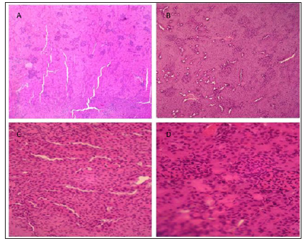
Hispathological studies (Figure 1) revealed that treatment with NS or ZE (Figures 1a & 1b) result in normal glomeruli and tubules structures. CIS-treated group (Figure 1c) has glomerular and peritubular congestion, RBC, mass edema and inflammatory cells, vacuolization and necrosis in the proximal tubular epithelial cells which were not observed in normal groups. Pretreatment with ZE (Figure 1d) reduce these changes associated with CIS treatment
Figure 1: Specimen of kidney showing effects of NS (A), ZE (B), CIS (C), and CIS+ZE (D) (H&E, X 400).
biomedres-openaccess-journal-bjstrBiochemical Tests
Creatinine: Administration of CIS produced a significant increase (P>0.005) in serum creatinine levels when compared to normal group (NS) indicating nephrotoxicity. Administration of ZE in (ZE+CIS) group result in a significant decrease (P>0.001) in serum creatinine compared to CIS group (Table 2).
Serum Urea: Administration of CIS produced a significant increase (P>0.005) in serum urea levels when compared to normal group (NS) indicating nephrotoxicity. Administration of ZE in (ZE+CIS) group result in a significant decrease(P>0.001) in serum urea compared to CIS group (Table 2).
Table 2: The effects of each group in Serum creatinine and urea levels.

biomedres-openaccess-journal-bjstrNote: Significant at α=0.05; a: Significant when compared with NS control group; b: Significant when compared with CIS group.
Activities of Antioxidant Enzymes
The activities of different antioxidant enzymes have been measured in all experiment groups.
Glutathione S-Transferase (GST): In rats treated with CIS, GST activity found to be lower than in NS group. GST activity was significantly increased (P<0.005) by pretreatment with ZE in (CIS+ZE) group (Table 3).
Catalase (Cat): In rats treated with CIS, Cat activity found to be lower than that in NS group. Cat activity was increased but non significantly by pretreatment with ZE in (CIS+ZE) group (Table 3).
Table 3: Mean values of Glutathione S-transferase (GST) and Catalase (Cat).

biomedres-openaccess-journal-bjstrNote: Significant at α=0.05; a: Significant when compared with NS group; b: Compared with CIS group.
Lipid Peroxidation
Malondialdehyde (MDA) levels in the CIS-treated animals were significant higher than those in the control group. Pretreatment with ZE in (CIS+ZE) group reduced these increases (Table 4).
Table 4: The average mean values of the specific activities of lipid peroxidation (MDA) for each experimental group.

biomedres-openaccess-journal-bjstrNote: Significant at α=0.05 a: Significant when compared with NS group; b: Compared with CIS group.

Discussion
In the present study, we showed that administration of cisplatin in rats resulted in perturbation of renal function as indicated by a significant increase in plasma levels of creatinine. This result is consistent with the previous studies performed on cisplatin induced nephrotoxicity in both experimental animals and human being [15]. Pretreatment of animals with ZE significantly reduced the increase in creatinine levels. These findings revealed that ZE has a protective potential on cisplatin-induced nephrotoxicity. It has been shown that chemotherapy-induced nephrotoxicity is largely mediated by lipid peroxidation. The later was found to be well correlated with depletion of GSH and impaired antioxidant defense modalities [16]. Measurement of MDA has been utilized as an indicator of lipid peroxidation [17,18]. In here, levels of MDA were significantly increased in cisplatin-treated group compared to those of saline group. In parallel, GSH was critically depleted in cisplatin-treated group. This oxidative stress induced by cisplatin might be due to decreased cellular Co-A, acetyl-CoA content and cellular ATP levels [19], or increased lactate dehydrogenase [20].
Previous studies indicate an important role of ROS in the development of nephrotoxicity by cisplatin [9,21]. Cisplatin induced free radical production causing oxidative renal damage. Results in the present study indicated that ZE significantly reduced MDA concentration in kidney tissue. This is probably due to free radicals scavenging and antioxidant properties of ZE. Glutathione S-transferase is believed to facilitate conjugation of GSH with free radicals leading to formation of thioether bond masking their reactivity. It functions as a scavenger of ROS, including hydroxyl radicals, single oxygen, nitric oxide and peroxynitrite [22]. Data of our study indicate that GSH increased when animals received ZE. Animals in the CIS group showed a significant loss in body weights. This reduction in body weight is attributed to reduced food intake and inhibition of protein synthesis induced by CIS. This is a welldescribed side effect in patients treated with chemotherapy [3], and was also reported in cisplatin-treated rats due to inhibition of appetite [9,11].

Conclusion
We conclude, based on our biochemical findings and histopathological evidence, that administration of ZE minimizes the nephrotoxic effects of cisplatin. Consequently, ZE should be considered as an important potential candidate in clinical trials designed to minimize cisplatin-induced nephrotoxicity.

Clinical Evaluation of Tachosil® in Hemostasis and Aerostasis in Abdominal and Thoracic Operations-https://biomedres01.blogspot.com/2021/01/clinical-evaluation-of-tachosil-in.html

More BJSTR Articles : https://biomedres01.blogspot.com

Saturday, January 2, 2021

Clinical Evaluation of Tachosil® in Hemostasis and Aerostasis in Abdominal and Thoracic Operations

Clinical Evaluation of Tachosil® in Hemostasis and Aerostasis in Abdominal and Thoracic Operations


Introduction
Traditionally, methods such as suturing, cauterization or Argon beam laser were used in order to control non-active bleeding in hepatic and splenic injuries which due to fragility of these tissues, may lead to further laceration and bleeding [1,2]. On the other hand, Argon beam laser is not available in the most hospital settings and in lung tissue, due to its distinct structure, The use of sutures is quite problematic and frequently results in lung tearing, especially in underlying parenchymal diseases such as emphysema. In addition, cauterization has no place in lung surgery in this regard [1].

The use of a surgical product that can cause rapid hemostasis has the following advantages: availability, ease of use in tissues which are easily ruptured such as spleen, pliability (to be tailored to the preferences of the relevant surfaces), usability in both open and laparoscopic surgery, good adhesiveness in the presence of body fluids, blood, absorption into the body without leaving a side effect and ability to be maintained in the room temperature without refrigeration [2]. Many products with different approaches and targets like collagen, gelatin are available on the market [2,3]. One of the most recent developments are fibrin-based haemostats such as Tachosil® [3]. Tachosil® surgical patch is the only product with the above-mentioned characteristics that contains a combination of collagen, fibrinogen and human thrombin. It has the ability to be used in a wide variety of open and laparoscopic surgical procedures and various tissues such as the spleen, liver, kidney and lung [4,5].

Additional significant application of Tachosil® includes preventing air leak in various thoracic operations [6]. The efficacy and safety of Tachosil® for preventing air leaks after pulmonary lobectomy has been reported in several trials [7,8]. Regarding the high burden of traumatic injuries as a national problem and taking into consideration the large number of its related surgeries, overall benefit/risk ratio of using a product to accelerate the creation of hemostasis and reduce the operative time, is quite high. Accordingly, we designed a research project to investigate the clinical benefits and outcomes of such a product and in the meanwhile, to teach and make surgical residents become familiar with a new method.

Methods
In this trial, 30 patients (18 men and 12 women, 14-73 years old and mean age of 38) were studied. They underwent thoracic and abdominal surgeries in Rasoul Akram General Hospital, Tehran, Iran, during a three-month period from June till September 2012 . This study were done by application of Tachosil® (Ferric Chemical Co.Tehran, Iran) in prevention and treatment of bleeding and air leak during abdominal surgeries (such as hepatic, splenic and renal operations) and thoracic surgeries including lung decortication and traumatic lung repair.

Tachosil® is a medical sponge containing 5.5 mg of fibrinogen and two unit’s thrombin per cm2 in which the active area is marked with yellow. The size used in the majority of surgical procedures including laparoscopic operations is 4.8 * 4.8 cm. This product, like other sealants used, undergoes denaturation when exposed to alcohol, iodine or heavy metals (antiseptic solution), so contact with povidone iodine solution is to be avoided. This product is stored at room temperature (less than 25 Celsius degree) and its shelf life is 36 months. When using this product, the wound was cleaned so that it contained no blood or other fluid, and Tachosil®, after getting wet, was immediately used. The yellow side was put on the wound and pressed for 3 minutes with a wet gas after which the wet gas was removed and Tachosil® with a super adhesive strength remained in the organ or surgical scars.

In thoracic surgeries, air leak control is achieved during surgery by increasing the air way pressure to 30 cm H2O and putting the repaired lung under a sterile solution to observe the possible bubbling from pulmonary parenchyma, which requires additional treatment. In order to identify whether patients with postoperative air leak need any invasive or surgical intervention, this observation was classified as minor (bubbling only following cough i.e., increased airway pressure), moderate (streamline bubbling) and severe (continuous bubbling with normal breathing). Moderate and severe air leaks were considered a surgical complication that frequently need re-operation.

Inclusion Criteria
The following procedures was included in the efficacy assessment study of the Tachosil®: laparotomy for abdominal trauma with grade I or II splenic and hepatic injuries, partial splenectomy, resection of a liver mass such as metastasis, open and laparoscopic cholecystectomy, renal trauma, surgical resection of a superficial renal mass, thoracotomy for traumatic lung lacerations, air leak control in lung decortication and wedge resection.

Exclusion Criteria
Neurological surgery, vascular surgery, gastrointestinal anastomosis, hemodynamically unstable patient, damage to the spleen, liver and kidneys more than grade II-pregnant and lactating women who are undergoing surgery. The resulting information was recorded in an appropriately designed questionnaire.
Statistical Analysis: Frequency (percentage) of the patients in each group is reported if relevant. In addition, quantitative variables are described using mean ± Standard Deviation (SD).

Results
In this study 30 patients undergoing thoracic and abdominal surgeries, were assessed using Tachosil®. The patients included 18 men (60%) and 12 women (40%), with age range of 14-73 and mean age of 38. The types of surgeries included 13 various operations including thoracotomy (for 9 different diseases), cholecystectomy, splenectomy, nephrectomy, and liver laceration repair. The number of surgeries included 19 cases of thoracotomy, including 8 cases of decortication, 3 cases of spontaneous pneumothorax repair, 2 cases of pneumonectomy, 1 case of lobectomy, 1 case of bullae resection, 1 case of mediastinal tumor resection, 1 case of wedge resection in diffuse lung disease, 1 case of pulmonary laceration repair due to gunshot, and 1 hydatid cyst surgery. Laparoscopic cholecystectomy was performed in 6 cases, among which 1 was acute cholecystitis. Splenectomy was performed in 3 cases, 2 of which were laparoscopically done. There were also 1 case of nephrectomy and 1 liver laceration repair.

Postoperative infection rate was zero and no patient was complicated with surgical site infection. The mean time needed for hemostasis was 2.5 minutes (range from 1.5 to 3 minutes). Postoperative bleeding occurred in 2 patients (in liver laceration repair and cholecystectomy due to acute cholecystitis), which was 6.6% of all patients. Considering only the abdominal surgeries this would be 18%. All cases of postoperative bleedings ceased spontaneously with no invasive or surgical intervention on second or third postoperative day, i.e. failure rate of zero (Table 1).

Table 1: Result of using Tachosil® in 11 cases of abdominal surgery.
biomedres-openaccess-journal-bjstr
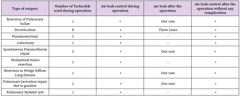
In thoracotomies 16 out of 19 patients required Tachosil® for air leak control, which included decortication, bullae resection, lobectomy, spontaneous pneumothorax repair, wedge resection in diffuse pulmonary disease, pulmonary repair due to gunshot, and hydatid cyst resection. postoperative minor air leak occurred in seven (36%) cases including decortication, spontaneous pneumothorax repair, wedge resection, and pulmonary laceration repair due to gunshot (Table 2). Spontaneous cessation of minor air leaks, which are not considered as complication, was observed in all patients. Thus, failure rate of air leak control was zero.

Table 2:Results of using Tachosil® in 19 cases of thoracotomy.
biomedres-openaccess-journal-bjstr
Discussion
The success of any surgical procedure is based on adequate hemostasis because ineffective local hemostasis is the major cause of bleeding during surgery. Many different biomaterial products can be used to achieve the same goal with less tissue injury and reduced cost and in a more rapid and easier manner [9,10]. Tachosil®, a collagen patch coated with human fibrinogen and thrombin, is used to improve postsurgical haemostasis [9,11,12]. In our study, during abdominal surgeries the time needed to achieve hemostasis was about 2.5 minutes and postoperative minor bleeding was noted in two patients (6.6%) undergoing acute cholecyctitis and liver trauma repair who did not required additional intervention.

Kawasaki showed that Tachosil® provided effective homeostasis within 5 min of application in 64 patients [5]. Their results of studies showed Tachosil® has noninferior hemostatic efficacy compared with TachoComb, and a similar safety profile, in patients undergoing liver resection. Their results therefore suggested that the beneficial effects of Tachosil® are similar to those of TachoComb and that Tachosil® is an effective mean of providing hemostasis in Japanese patients during surgery [5]. The clinical efficacy of Tachosil® was shown by a clinical study in hepatic surgery [13]. In this the study, Tachosil® were proved to be better than argon beamer in obtaining effective and fast intraoperative hemostasis [13]. In the field of hepatic surgery, a trial demonstrated the efficacy and safety of Tachosil® versus argon beamer in liver resection [14]. In the two groups of patients, the mean hemostasis time was less when Tachosil® was used (3-6 minutes) compared with argon beamer (5.0 minutes P = 0.008). Postoperative drainage volume, drainage fluid, and drainage duration did not differ between the two groups. The study confirmed that Tachosil® hemostasis is significantly faster than argon beamer after liver resection.

Briceño in a clinical trial, used Tachosil® in which 115 patients. These patients were distributed into two groups for major and minor hepatectomies, with or without application of a carrierbound collagen sponge on the raw surface of the liver. Their results showed that Tachosil® was effective in decreasing drainage volume, postoperative blood transfusion, complications and hospital stay [15]. Grottke investigated the efficacy of Tachosil® in stopping severe bleeding in a coagulopathic pig model with blunt liver injury [16,17]. The study analyzed two groups of pigs that were previously prepared by splenectomy and cystotomy and by inducing coagulopathy. Subsequently, a grade III liver injury was induced. At this point, animals were randomly assigned to receive a placebo patch or Tachosil®. Coagulation parameters and hemodynamic variables as well as the chosen treatment were monitored for 2 hours after injury and patch placement. All animals treated with Tachosil® survived, whereas 100% of the control group died before reaching the end of the observation period (P < 0.001).

Additional significant application of Tachosil® includes preventing air leak in various thoracic operations. In thoracic operations, air leak control was obtained in all patients, but postoperative minor air leak was observed in 7 patients (36%) which disappeared on the third day with no intervention required and therefore is not considered a surgical complication. In this study, bleeding and air leak control failure rates were zero and surgical site infection or any other complication attributed to Tachosil® was not observed. Air leaks have been reported to occur in 48–88% of patients undergoing lung resection [6].

Benefits of Tachosil® application for splenic trauma or fragile or atraumatic damaged spleen have been reported. In hematologic diseases, Tachosil® may be useful in clotting and for blood composition disorders. In many fields of abdominal surgery such as pancreatic and splenic procedures, fragile tissue is a major concern and use of this sealant is effective in hemostasis and also prevents leakage. In fact, pancreatic surgery is one of the most difficult surgical fields due to the risk of pancreatic fistula. We had no case of pancreatic lesion to be included in our study but the results of Tachosil® use in three cases of splenic resections, including two laparoscopic splenectomies, were quite encouraging and helpful. In our study Tachosil® was used in six cases of laparoscopic cholecystectomy and two cases of laparoscopic splenectomy with no adverse or undesirable outcome.

Our study also demonstrated the efficacy of Tachosil® in reducing postoperative air leaks in patients undergoing various thoracic operations, especially in a group of high-risk patients in whom intraoperative and/or postoperative air leaks are expected. It is to be noted that our study presents some limitations. The number of patients was limited by the nature of the study itself, the aim of which was to enroll diverse surgical patients in whom a clear indication to use Tachosil® could be demonstrated. In addition, this was intended as a pilot study with no formal calculation of the sample size needed to achieve a statistical power for confident evaluation of our results.

Conclusion
Tachosil® is a fast, safe and effective modality to achieve hemostasis and aerostasis in various abdominal and thoracic operations with no imposed tissue injury which eliminates the use of invasive suturing techniques in delicate tissues.

New Technologies for Treatment of Coronary Artery Disease-https://biomedres01.blogspot.com/2021/01/new-technologies-for-treatment-of.html

More BJSTR Articles : https://biomedres01.blogspot.com

New Technologies for Treatment of Coronary Artery Disease

New Technologies for Treatment of Coronary Artery Disease


Introduction
Despite tremendous advances in therapy and prevention, cardiovascular diseases (CVDs), including acute myocardial infarction (AMI) and its complication heart failure, are the leading causes of death worldwide [1-3]. Therefore, translational research and basic science becomes necessary to discover the novel mechanisms responsible for cardiovascular repair during athero-thrombosis which results in to AMI [4-6]. The inflammatory tissue in the myocardium and the intensity of neuro-humoral dysfunction after AMI are important determinants of complications as well as subsequent healing process. The increase in leucocytes; neutrophils, macrophages and lymphocytes in the myocardium contribute to the clearance of dead cells from the myocardial tissues while activating reparative pathways necessary for myocardial remodeling [5,7]. The thinning of the wall occurs due to apoptosis of cardiomyocyte with triggering of ventricular dilation and subcellular remodeling, leading to left ventricular dysfunction [7-9]. The underlying mechanisms appear to be increase in cortisol and vasopressin causing neuro-humoral dysfunction and oxidative stress, leading to inflammation and apoptosis [8-10].

Activation of angiotensin II signalling in different brain sites; the paraventricular nucleus (PVN), rostral ventrolateral medulla (RVLM), and area postrema (AP) may cause increased cardiac contractility with worsening of HF [8].The increase in angiotensin II signalling in the brain, enhances sympathetic nerve activity through actions on both central and peripheral sites during chronic HF. Stem cell therapy to target these brain areas may be an important technology to benefit patients with HF. Advances in new technology are; delivery of anti-inflammatory hormones thereby reducing arterial wall inflammation by using it in the form of liposomal glucocorticoid carrier [7]. The lipid nanoparticles are used to deliver siRNA antagonistic to the C-C chemokine type 2 pro-inflammatory receptor, and HDL nanoparticles examined to deliver simvastatin to inhibit monocyte recruitments [12-14]. In percutaneous coronary interventions, nanotechnology has demonstrated potential benefits by stent coating, gene therapy and stem cells [15-18]. They have been studied for their ability to release drugs as well as promote healing and reduce the rate and extent of restenosis [11]. This selected review aims to examine the role of new technologies in the management of CAD

Utility and Necessity of New Technologies
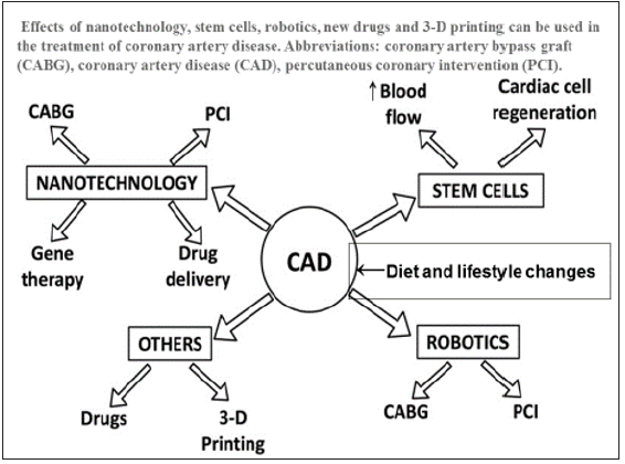
Current methods of treatment for heart failure after myocardial infarction are limited to palliation without any cure. Conventional surgical interventions such as coronary artery bypass graft or percutaneous coronary interventions are only able to partially restore myocardial function. There is only a minor improvement in the left ventricular ejection fraction due to reperfusion injury after these interventions [1-3]. The ultimate goal of cardiac repair is to regenerate functionally viable myocardium after myocardial infarction to prevent cardiac death. Identification of the innovative factors is essential in cardiac healing as well as in improving endothelial function of the coronary vasculature via using specialized pro-resolving mediators because the emerging factors provide the key molecular signals for the activation of the reparative cells in both endothelium and myocardium [7,11,12- 15]. There have been several advancements in the technological care of CAD in the last 3 decades. Of these technologies, stents, gene therapy, stem cells, drug delivery and 3-D printing are most important (Figure 1). Robotic angioplasty and coronary bypass grafting are also emerging to improve the clinical results.

Figure 1: Effects of nanotechnology advancements; stem cells, robotics, new drugs and 3-D printing in the treatment of coronary artery disease. Abbreviations: coronary artery bypass graft (CABG), coronary artery disease (CAD), percutaneous coronary intervention (PCI).(modified from Kandaswami et al. [7].
biomedres-openaccess-journal-bjstr
Stents for Coronary Angioplasty
Stents are used in coronary thrombosis to allow patency of the vessel in patients with CAD. In an animal model, gelbased nanoparticles combined with anti-proliferative and antiapoptotic agent rapamycin, were studied, which were found to re-endothelialize injured arteries and reduce hyperplasia [15]. Research in drug delivery has shown promise in treating cardiac diseases by using smart nanoparticles such as a pH-dependent delivery of antioxidants [16]. In clinical studies, nano-sized hydroxyapatite coating for controlled release of sirolimus, an immune- suppressive drug coated stent, revealed satisfactory findings [17]. A new gene-eluting metal stent fabricated by bioinspired surface modification with hyaluronic acid and deposition of DNA/PEI polyplexes has also been developed for clinical use [17]. Similarly, the release of sirolimus was studied using carbon nanoparticle coated stents with consistent drug release, as reported in an in-vitro study [18]. A comparison of the sirolimus-releasing stents with pitavastatin nanoparticle-eluting stents revealed that latter were found to be more efficient in terms of faster endothelial healing while being comparable in other parameters [22].

Endothelial healing and re-endothelialization can restore the injured vessel back to health. Magnetic silica nanoparticles were loaded with rapamycin, coated onto the stent which showed rapid endothelialization in the in-vivo studies [23]. Experimental studies reveal that liposome encapsulated alendronate (a bisphosphonate) can reduce restenosis and neo-intimal formations [39]. Polycaprolactone was found to be an effective carrier for nitric oxide to prevent restenosis [24]. Similarly, paclitaxel (antimitotic drug) in the form of albumin-based nanoparticles have shown to have significant anti-proliferative and restenosis effects without significant toxicity even when administered systemically [11,25,26]. The nanoparticles in these cases were either used to improve cell membrane permeability (alendronate) or binding capacity to the targeted tissues (paclitaxel) [1,24,25]. Polymeric stent coatings in the form of poly (lactic-co-glycolic acid) were proven to have a controlled release of the drug paclitaxel (nanocoatings-64) and polyethylene glycol was proven to reduce platelet adhesion [24]. It has been reviewed that nano-modifications can also help scientists in targeting specific delivery of medications such as collagen IV, chondroitin sulfate, tissue factor, or stents [7].

The promotion of healing by inducing endothelialization of the stent is also possible by nanotechnology in the form of nanofibrous matrix because it can attract endothelial cells, polyhedral oligomericsilsesquioxanepoly-(carbonate-urea) urethane which improve adherence and proliferation of human endothelial cells [7,11]. Other healing promoting agents are peptide amphiphilenanofiber coating, for promotion of endothelial cell adhesion and magnetic nanoparticles that cause preferential movement of cells towards the stent [7,11,24-27] (Figure 1). Nanotechnology also has potential applications in finding synthetic alternatives for coronary artery bypass grafts. Researchers have studied the potential of electro-spunnanosized fibrous scaffolds, which may prove to be an alternative synthetic graft for coronary artery bypass graft procedures [7,11]. Another area to choose technology may be targeting drug-eluting stents via gene therapy, because gene eluting stents can be used to overcome restenosis, in-stent thrombosis, and delayed endothelialisation [28,29]. In this connection, multiple nano-coatings in the form of hyaluronic acid as a vehicle for pDNA, nanobiohybrid hydrogel, a Tat peptide and DNA, and poly lactic-coglycolic acid nanoparticles as vehicle for PDGF receptor-β antisense RNA, have been studied in animal models and have shown promising results [30].

The gene targets have been studied extensively are; antisense oligonucleotide, chitosan-plasmid DNA, Akt1 siRNA, vascular endothelial growth factor, prostacyclin synthase, and endothelial nitric oxide synthase [28]. Recently, magnetic resonance imaging of acute thrombosis has become possible to visualize thrombininhibiting perfluorocarbon nanoparticles that provide a novel strategy for the treatment by using modular, multifunctional micelles [19,20]. In an experiment in hyper-cholesterolemic mice, targeted nanoparticles containing the pro-resolving peptide Ac2- 26 has been reported to protect against advanced atherosclerosis [21]. Recently, activation of angiotensin II signaling in different brain sites such as the paraventricular nucleus (PVN), rostral ventrolateral medulla RVLM), and area postrema (AP) has been found to increase the release of norepinephrine, oxidative stress, and inflammation leading to increased cardiac contractility [8,9]. It is possible that blocking angiotensin II type 1 receptors decreases sympathetic nerve activity and cardiac sympathetic afferent reflex when therapy is administered to the PVN. The administration of an angiotensin receptor blocker by injection into the AP activates the sympatho-inhibitory baroreflex indicating that receptor blockers act by increasing parasympathetic activity. In chronic HF, in peripheral regions, angiotensin II elevates both norepinephrine release and synthesis and inhibits norepinephrine uptake at nerve endings, which may contribute to the increase in sympathetic nerve activity [8-10]. Of all above technologies, stem cell therapy is most important because it is being used for treatment of both, myocardial dysfunction as well as for CAD.

The Stem Cells
Of all technologies that are in way for the managements of CAD, stem cells therapy is most interesting along with stenting. It has been suggested to repair myocardial damage and increase blood supply in ischemic conditions of the heart, thereby reversing the effects of CAD [31-38]. In this respect, both vascular growth factors and stem cells have generated a lot of interest as a mode of treatment in patients with CAD [31]. Such therapies aim to improve the blood supply to ischemic areas of the heart by stem cells, as well as promote cardiac cell regeneration (Figure 1). Stem cells provide benefits in one of two ways: by a direct effect of the stem cells, or by paracrine factors secreted by these stem cells [32]. Studies using hematopoietic cells; mononuclear cells and endothelial progenitor cells for various forms of CAD have been contradictory, although some studies have demonstrated a beneficial effect in these patients [33-36]. A new alternative is the creation of induced pluripotent stem cells, for which adult cells are transformed into pluripotent stem cells, similar to embryonic stem cells, but cancer may be an adverse effect [32,37,38].

There is physiological and metabolic evidence that many tissues and cells in the body have intrinsic regenerative potential and undergoes constant turnover throughout adult life [39-42]. It has also been demonstrated that the heart possesses cardiac stem cells that could be responsible for the intrinsic regeneration and turnover throughout adult life [42]. There is physiological cardiac remodeling in response to endurance exercise training in all cardiac cells [43]. These cells are greater in the apices of the atrium and ventricles and are known to be involved in tissue homeostasis [44- 46]. The reparative potential of these cells is limited, especially in conditions with extensive damage such as myocardial infarction and chronic heart failure [44-48]. There is also an interest to develop and inject multiple stem cells that can communicate with each other, named as a cardio-cluster which are cocktails of cells that include cardiac progenitor cells, mesenchymal stem cells, endothelial progenitor cells and fibroblasts. They have the potential to promote cardiac cell regeneration in disease states where cell function is reduced such as CAD [49].

The stem cells studied in clinical research are ranged from bone marrow to adipose tissue to skeletal muscle stem cells. Bone marrow-derived mononuclear cells are the most readily available cells for transplantation in the body. They are easy to identify based on their cell surface markers and can be isolated from the bone marrow [32]. However, their therapeutic potential is low since the harvested cells contain a multitude of cells with a small proportion of stem cells [35,39]. The bone marrow-derived mesenchymal stem cells are found in even lower concentrations than that of mononuclear cells thus requiring several weeks of maturation with different growth factors in the laboratory, prior to therapy. The adipose derived stem cells can be surgically harvested from adipose tissues. They are more abundant in comparison to the bone marrow-derived cells. This drastically reduces the time and cost involved in laboratory procedures to culture them for clinical use [39]. The pluripotent stem cells have a high potential for transformation. Although embryos represent the most obvious source of stem cells, their use has ethical concerns and is in debate.

Additionally, these cells could potentially face rejection when transplanted to a recipient. However, it is possible to reprogram adult cells and transform them into pluripotent cells which have similar properties as embryonic stem cells, thereby being called induced pluripotent stem cells. These cells can be auto-transplanted and therefore would not be rejected but may predispose cancer [37,41]. In view of the risk of teratomatous changes, this area requires more research before they can be considered safe for human trials. Another interesting source of stem cell are cardiac stem cells [42-44]. The clinical data for stem cell therapy is in its early days with reported literature covering both non-randomized and randomized trials [50-52]. There is improved left ventricular ejection fraction (LVEF) function following injection of mononuclear stem cells in patients with MI within three months. Improvement in exercise capacity, reduced mortality and scar tissue are shown in a 5-year follow up [50]. Several other studies showed similar effects following treatments with mononuclear stem cells after myocardial infarction [36]. An earlier meta-analysis reported an improvement in LVEF function by 2.99% following bone marrow stem cell transplantation in patients after MI [51].

However, the meta-analysis did not include recent studies that reported no improvement in left ventricular function [33,34]. In patients suffering from chronic ischemic heart disease, there is reported evidence towards improved cardiac function following the use of bone marrow derived-stem cells [52]. There have been several trials that have studied the clinical efficacy of mesenchymal stem cells. They have reported an improvement in cardiac function and relative safety in the use of mesenchymal stem cells [53-56]. Cardiac derived stem cells have also undergone clinical testing and have shown promising results with an improvement in LVEF [57], an improvement in the left ventricular mass that was viable [58,94], improved quality of life [57], reduced scar mass, improved and safety of the procedure [57,58] due to better regional contractility [59]. In a meta-analysis, a critical evaluation of clinical evidence on the safety and efficacy of autologous adult bone marrow-derived stem/progenitor cells has been examined as a treatment for chronic CAD and congestive heart failure [4]. This meta-analysis included, 38 randomised controlled trials involving 1907 participants; 1114 cell therapy, 793 controls.

Twenty-three trials were at high or unclear risk of selection bias. Treatment with stem cells, was associated with significant decline in the incidence of long-term mortality (≥ 12 months) (risk ratio (RR) 0.42, 95% confidence interval (CI) 0.21 to 0.87; among 491 participants. Cell therapy was also associated with a long-term reduction in the incidence of non-fatal myocardial infarction (RR 0.38, 95% CI 0.15 to 0.97; among 345 participants. The incidence of arrhythmias (RR 0.42, 95% CI 0.18 to 0.99; among 82 participants was also decreased with this treatment. However, there was no evidence that cell therapy has any influence on the risk of rehospitalisation for heart failure (RR 0.63, 95% CI 0.36 to 1.09; among 375 participants, or composite incidence of mortality, non-fatal myocardial infarction, and/or rehospitalisation for heart failure (RR 0.64, 95% CI 0.38 to 1.08; among 141 participants = 141, or long-term left ventricular ejection fraction when measured by magnetic resonance imaging (mean difference -1.60, 95% CI -8.70 to 5.50; among 25 participants[4]. The authors suggested that there is low quality evidence indicating that treatment with bone marrow-derived stem/progenitor cells reduces mortality and improves left ventricular ejection fraction over short- and long-term follow-up and may reduce the incidence of non-fatal myocardial infarction and improve chronic CAD and CHF. These findings should be interpreted with caution, as event rates were generally low, leading to a lack of precision.

Interestingly, some patients treated with cardiac stem cells, 10-14 months after myocardial infarction may have similar therapeutic benefit as someone treated earlier, suggesting that cardiac stem cells could be beneficial in chronic ischemia patients [32,50-58]. However, it should be noted that the observed clinical benefit was less than the expected clinical benefit based on prior in vitro and animal studies [32]. Stem cell therapy continues to be a promising treatment modality for CAD. The experimental and clinical studies have shown promising results. However, further research is needed to understand the exact mechanisms of action and the ideal source of stem cells to derive optimum benefit and to further our understanding. Several challenges such as long-term safety and route of administration, have to be overcome. However, the direction of current research looks promising.

3-D Printing
There are other technologies such as 3-D Printing Cardiac conditions often need 3-D imaging like magnetic resonance imaging, computerized tomography, and 3-D echography to diagnose and treat myocardial damage and athero-thrombosis. Although these images are in 3-D but they are viewed on a 2-D computer screen or films which could be sufficient for some cardiac procedures. However, the current imaging modalities are not effective for more complex interventions. It seems that 3-D printing has a potential role in CAD because it overcomes these limitations as well as allow for complete visualization, tactile sense, education and surgical planning as well as simulation [60]. It needs additive manufacturing of a model using 3-D data from imaging modalities. It has been observed that 3-D printing could be more effective in planning and treating complex situations (bifurcation lesions) that require stent placement. Radiologists and scientists are trying to see the full potential of 3-D printing, because in cardiology it has tremendous potential in the treatment of congenital defects, cardiac tumors, cardiomyopathy, functional flow models, valvular heart diseases, stent placement for CAD and other cardiac surgeries [60-64].

These models can be used in a pulsatile flow loop environment, not only to visualize and understand complex flow patterns but also to simulate interventions [63]. It is possible to visualize 3-D printed heart with coronary arteries by this technology, for examination of the extent of occlusion and stenosis in CAD. Compared to imaging and treatment modalities, 3-D printed models are also useful in CAD research [63]. Models of tissue engineering are examined to fabricate stem cells along with extracellular matrix (tissue printing) for implantation in the body. In vitro studies have been successful in tissue printing cardiac cells in different scaffolds. The implantation of printed tissue in epicardial tissues showed beneficial effects including decline in adverse remodeling and improved perfusion in the models of myocardial infarction.

In brief, it seems that nanotechnology has led to an interesting and promising direction in the treatment of CAD and CHF. It has valuable potential in delivering drugs that are otherwise limited by their pharmacokinetics. Its applications in stent and gene therapy are potentially useful for future therapeutics based on these modalities. Randomized controlled trials are needed to establish potential evidence to support the use of these newer technologies for therapy of CAD. This needs to be carried out with potential collaboration between researchers, engineers, biomedical engineers, nanotechnologist and physicians. and clinicians.

The Connection Between Alzheimer’s Disease and Prion Diseases: A Mini-Review-https://biomedres01.blogspot.com/2021/01/the-connection-between-alzheimers.html

More BJSTR Articles : https://biomedres01.blogspot.com
The Connection Between Alzheimer’s Disease and Prion Diseases: A Mini-Review

The Connection Between Alzheimer’s Disease and Prion Diseases: A Mini-Review


Introduction
Alzheimer’s disease (AD) is a neurodegenerative disease with a slow progression that worsens over time [1]. AD is the main cause of dementia and this syndrome can be characterized by any reduction in cognitive capabilities that interferes with daily life [2]. There are two main forms of AD: early-onset AD, which is rare (≤ 1%), occurs in individuals under 65 years of age, and is caused by genetic mutations. The other form is late-onset AD and this is the most common form. Late-onset AD occurs in individuals 65 years of age and older and it is multifactorial [1]. Prion diseases, also known as transmissible spongiform encephalopathies, are a group of rare infectious neurodegenerative and fatal illnesses that includes Creutzfeldt-Jakob disease, variant Creutzfeldt-Jakob disease, Gerstmann-Straussler-Scheinker disease, fatal familial insomnia, and kuru [3]. In addition to the loss of synapses and neurons in specific brain areas, prion diseases, AD, and other neurodegenerative diseases such as Parkinson’s and Huntington’s disease have several other common features such as the progressive accumulation of misfolded proteins that form amyloid [4].

The main proteins involved in the accumulation of misfolded aggregates in AD are extracellular amyloid beta 42 (Aβ42), which forms amyloid plaques, and hyperphosphorylated tau (hp-tau), which forms intracellular neurofibrillary tangles [5]. In contrast, in prion diseases, the aggregates are formed by extracellular abnormal prion protein (PrPSc) [3]. These proteins, in their native form, undergo a misfolding process that produces a structure with β-sheet-rich structures [4]. Several studies support the hypothesis that misfolding, oligomerization, and accumulation of these proteins are the main events that trigger the pathological events in neurons in both AD and prion diseases [6]. Aβ42, hp-tau, and PrPSc aggregation seems to adjust better to the seeding-nucleation model first proposed by Jarret and Lansbury [7].

In their current form, this model can be summarized as follows: the first event is the formation of a nucleus or seed from the misfolded monomeric protein; then, this structure evolves to oligomers, protofibrils, and mature fibrils, which eventually suffer fragmentation and the fragments produced can migrate and function as a new seed which can recruit soluble normal protein and the cycle repeats [4,8]. Of all the structural conformations previously mentioned, the oligomers are the more toxic form [6,9,10]. Experimental evidence shows that normal prion protein (PrPC) can function as an Aβ42 receptor and this interaction produces a decrease in long term potentiation in neurons of the hippocampus [11,12]. Other studies have shown that activation of PrPC stimulates hp-tau [13,14], which suggests that PrPC is an intermediary between Aβ42 and the hyperphosphorylation of tau.

Conclusion
Strong evidence supports the hypothesis that AD and prion diseases have some similar mechanisms for misfolding, aggregation, and propagation, and also that PrPC can function as an Aβ42 receptor which is an intermediary for the hyperphosphorylation of tau. This evidence may suggest a new method for finding new therapeutic agents.

Problems of Obesity in Drivers and Road Safety-https://biomedres01.blogspot.com/2020/12/problems-of-obesity-in-drivers-and-road.html

More BJSTR Articles : https://biomedres01.blogspot.com
Friday, January 1, 2021

Happy New Year: BJSTR


                                                    Wishing you a Happy and prosperous New Year

Effect of Nutritional Intervention on the Temporal Dynamics of Muscle Strength Adaptation

  Effect of Nutritional Intervention on the Temporal Dynamics of Muscle Strength Adaptation Introduction Muscular adaptation is determined b...