Friday, August 6, 2021

Teaching Surgical Skills During the Present Explosion of Knowledge and Technologies


Teaching Surgical Skills During the Present Explosion of Knowledge and Technologies 

Opinion

The continuous introduction of new technologies into surgery mandated the introduction of new ways of training to cope with it. Mastering the new skills to use the new technologies is mandatory for the success and safety. For example, introduction of laparoscopic surgery needed new types of skills to be taught. Even the different types of laparoscopic operations which were developed by time needed different types of skills. Microscopic, robotic and remote surgeries are, but a few other examples. Teaching these new skills during the postgraduate training program makes it inflated and redundant. To deflate it either the time for postgraduate training increases or some of the skills is taught before entering the training programme. Increasing the time for training is not desired by any. Teaching some skills somewhere else is possible as suggested later.

On the other hand, explosion of knowledge in all surgical fields makes the undergraduate curriculum overloaded. Because of this, the surgical skills which will not be used independently in houseman ship should not be taught in the undergraduate curriculum. Now, in both levels of training we have a problem of time. The aim is to have a well-trained surgeon. The base of this surgeon lies in the undergraduate teaching of surgical skills. The actual making of a surgeon is in the postgraduate training. The solution lies in defining all the surgical skills needed to start the postgraduate training and then find the most suitable place for them. Most of the surgeons who train the postgraduates also teach the undergraduates. These surgeons should be actively involved in revision of both under and postgraduate curricula. These surgeons also supervise house officers and know the skills they need to start houseman ship. They know all the surgical skills that are needed to start the postgraduate programme. The surgical skills which are needed to start the houseman ship should be taught for the undergraduates. Those which are not needed to start the houseman ship should be taught during the houseman ship.

My opinion is that there is no need for medical students to attend surgical operations. The skills gained from attending operations can be taught in the skill lab. Their presence in the operating room will crowed it, putting them in the way of the real staff. They increase the hazard of infection and mostly they do not participate really in operations, so, they learn nothing. This can decrease the load on the students and give them more time to master the important surgical skills. In my opinion, the distribution of skills should be as follows:

The surgical skills which are needed to start the houseman ship and should be included in the undergraduate curriculum are: (They are NOT written in order of preference)

a. CPR.

b. Surgical Audit.

c. Surgical ethics.

d. Professionalism.

e. Communication skills.

f. Scrupping.

g. Glove wearing.

h. Simple suturing.

i. Basic knot tying.

j. Urethral catheterization.

k. Naso-gastric intubation.

l. Intravenous injection and cannulation.

m. Pharyngeal airway placement.

n. Cervical collar application.

o. Suture removal.

p. Bag mask & nasal prongs ventilation.

q. All clinical skills needed for examination of a surgical patient including digital examination of the anus/rectum and vagina.

The surgical skills to be taught during the houseman ship: (They are NOT written in order of preference)

a) ATLS.

b) Deep knot tie.

c) Chest intubation.

d) Tracheostomy & Cricothyroidotomy.

e) Mattress suture.

f) Subcuticular suture.

g) Surgical incisions.

h) Positioning of patients on the surgical table for different operations.

i) Simple skin excisions.

j) Anoscopy/proctoscopy.

k) Abdominal paracentesis.

l) Plaster cast/splint limb immobilization.

m) Handling and use of different surgical instruments.

n) Application and removal of skin staples.

o) Removal of drains.

p) Arterial blood sampling.

q) Application of local anesthesia.

r) Skills for laparoscopy and other new technologies.

The last skills should be gained by simulation courses which are designed by the postgraduate trainers and conducted by recognized bodies such as the Continuous Professional Development. They can be considered a prerequisite for admission into the postgraduate surgical training programme. It is really annoying when you, a postgraduate trainer, discover that many of your trainees do not know many of the basic surgical skills. Evaluation for these skills should be included in the assessment for completion of medical school and houseman ship.

For more Articles: https://biomedres01.blogspot.com/


Lab Grown Meat: The Future Sustainable Alternative to Meat or a Novel Functional Food?


Lab Grown Meat: The Future Sustainable Alternative to Meat or a Novel Functional Food?

Introduction

The rising world population is expected to reach 9.5 billion by 2050. This population growth coupled with factors such as social, economic and demographic changes (urbanization, rising incomes in emerging economies, etc.) and the resulting changes of consumer patterns will exert increased pressure on global resources for the production and availability of more food but also different food items associated with different diets [1,2]. According to an analysis conducted by Henchion et al. [3], an increase of meat consumption by about 60% has been estimated between 1990 and 2009 and this trend is expected to continue due to increased income in Asian, Latin American and Middle Eastern countries. Increased demand for proteins, mainly of animal origin, is expected to have a negative impact on the environment causing greenhouse gas emissions (GHG), requiring even more water consumption and land exploitation for their production [4]. In order to address this critical problem, an even more sustainable production is required, with the use of existing protein sources as well as additional alternatives for direct human consumption minimizing as the possible intervention of animals.

In order to support sustainable production and disposal of proteins, there are several possible scenarios regarding both the exploitation of existing sources of protein but also the discovery and development of new ones, always taking into account the nutritional, environmental and technological challenges as well as the consumer’s and market response. Lab grown meat although still at an early stage, seems to be a promising alternative technology for the production of proteins of animal origin and the availability of products traditionally produced through animals in a way that requires significantly reduced animal participation or not even at all. This technological innovation has also an ambitious objective to offer the possibility of reducing the negative impact of today’s meat production and consumption on humans, animals and the environment. Consumers need also to respond to various questions about the cost of production, the possibility of mass production, food safety and the potential ethical dilemmas posed by different population groups.

Technology and Cost of Lab Grown Meat Production

A 43 year old male presented with NSTEMI, which was treated conservatively, with an uneventful recovery. He was of normal height and weight and was of South Indian origin. Risk factor for CAD was smoking for 5 years only. There was no history of previous chest pain, unusual childhood illness or any inflammatory disease. Echocardiography showed an ejection fraction of 55%. Coronary angiography showed dissection in all the three vessels, with the LAD occluded following the dissection (Figure 1). Autoantibody screening, inflammatory screening and CT scan of chest and abdomen was normal. He underwent CABG with uneventful recovery.

The rapid growth rates of the stem cells achieved in this way involve a shorter period of tissue growth compared to that required for animal husbandry and consequently the required nutrient and energy inputs are reduced. The cultivation of cells and tissues in the laboratory is not currently effective in energy, water and raw materials’ demand, and so far has been used only in scientific and medical applications [8]. Economic benefits as well as sustainability benefits are also unclear because reductions in some inputs may be offset by the extra cost due to a stricter sanitation regime and other energy inputs required [7]. The cell culture medium can be produced either from animal materials, such as bovine and horse serum, which reduce many viability benefits of the cultured meat [7], or from a suitable culture medium produced from non-animal origin, such as hydrolyzed cyanobacteria referred as blue-green algae [9] and Maitake mushroom extract [7]. No significant progress has yet been made for large-scale production and decisive factors and parameters of this process still need to be researched in depth. Among these, the production of cultured meat requires appropriate cells and growth media, preferably of non-animal origin, in order to avoid animal components containing agents for communicable diseases, and edible materials suitable for cell growth matrices to produce thicker and more continuous meat pieces such as steaks. A such efficient process for the production of non-animal culture media is still considered as a great challenge and a very important step towards the acceptance of the cultured meat [10].

Impact on Public Health

Lab grown meat could be an excellent functional food since can be modified to alter the profile of essential amino acids and fats, to be enriched in vitamins, minerals and bioactive compounds so that not only is it in proportionate amounts of natural meat but also exceeds it to cover specific dietary needs for people with various ailments [11]. Additionally, after the appearance of functional and fortified foods, consumers are more willing to test products that have been modified to have particular functional and nutritional characteristics [12,13]. Strictly controlled hygiene conditions in sterile systems applied to the production of lab grown meat contribute significantly to improving its safety by minimizing the risk of zoonotic and food-borne pathogens, viruses such as avian influenza and swine flu or prions for transmissible spongiform encephalopathies [14]. Scientists also hope that the need for pesticides, fungicides, growth factors and antimicrobials which are used in excess for conventional meat production, may be significantly reduced as the consumption of culture meat increases [15-17]. In the future, the ever-growing production and cost reduction of lab grown meat, possibly below traditional animal husbandry, would make its consumption more affordable and could increase access to meat even in developing countries. In this case, the cultured meat could help alleviate certain nutritional deficiencies in these populations and support the physical and mental development of children [17].

Environmental Consequences

The potential benefits of developing and expanding the production and consumption of lab cultured meat are referred to in some Life Cycle Assessments although they are based on hypothetical models of the form that cultured meat can take. Replacing conventionally produced meat with cultured meat could potentially help mitigate greenhouse gas emissions, because instead of using larger land for the necessary agricultural crops required for livestock farming, large areas could be released and redeveloped or used for other purposes such as carbon capture. Tuomisto and de Mattos [9] reported that if the cultured meat is grown in algae culture medium, although energy consumption will not decrease dramatically, GHG emissions will fall by 78- 96%, land use by 99%, water consumption by 82-96% and energy consumption by 7-45% compared to those from the conventional farming depending upon the type of meat, except conventional poultry meat that requires less energy. In a similar research [18] it was reported that the cultured beef has a lower heating potential than the conventional, on the contrary cultured pork and poultry meat may involve significant energy use leading to higher heating potential than the conventional products.

Using a different comparison field, in another survey [19] cultivated meat was compared with a range of meat protein alternatives (plant, mycoprotein, dairy and chicken). The researchers found that the lab grown meat can have less environmental impact than conventional beef and possibly pork, but higher than for chicken and plant protein production, mainly due to high energy requirements, with only exceptions the effects of land use and ecotoxicity of terrestrial soils and freshwater. However, in all the aforementioned cases, there are significant environmental benefits in all types of cultured meat in terms of land use. Production of lab grown meat could also have potential benefits for the conservation of wildlife by reducing pressure to convert natural habitats to farmland and also providing an alternative way of producing meat from endangered and rare species that are currently at high risks by over hunting or fishing for food.

However, according to Tuomisto and de Mattos [9] the largescale replacement of conventional meat production may have some negative effects on rural biodiversity due to a reduction in the need for meadows and pastures. The possible conversion of the meadows into forests may benefit certain species, but others may be threatened. Potential additional benefits are the reduction of large direct and indirect emissions from agriculture (e.g. digestion gasses from livestock, production of fertilizers and other agrochemicals, use of fossil fuels by tractors, etc) [20]. According to FAO [21], the livestock sector is a significant contributor to GHG emissions with an estimated total of 8.1 gigatonnes CO2 -eq in 2010 (using 298 and 34 as global warming potential for N2O and CH4 respectively). The production of lab grown meat also results in significantly lower losses of nutrients in waste compared to conventionally produced meat because the effluents from the production of cyanobacteria can be controlled more effectively in relation to the runoff from the agricultural fields [9].

The effects of transporting the lab grown meat are likely to be lower because animals will not be transported to the slaughterhouses (from long or even short distances) as well as carcasses or large cuts of meat from slaughterhouses will not be transported to the market or to installations for further processing under cooling, and with an appropriate design, its production sites may be closer to the markets and the points of consumption. As a result, there would be a significant reduction in energy consumption for chilled transport, as the cultivated meat has a lower mass because there are no nonedible parts (bones, blood, etc.). In addition, slaughterhouse waste and the environmental and economic consequences associated with it are significantly reduced [9]. However, further research is required to assess the overall environmental impact of cultured meat production during the life cycle from production to the final consumer

Ethical, Socio-Political and Economic Aspects

Consumers’ perceptions and ethical dilemmas are crucial and also a potential barrier to the acceptance of the lab grown meat and its commercial success [22]. The product should have as much as possible similar sensory and organoleptic characteristics (taste, texture and appearance) with the natural meat, which is widely accepted by consumers; however, this is currently difficult to achieve [8]. The extent to which muscle biology can be copied will determine the complexity of the tissue production process. The production of a whole muscle piece is the long-term goal. This requires a complex system that involves multiple types of cells grown together in an organized manner, and a structure that will require a reproducible blood vessel network. A simpler and most feasible goal in the near future is the production of a muscle protein component based on muscle cells alone. Among the serious benefits of lab grown meat, the resulting reduction in the population of meat-producing animals is reported in the international literature [5]. Although it is an exaggeration to see that an animal is sufficient to meet world meat needs, it is feasible and understandable that the reduced populations of animals required for conventional meat production would make it possible to obsolete intensive and industrialized livestock farming and would contribute to improving livestock conditions that will still be needed [6].

Another important moral issue is the treatment of stem cell donors as these cells have to be collected from an animal source (live or not). In that case, the necessary invasive technique to obtain the right type of muscle tissue may be painful. Also, the necessary serum for the growth of cell cultures should also be taken from adult animals, newborns or fetal sources raising also ethical concerns. In the future, this substrate of animal origin could be replaced by sources of plant origin such as mushroom extract which achieves higher growth rates compared to fetal bovine serum and is more cost-effective [23]. Vegetarians and other people such as members of certain religious groups, opposing the use of animals, and consider the consumption of meat or other foods of animal origin is not necessary for human health and thus these issues could eventually be an obstacle. Acceptance criteria are mainly ethical in terms of technology and its application, as well as its relationship to the natural product, the expected qualitative characteristics and the potential benefits or risks. Consumer knowledge of the lab grown meat is currently very poor. Although surveys conducted so far show that most consumers are reluctant to answer when asked if they are willing to test cultured meat in the future, only a small minority categorically rejects the idea [24].

The willingness of consumers to consume lab grown meat is also related to a great degree to the viability benefits in relation to conventionally produced meat and the information provided about these benefits seems to be critically important to increase consumer’s confidence and acceptance. Another key challenge for the socioeconomic status of this novel technology, as it happens with all innovative technologies entering the market, is the disruption of the livestock industry, where millions of people are directly or indirectly involved [25]. What will be the reactions of the stakeholders of this huge and very important economic field of global economy? To which extend it will replace the conventional meat production or instead it will increase the meat consumption globally? Who will produce the cultured meat and profit from it? Will it provide a new frontier for capital accumulation for multinational corporations or a shift towards localized production? All these difficult questions remain unanswered and will soon need to be addressed by governments and society.

Legislative Regulatory Framework

Some start-ups companies in the US and other countries claim that the lab grown meat (or clean meat or artificial meat or laboratory meat) may be placed on retail shelves perhaps before 2021. However, before this happens, a legislative regulatory framework for this innovative food should be introduced. This legislative framework should specify, among other things, which controls are to be carried out and which bodies are responsible to carry them out at the production and disposal level. In the EU, lab grown meat should be adopted as a novel food and comes under the Regulation EU 2015/2283 [26] regime for novel foods. In order to be approved as a novel food, growers are required to submit an application which must include a complete dossier with all relevant data (production process, product safety issues, ethics, labeling, etc.). If the product is considered safe after a scientific assessment by the European Food Safety Authority (EFSA), the Commission may adopt a regulation approving this product.

The Commission has recently confirmed that no application for the approval of lab grown meat has been submitted so far and that such a product cannot yet be placed on the market and any such meat will be seized by the authorities. The Novel Food Regulation stipulates that approved novel foods on the list may be subject to labeling requirements in order to fully inform the consumer, for example, of the description of the food or its composition. It is therefore very likely to add specifications for the lab grown meat. In any case, the EU regulation Regulation (EU) 2011/1169 [27] on consumer information on food will also apply to lab grown meat as soon as it is approved, but its implementation may prove difficult. For example, there is an obligation to indicate on the label the name of the food, but at present there are unresolved issues regarding the name of the lab grown meat. Lab grown meat or cultured meat has not yet been released in the market and there is neither a registered name nor even a common name. Many names already exist, but the choice of the final name is a very sensitive affair and not at all easy.

A serious problem for its final name is the fact that consumers must be provided with clear and precise information about their method of manufacture or production, so producers should ensure that the name of the product makes it clear that the meat has been grown in laboratory. In accordance with the current regulations [28,29], meat means “all parts of domestic bovine animals, swine, sheep, goats and solipeds which are suitable for human consumption”. Therefore, the process towards a regulatory framework will actually start in the EU as and when an application for approval to EFSA. In the United States the there is no any regulation yet, although most cultured meat companies are based there. The definition of “meat product” for lab grown meat does not comply also in the US under the Federal Meat Inspection Act [30], in which meat and meat products should come from carcass.

Conclusion

Lab grown meat appears to be a particularly interesting alternative to conventional meat from animal carcasses, the problems caused by its production methods and its shortcomings in meeting future emerging global demands for protein availability. In addition, the possibilities offered by the flexible production process and the modification of its composition, make it a promising functional food with the capability to meet the specific needs of many different consumer groups. However, so far it is still at an embryonic stage without large-scale production technology being developed. Important questions, especially moral ones are also unanswered yet. Information and involvement of social stakeholders and consumers in any decisions to be taken is necessary in order to build acceptance through a transparent process. The big challenge of a sustainable future food supply can only be achieved by pursuing a number of viable solutions that will only become effective when combined. Such solutions include the prudent meat consumption, the abolition of “industrial” livestock farming and the promotion of organic farming as well as support for the development and exploitation of plant or other protein sources. Cultured meat is among the many possible factors that can contribute to solve the problem.

 For more articles:  https://biomedres01.blogspot.com/

Radiology Being Digital


Radiology Being Digital

Opinion

It is true that it is almost a commonplace to say that the digitization of both the radiologic images and the clinical and demographic data provides an increase in the quality of health care and procedures. But not to declare her victorious we must bet on her in an uncritical way. Because, in addition, technological advances have not been free. The price we have paid in western countries for this transformation is to have a health system that is more impersonal, which is colder, and, of course, more distant in human relationships, a system in which the most difficult to obtain is personal attention. Perhaps now we can reflect on what we have lost and what we have gained, even without the intention of undoing the path. For the prophets of the digitization the path was already traced: since they have long preached that the analogical past is something that seems old fashioned, and that getting carried away by the path of the zeros and ones and, excited, observing on the screens all the details in detail, is sure to reach a place among the chosen ones of the modernity. Zygmunt Bauman raises an interesting discussion between the solid and the liquid to give visibility to this landscape of modernity. However, the reality is much more convoluted, especially in relation to important details, some of them from the social point of view: with the digital era, inequalities in our societies have increased.

Even more: it seems that the technological revolution does not provide an increase of productivity like that of the previous industrial revolution, and this makes the economic growth of this century proportionally inferior to that of previous times. A good question for the current managers of some public services. For the moment, what is the balance of the debate that is often understood as medicine of machines versus medicine of words? Well, provisionally: good in the aspects most related to management (its great merit is to have achieved that place under the complex and difficult relationships that have always been the hospital ones: its information system will come to adapt as it has done to all this process). Moderately good, although with a division of opinions, in those aspects that are more related to daily work (in my opinion, the change has been partly a partial renewal and in part a radical break and has been done without collapsing the whole building). Bad or nonexistent in those aspects that have more relationship with citizens: today, just as yesterday in the analog world, users find it very difficult to access to their medical records, however electronic. In relation to these social aspects, the change from which everyone expected so many things, the analogue-digital transition, and what has been called “technomedicine”, has represented very little. All of the above, with the advantages (and doubts), generates an unwanted consequence, at the same time that it raises another reflection that still being collateral, does not cease to be proper of our days, and that must be part of this debate: the arguments, although most of them favorable, should not conceal the damage resulting from poor utilization of some of the technology, and more when compared to the situation prior to the massive implementation of these techniques.

At present, there is a disproportionate increase in demand, and there are many more radiological tests than necessary: about 30% could be avoided, as the Spanish Society of Radiology (SERAM) has recently indicated. Twenty years ago, there was the feeling that professional habits could be changing according to the dazzling results of the different developments that arise. But there are motivations of a more technical etiology. Of course, there are differences between analog radiography and digital radiology. To put it philosophically, where analogical radiology ends, digital radiology begins. The digitization strategy in the health environment rests on two strength ideas: the logical integration of the image data (PACS) with the clinical and demographic data of the patient; and the transmission of data and images. The advantages of the digital format (reconstruction of the images in the different planes of the space, in three dimensions and video (4D), transmission to remote places and through the network of the medical images accompanied by pertinent information of the patients, etc), partly explain the primacy of the process. It has always been said that when the images have become digital has risen a step in its manipulation. But the clearest impression of the uniqueness that the digitalization can produce to the medical doctors takes place when, seated in front of the screens, absorbed in the reading or the interpretation, they receive the report of the last examinations. That is the nature of the task.

 For more articles: https://biomedres01.blogspot.com/
Extremity Soft Tissue Sarcoma Mimicking Traumatic Intramuscular Hematoma: A Case Report and Review of The Literature


Extremity Soft Tissue Sarcoma Mimicking Traumatic Intramuscular Hematoma: A Case Report and Review of The Literature 

Introduction

The management of large intramuscular hematoma in the extremities is sometimes difficult, especially when there is no clear etiology. It can be spontaneous or secondary to trauma, bleeding disorders, tumor, or recent surgery in clinical practice [1,2]. Patients usually complain pain, swelling, and frequent ecchymosis over the involved area after trauma. However, if a patient presents with an expanding traumatic hemorrhagic mass without bleeding disorder or bruise, several differential diagnoses should be considered including chronic expanding hematoma, aneurysm, abscess, and soft tissue sarcoma. Undetected sarcoma will cause catastrophic result. Dealing with these cases are quite challenging since both the intramuscular hematoma and soft tissue sarcoma share the similar clinical symptoms and imaging findings.

Case Report

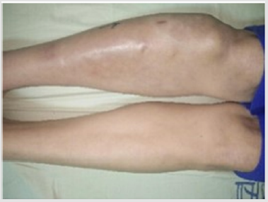
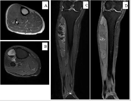
A 60-year old female kicked on the motor vehicle with the right lower extremity and developed mild pain at the right calf in September 2017. Five months later, without improvement in her symptoms and persistent swelling in the lateral aspect of calf, she came to the out-patient department (OPD) in Kaohsiung Medical University Hospital. Physical exam revealed pain to palpitation and slight swelling of right lateral calf. Based on the trauma history with no other specific findings, the physician diagnosed a muscle sprain with concomitant bleeding and treated her with pain medication. For further survey of prolonged leg swelling, an MRI was obtained weeks later that showed multilobulated mass (about 16 x 4cm) in anterior tibialis muscle, heterogeneous signal intensity on both T1 and T2 weighted images. Both the radiologist and the attending physician interpreted the MRI as intramuscular hematoma with multiple stages. So conservative care with continued physical therapy was recommended. Three months later, the mass continued to increase in size and symptoms became worsened. The patient visited our OPD again and was thus referred to the Orthopedic oncology surgeon for evaluation. Clinical exam showed a 18 x 4.5cm, firm, irregular, immobile mass extending from the anterior to the lateral aspect of right calf (Figure 1).

Figure 1: Photograph of the right tibia before surgery. Right calf is severely swelling.

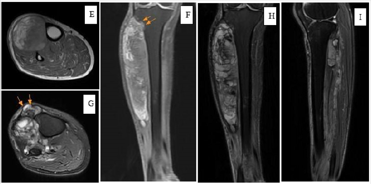
There was no numbness or weakness or sensory defect in the affected lower limb. Laboratory data including bleeding time and blood coagulation was normal. Fine needle biopsy was carried out, and the analysis proved the fresh with old unclotted blood. Due to symptoms progression, a repeat MRI was performed. The finding reported by the radiologist revealed as increasing size (20 x 5cm) of intramuscular hemorrhagic lesion in anterior tibialis, with involvement of pretibial fascia and cortex destruction at the proximal tibia (Figure 2), with high possibility of malignancy. Angiography also revealed a highly vascularized mass with malignancy characteristic. Due to the high possibility of malignancy, she underwent wild excision of the tumor on May 2018. At the surgery, a large encapsulated mass containing thick gelatinous gray material was found (Figure 3), involving pretibial fascia as well as the lateral and posterior component of the calf. The resected specimen was sent for frozen pathology and demonstrated highly suspect myosarcoma. It revealed the undifferentiated, spindle to multinucleated polygonal neoplastic cell with abundant of eosinophilic cytoplasm. The final histopathology revealed pleomorphic rhabdomyosarcoma. After the surgery, the pain resolved significantly. The patient received adjuvant radiation therapy. The recovery was good. No lung metastasis was found at the time this report was made.

Figure 2a: (First MRI, A: axial T1-weighted, B: axial T2-weighted, C: coronal T1-weighted, D: coronal T2-weighted).

Note: A 60-year-old female developed a painful lump at the anterolateral side of calf after minor trauma. MRI revealed intramuscular hematoma with multiple stage and no evidence of bone and fascia invasion.

Figure 2b: (Second MRI, E: coronal T1-weighted, F: axial T1-weighted, G: axial T2-weighted+FS, H: coronal T2-weighted +FS, I: sagittal T2-weighted+FS).

(E, F)- an increased size of intramuscular mass with a mix of iso-to-high signal

intensity in the anterolateral aspect of calf. Two arrows indicate cortical destruction at the

proximal lateral tibia (not seen in the first MRI)

(G)- tumor invasion with pretibial fascia involvement (two arrows) (not seen in the first MRI)

(H, I)- an increased mass size with heterogeneously distributed iso-to-high signal intensity in the anterior, lateral, and posterior aspect of calf.

Discussion

Soft tissue sarcoma accounts for about 1% of adult malignancy. It usually presents as a painless lump and can occur at any site over the whole body. Almost 45% of the soft tissue sarcoma appears adjacent to the long bone of the extremities, especially in the lower limb [1]. However, few people know that soft tissue sarcoma occasionally accompanies large hematoma [2]. In certain situation, soft tissue sarcoma may be initially misdiagnosed as deep intramuscular hematoma. Clinicians should be aware of the possibility of soft tissue sarcoma masquerading hematoma after trauma. MRI is a powerful tool for diagnosing soft tissue masses due to high contrast tissue resolution [3]. It remains the gold standard to distinguish the soft tissue tumor from hematoma. The hematoma revealed various signal intensity at difference stages [4]. Acute hematoma shows low-to-intermediate signal intensity on T1 weighted image (T1WI) and high signal on T2 weighted image (T2WI) [5]. In the following days, the hematoma may appear intermediate signal intensity in T2WI. Chronic hematomas are usually surrounded by the fibrous capsule demonstrating hypointensity in both T1 and T2 weighted images.

Mixture of heterogeneous signal may occur in recurrent blooding event as a result of blood products in different metabolic stage. For the soft tissue sarcoma, it generally contains a solid component which often reveals iso-hypo intensity to muscle on T1WI and heterogeneous hyperintensity on T2WI [6]. However, variation in image finding are occasionally observed. Contrast enhanced MRI is helpful but overlapping of features will lead to uncertainty. For instance, diagnosis can be challenging if large portion of the tumor is occupied by hematoma without a solid mass lesion on the MRI [7]. Ogose et al. [8] reported an extra-skeletal Ewing sarcoma mimicking a traumatic hematoma in a 16-year old baseball player with a history of recurrent hematoma of the thigh. Therefore, a detailed survey of each patient’s history, clinical course, and radiography including MRI plays an important role in making differential diagnosis. After reviewing current studies, we concluded that the soft tissue sarcoma mimicking as hematoma should be considered in the following situation:

a) The lesion doesn’t correlate with the expected clinical course of blood clot degeneration. If the hematoma enlarges or pain deteriorates, malignancy should be warranted.

b) If the large intramuscular hematoma presents, and the patient denied bleeding disorder and prior trauma, soft tissue sarcoma should be highly suspected.

c) In chronic hematoma with peripheral wall enhancement on T2WI, awareness should be raised because its appearance is much more likely a sarcomatous lesion [1].

d) The mechanism of the injury reveals disproportion with clinical severity such as pain, swelling, and functional disability [9].

e) Traumatic hemorrhage usually causes subcutaneous ecchymosis, while intratumoral hemorrhage often is constrained by a pseudo capsule and less likely to have ecchymosis [10].

In complicated cases, it poses great challenges for clinician in making correct diagnosis after history taking, physical examination, and initial radiographic assessment. Biopsy are recommended and can provide more information for making diagnosis. Fine need aspiration was traditionally considered a useful method for the diagnosis of bone and soft-tissue tumor. Nonetheless, in sarcomarelated hematoma, it involves the risk of sample error and yields very low diagnostic accuracy because of insufficient tumor cells from aspiration. Imaizumi, reported six cases with soft tissue sarcoma that were initially diagnosed as traumatic hematoma. In five of the patients, the cytology revealed no malignant cell. In our case, the report for fine needle biopsy also showed negative for malignant cell. For entities of unknown aggressiveness that may represent a sarcoma, core needle or open biopsy is preferable and has higher diagnostic accuracy. In most published studies, MRI was performed only once before preceding to the biopsy and surgery. We recommended to repeat MRI before the next procedure. It might be helpful to detect the changes of focal lesion. In our case, the first MRI was not in favor of malignant tumor. Several months later, a repeat MRI showed high possibility of malignant tumor. We propose that repeat MRI before biopsy and surgery may be beneficial to provide more information to make differential diagnosis.

Conclusion

If a patient presents with expanding traumatic intramuscular hematoma, several differential diagnosis should be considered including soft tissue sarcoma. Careful investigation of trauma mechanism, clinical symptoms, and MRI findings are helpful to avoid misdiagnosis. Repeat MRI may provide more information on the new development or elapse of the soft tissue mass and reevaluate the possibility of soft tissue tumor mimicking hematoma. Core needle or open biopsy can be helpful to make more accurate diagnosis and treatment.

Acknowledgement

Yu-De, Su carried out the patient database and drafted the manuscript. Po-Yin, Shen, conceived of the study and helped to draft the manuscript.

For more Articles: https://biomedres01.blogspot.com/
 

Thursday, August 5, 2021

Causes of Back Pain. How to Treat?


Causes of Back Pain. How to Treat? 

Introduction

In article we describe the causes of the back pain [8-19]. The same opinion was S Malawski et al. [20,21]. We describe also the possibility and effectiveness of physiotherapy of the back pain syndrome. We present details of ours physiotherapy methods.

Hiperlordosis of lumbar Spine

The deformity is connected with the hips flexion contracture, in result anterior tilt of pelvis and next “hiperlordosis of lower part of spine” what is the frequent cause of “back pain”. The primary cause of “the back pain” is Minimal Brain Dysfunction (MBD) in newborn, babies and next in older children. When this disorder is not treated in adolescents period of life in adults causes the back pain. In some patients the hiperlordosis of lumbar spine can be the compensatory deformity of ante-pulsion position of shoulders. Such causes are mostly in women when in girls’ period of life they had the habit to put shoulder forwards.

Degenerative Lumbar or Lumbar-Thoracic Left Convex Curve

In this group of patients the “back pain” in causes by so-called idiopathic scoliosis in second group in Lublin classification. There are “C” or “S” curves of the lumbar spine are connected with the special “model of hip movements” and next with “permanent standing ‘at ease’ on the right leg”. This asymmetry of hips movement is connected with the Syndrome of Contractures and Deformities [21,22]. The adduction of the right hips in straight position of joint is limited, because of this the hip joint is more stable and right leg is taken for permanent standing ‘at ease’ [23].

Stiffness of the Spine

In this group “the back pain” is connected with the stiffness of the spine. This deformity belong to scoliosis in 3rd epg group in Lublin classification. The spine deformity is connected with the special model of hip movements and next with “gait”. In situation of maximal limited adduction of movement of right hip and limited movement of left hip (examination in straight position of joint)- during gait the “absent movement of hip or hips” is transmitted to the pelvis and to the spine as bigger than normal rotation movement. In results of this rotation movement appears distortions in intervertebral joints and in next stiffness of the spine. The stiffness of the spine disturbed this activity and if is not present come to “pain’s distensions symptoms”.

Spondylolisis or spondylo-listhesis: In this group the pain is because of dislocation of vertebra bodies mostly in the connection of L5 and S1, in some cause between L4 and L5, in other cases in two levels L4 - L5 - S1. The cause of spondylolisthesis can be congenital or acquisitioned. This second form is mostly after overloading of lumbar part of spine in special hard form of job in industry, in farmers work at cetera.

Other - Rare Causes - Urgent and Acute “Prolapsed Nucleus”

Patients in this group belong to the group with “hiperlordosis of lumbar spine” and to group with “degenerative left convex lumbar scoliosis”. In older age, in special hard job situations, can appear “acute back pain syndrome” in lower part of spine. The annoying pain can be in lumbar or sacral - lumbar part of spine. The movement in this part of spine “in pain time” is totally limited. The urgent and acute “prolapsed nucleus” can appear in cervical part of spine. Other causes of “pain syndromes in cervical part of spine” is endezopathies.

Material

The whole material count 286 patients with the age 40-70. All patients suffered because of pain in lower part of spine. Precise diagnosis found the causes described in subchapters and mostly there were “hiperlordosis” and “degenerative scoliosis” of lumbar part of spine.

Physiotherapy

Our Treatment in Points:

a) Chair extension for the lumbar spine” for 20-30 minutes, five or more times every day. When heavy pain- extension should be longer time, even whole day.

b) Rest and sleeping in embryo position- mostly on the right side of the body.

c) Important in the therapy is to avoid the “standing ‘at ease’ on the right leg” in every day situations. We recommend to stand on the left leg or on both legs in abduction of 20-30 degree and in internal rotation of hips 15-20 degree.

d) Very beneficial is thermotherapy of lumbar spine, gentle hand massage, laser, local criotherapy, magnetic field, diadynamic.

e) Flexion exercises for the spine in the pain-free phase of the illness.

f) Stretching exercises to correct the position of the pelvis and to lengthen flexors of knees and of Achilles tendons, plus m. triceps surae.

Conclusion

a) The back pain causes are:

1. Lumbar hiperlordosis,

2. Scoliosis “C” or “S” in 2nd group of spine deformity in Lublin classification,

3. Scoliosis in form of stiffness in 3rd group of spine deformity in Lublin classification.

4. Spondylolisthesis-congenital or acquired. This group (points 1, 2, 3, 4) counts / embrace 95 % of patients.

b) Prolapsed nucleus pulpous can be the cause of acute pain in patients from various groups described above. This group counts / embrace 5% of patients.

c) We recommend the prophylaxis and treatment of scoliosis by stretching exercises to receive full movement of hip, proper position of pelvis and full flexions and rotation movement of spine.

d) Important are: standing on the left leg, sitting relax-never straight up, rest in embryo position, stretching form of sport like karate, taekwondo, aikido, kung fu, yoga.

 For more Articles: https://biomedres01.blogspot.com/
 

Light Emitting Diodes as Alternative Light Sources: Effects of Ultraviolet Frequencies on Microbial Replication


Light Emitting Diodes as Alternative Light Sources: Effects of Ultraviolet Frequencies on Microbial Replication 

Introduction

Hospital acquired infections (HIAs) are a major global problem as they increase morbidity, mortality, hospitalization length and the cost of care for hospitalized patients [1,2]. Several pathogenic microorganisms including Pseudomonas aeruginosa, E. coli, Enterococcus spy (including VRE) and methicillin-resistant Staphylococcus aureus are able to remain on medical surfaces and devices for long periods [3,4]. Studies carried out by Weber et al. [5] show that about 20-40% of HIAs are due to the lack of hygienic attention of health care workers who may have become contaminated through direct infection with the patient in hospital or indirectly by touching the contaminated environmental surfaces. In fact, the environment is increasingly recognized as a significant element for microbial cross-contamination [6]. For this reason, a report by the CDC highlights the need to not underestimate the standards of good hygiene practice, which, although obvious, remain a reference element [7]. Among the methods of disinfection, the use of disinfectants is another element to be considered in order to implement a proper sanitization of environments and objects. In addition, the use of ultraviolet radiation is also becoming more and more emerging [8].

The radiation with the most germicidal effect is represented by UV-C (200-280 nm). It has long been known that the damage that these determine is given by the formation of bonds between the pyrimidine nitrogenous bases present in the DNA of microorganisms that combine to form dimers of thymine that if they remain prevent microorganisms from being able to replicate [9]. Such radiations are artificially produced by the classic germicidal lamps. Recent researches have shown that other electronic frequencies can have a biocidal effect. Also, the appearance of alternative light sources: Light Emitting Diodes (LEDs) allows you to select the frequencies of interest to capture the effects of these on various species of microbes [8,10]. In view of this, it is necessary to examine the issue in greater depth in order to be able to consider its application in the health environment. The purpose of this work is to verify the biocidal effect of the various UV frequencies (A-B-C) emitted by LEDs on some microbial species, relevant in HIAs, at different exposure times (10-30-60 minutes).

Methods

This research is a pilot study in which pre and post exposure phases were performed at different LED sources, with the following frequencies: 276 and 279 nm (UV-C) by CUD7GF1A; 306 and 308 nm (UV-B) by LEUVA66G; 305 and 306 nm (UV-B) by CUD1AF4C; 343 and 354 nm (UV-A) by CUD4AF1B. The survey was conducted in January and February 2018 in the Laboratory of Environmental Hygiene (microbiological section) of the University of Siena.

Experimental Setting

In order to conduct and standardize the experiments it was necessary to build a setting that would allow a uniform distribution between the light source and the Petri dishes. The setting to conduct the study was designed with the help of the 3D modeling program, Solid works, and then realized with a 3D lithographic printer, Form lab Form 2. Our setting had two supports, shaped as an inverted cone, in which the vertex had a hole specifically created to allow the insertion of the various LEDs. Those LEDs were soldered on PCBs and powered in order to generate 3 mW. To standardize this power and measure the different wavelengths emitted by the LEDs, an integrating sphere connected to a spectrophotometer was used.

Selection of Microorganisms and Preparation Protocol

The microorganisms were chosen on the basis of the following features

a. Morphological characteristics

b. Ability to produce spores (forms of resistance)

c. Causation of HIAs.

They were: Staphylococcus aureus ATCC13150; Pseudomonas aeruginosa ATCC27853; Escherichia coli (not ATCC); Bacillus subtilis (not ATCC); Enterococcus faecalis ATCC51299. In the case of Bacillus subtilis the inoculation was on Nutrient Agar with the addition of a Manganese chloride solution. The incubation of the Petri dishes took place at a temperature of 30°C and lasted for about 10 days until the complete transformation of the microorganisms in sporigenous form. For the spores of Bacillus subtilis and for all other microorganisms in the study, bacterial suspensions were set up in 10 ml of sterile Phosphate Buffered Saline (PBS) in order to obtain a concentration of 0.3 McFarland. For each bacterial and sporigenous suspension dilutions were made and for the present work a dilution of 10-2 was used.

Rates of 40μL of these dilutions were sown in duplicate by spatula in 55 mm diameter Petri dishes in the agar specific medium. For Staphylococcus aureus (Mannitol Salt Agar); Pseudomonas aeruginosa (Cetrimide agar); Escherichia coli (Brilliance E. coli/ Coliform Selective Medium); Bacillus subtilis (Nutrient Agar); Enterococcus faecalis (Slanet And Bartley Medium). The Petri dishes thus contaminated were exposed to the radiation of the various LEDs. Each Petri dish was individually mounted in the cone inversed setting, and exposed to a specific UV wavelength at 10, 30 and 60 minutes. Subsequently, the irradiated plates were incubated in a thermostat at 36°C, for each of them reading was made at 24 hours. For each microorganism, four Petri dishes contaminated but not exposed to UV radiation were used as controls. They were also incubated in a thermostat at 36°C and then read at 24 hours.

Results

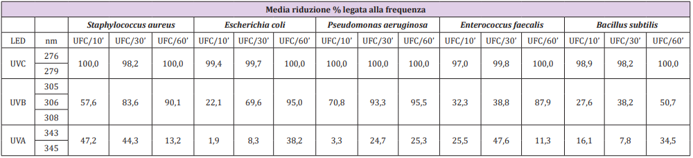
Differences in the effectiveness of LEDs in reducing the microbial load have been observed, from UV-C frequencies to UV-B and eventually to UV-A. The average reduction percentage linked to the frequency of UV-C LEDs was 100% already at 10’ for Sthaphylococcus aureus, Escherichia coli and Pseudomonas aeruginosa, while for Enterococcus faecalis there was a reduction of 97% and 99% for Bacillus subtilis. After 30’ of exposure for Sthaphylococcus aureus and Bacillus subtilis there was a reduction of 98.2%, for Escherichia coli of 99.7%, for Enterococcus faecalis and Pseudomonas aeruginosa of 100%. All the microorganisms tested after 60’ of exposure showed, instead, a reduction of 100%. The average percentage of reduction related to the frequency of UV-B LEDs was lower than the results obtained by testing UV-C rays, the microbial charge decreased as the exposure time increased. In particular, it emerges that Pseudomonas aeruginosa was most affected by their effectiveness, and at 10’ it was already reduced by 70.8% and 95.5% at 60’, while the microorganism that was less sensitive was Escherichia coli, with a reduction of 22.1% at 10’ and 95% at 60’. The average percentage of reduction related to the frequency of UV-A LEDs is the lowest compared to the others mentioned above, but still gave good results compared to the controls, although with certain critical issues, especially for Sthaphylococcus aureus, Enterococcus faecalis and Bacillus subtilis. Table 1 shows the results of the specific species-frequency pairings.

Table 1: Average percentage reduction linked to the frequency of the various LEDs tested.

Discussion

The results of testing LEDs at different wavelengths show that their effectiveness depends on the dissimilar sensitivity of microorganisms to ultraviolet light and on the time of irradiation, which results in a progressive reduction of the microbial load. This is consistent with most microorganisms [11]. Recent study shows that an effect that alters DNA replication is not only dependent on UV-C (200-280 nm), but also on UV-B (280-320 nm). [10] Our results have shown that UV-C, UV-B and UV-A have a reducing effect on the microbial load, albeit with important differences between them. The penetration capacity of UV-A and UV-C is different; the former does not induce direct damage to DNA, capable of forming thymine dimers, [12] but damage cellular proteins, induce the formation of reactive oxygen species such as singlet oxygen and hydrogen peroxide as also claimed by Hargreaves A et al. [13]. Noteworthy is the fact that while the DNA damage induced by UV-C can be repaired by a photolytic enzyme [14], those reported by UV-A cannot be healed in any way, because the enzymes used for the repair are damaged. Our results have shown that UV-A, although to a lesser extent, partially inhibits bacterial replication. In this case, such a reduction could be assumed to be due to this mechanism.

The possibility of associating the effects of both UV-A and UV-C with bacterial replication is currently a field in which several studies are being conducted. Akgün M.P. and collaborators have recently conducted a study where the combination of UV-C and UV-A has allowed a greater effectiveness in reducing the microbial load [10,15]. In our study for Staphylococcus aureus, Enterococcus faecalis and Bacillus subtilis, critical issues were found. The number of colony-forming units (CFU) was higher than the longest exposure times to UV-C and UV-A, probably due to a sampling problem, possible limit of this pilot study. Moreover, almost all the Petri dishes showed a contamination on the circumference borders of them, probably due to their direct contact with the cone rested in the on the medium. This was particularly evident with the experiments conducted with UV-C, in which these contaminations were very sharp due the inability of the radiation to reach the resting surface of cone circumference. However, this limit could be useful to confirm a positive sowing of the Petri Dishes. This observation leads to a probable underestimation of the results, which could have had a greater percentage reduction. This small systematic inconvenience was controllable and allowed us to think about possible improvements in the experimental setting. In fact, assuming to use of 90 mm Petri dishes, instead of 55 mm in diameter, we could have, for each plate treated, a matched control of itself, in the external part of the Petri dishes.

Conclusion

The results obtained in testing the efficacy of LEDs at different wavelengths are consistent with what has been reported in recent literature. In particular, it is stressed that the greatest action with a germicidal effect is performed by UV-C rays (200-280 nm) [10], but similarly other frequencies of the light can be useful to reduce the microbial replication although efficacy decreases at increasing wavelength, progressively, from UV-C, UV-B (280- 320 nm) and finally UV-A (320- 400 nm). Moreover, the hypothesis of being able to combine and control wavelengths emitted by UV-A, UV-B and UVC suggests the possibility of having a greater effectiveness in inhibiting microbial replication by exploiting its different properties. Also, the use of these innovative sources, although still very “young” and have a large margin for improvement, can be found in numerous disinfection application [6,16,17]. LEDs have many advantages over UV lamps, including small size; impact resistance; no need to heat up to operate; low energy consumption; longer life than germicidal lamps; no mercury content; but most importantly, they emit multiple individual wavelengths [10,15]. Such an eventuality opens in fact innumerable hypotheses of study and possible applicative relapses.

Acknowledgement

We want to thank EBV Elektronik (Dr. Pierluigi Rossetti) and Seoul Viosys for providing us samples of LEDs.

For more Articles:  https://biomedres01.blogspot.com/
 

Clavicle Fracture with Injury of Subclavian Artery and Brachial Plexus: Case Report

Clavicle Fracture with Injury of Subclavian Artery and Brachial Plexus: Case Report

Introduction

Clavicle fractures are extremely common, accounting for up to 2.6-12% of all fractures [1-4] and 44-66% of all fractures about the shoulder. Clavicle fractures have several classification schemes [2,5-8] and are generally classified into three groups based on their locations [6]. Fractures of the middle third, or midshaft of the clavicle, classified as group I, are the most commonly encountered, accounting for up to 80% of all clavicle fractures [1,9]. Fractures of the distal third or acromial end of the clavicle, consisting of 15% of clavicle fractures, are classified as group II, and fractures of the medial third or sternal end are classified as group III, making up 5% of injuries. Complications of clavicle fractures are relatively uncommon and include mal-union, non-union, nerve paresthesia, brachial plexus neuropathy, thoracic outlet syndrome, pneumothorax, and vascular injuries [10]. Damage to the neurovascular structure associated with closed fractures of the clavicle is rare [11,12] and usually related to penetrating traumas [13-16]. In this case report, we present a patient with the injury of both the subclavian artery and brachial plexus following a clavicle fracture. Our case report is unique as it presents a combination of two rare complications of clavicle fractures.

Case Report

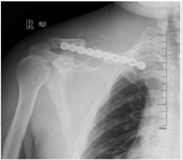
A 45-year-old man presented into the Emergency of our hospital after experiencing right shoulder pain with progressive motor and sensory dysfunction of right upper extremity. 12hours before the presentation, the patient had a fallen accident while riding a bike. Diagnosed right middle third clavicle fracture with a bone fragment downward which was shown on X-ray in the local hospital, the patient progressively underwent a sensomotor loss. Four hours after the accident, he appeared an aggravating numbness and motor dysfunction. Eight hours then, he lost the sensory and motor ability of the right upper extremity completely. Then the patient was transferred to our hospital for further treatment. Admission examination: swelling right shoulder, chest and neck, subcutaneous congestion and bruising, localized tenderness (+), and bone rubbing sign. Activity was limited in the right shoulder, right upper extremity sensation disappeared, right upper limb muscle strength level 0/5, right radial artery pulse unpalpated, right upper limb skin temperature normal. X-ray: right middle third clavicle fracture (Figure 1). Diagnosis: right clavicle fracture with nervous and vascular injury.

Figure 1: pre-operative X-ray (2013-3-18).

Treatment: Clavicular fracture with plate fixation, and subclavian artery, vein and brachial plexus repair. During the operation, we found right middle third clavicle comminuted fracture with four fragments and subclavian tissue swelling significantly, and we palpated the proximal arterial pulse while the distal arterial pulse was untouched. Thus, we deduced that the subclavian artery ruptured by the broken fracture that caused the distal artery stopped (Figure 2). We had proximal clavicular osteotomy to expose the blood vessels area. Once the blocked fracture was removed, the artery jetted out and blood pressure dropped sharply to 75/54mmHg, and the heart rate rose to 112beats/ min. Immediately gauzes were packed to stop bleeding. With the help of the autologous blood transfusion and homotypic blood transfusion, we took the comminuted fracture block out, finding subclavian artery ruptured and brachial plexus injured severely. After subclavian artery anastomosis, patient blood pressure rose to 80/68mmHg, heart rate dropped to 105 beats/min and the right upper limb artery fluctuations can be touched.

Figure 2: Ruptured subclavian artery during operation.

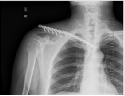
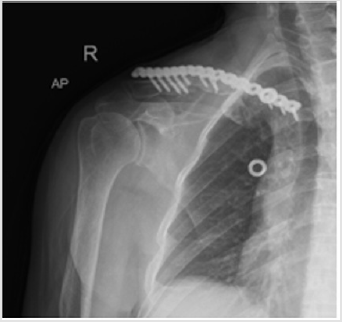
Then we reduced the fracture as a whole and fixed with clavicle locking plate. In total, blood loss was about 4500ml and blood transfusion were 3000 ml autologous blood transfusion and MAP 27U homotypic blood transfusion. Antibiotic, anticoagulation and neurotrophic drugs were used post-operatively. One month later, the patient’s follow-up revealed no sign of neural recovery with motor and sensory complete dysfunction of right upper extremity. Right clavicle X-ray was seen in Figure 3. Right upper extremity vascular ultrasound showed: subclavian artery and vein normal. Electromyography for right up extremity demonstrated : right brachial plexus root damage, C5-7damage, C8-T1 stem damage. Thus, plate fixation and bone graft was performed after admission, and he was discharged from our hospital after symptomatic treatment for 8 days post-operatively. Right clavicle X-ray was performed post-operatively (Figure 4).

Figure 3: Post-operative X-ray.

Figure 4: Post-operative X-ray.

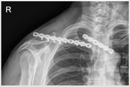
The patient sustained complete loss of sensation and motor power of his right upper extremity for six months after the second operation, and then the sensory and motor function was restored gradually. Follow-up X-ray of our hospital in November 2014 was performed (Figure 5). After that, he exercised his arm every day. He could hold some heavy stuff using his right hand in 2015 and his muscle strength resumed almost completely in 2016. He came to the out-patient clinic in our hospital for follow-up in November 2017 (Figure 6) without any apparent chief complaint. Physical examination: right upper extremity hypoesthesia, right upper limb muscle strength level 5/5, right radial artery pulse palpated, five proximal interphalangeal joints straightening limitation (Figure 7a- 7f). X-ray: right clavicle plate rupture (Figure 6). Electromyography:

Figure 5: X-ray of 18 months follow-up.

Figure 6: X-ray of 51 months follow-up.

a) Right upper extremity mcv: cmap amplitudes of right median nerve, right ulnar nerve, right radial nerve, right axillary nerve and right musculocutaneous nerve all decreased;

b) right upper limb scv: right median nerve and right ulnar nerve were not detected

c) needle electromyography: many positive sharp waves appeared in the right extensor pollicis brevis during resting state;

d) Conclusion: neurogenic deficit of right upper limb (all brachial plexus injury).

Discussion

Clavicle fracture is one common clinical fracture, and the comminuted fragment may pierce subclavian vessels and nerves, which makes small odds for life salvage. Vascular complications of clavicle fracture are rare and are generally recognized as an early complication due to transection of the vein by a displaced fracture of the mid-shaft or lateral end [16-18] or by a late complication [3,19], secondary to the compression caused by abundant callus formation. Brachial plexus injury following clavicle fracture is generally a delayed phenomenon occurring days, weeks and even months after the original lesion [20-23]. In this case, the neurological symptoms were progressing rapidly over a few hours. The fracture injured subclavian artery and the brachial plexus. Because the fragments inserted in the artery wall, vasomotoric symptoms were not that obvious at the time of injury. During the surgery, we found exposing the injured blood vessel and nerves directly was difficult, thus first step we truncated the proximal clavicular, therefore, finding the ruptured vessel and nerves could be easier. Secondly, we had subclavian artery anastomosis and then fracture reduction and fixation.

Some scholars reported that clavicle fracture with subclavian artery rupture needed exploratory thoracotomy [24], while in this article, we used the orthopedic incision to finish it with the help of vascular surgeon, yet with great difficulties and it required good skills and technique. It also avoided the great loss of blood and trauma from open chest operation, which was vitally important for life saving. Reviewing the treating process, we did not do the angiography exam before operation, thus blood loss was evaluated improperly, thanks for the sufficient preoperative blood preparation, and the utilization of autologous blood transfusion which provided important protection for saving.

For more Articles: https://biomedres01.blogspot.com/


Tuesday, August 3, 2021

Effects of Psychological Intervention on Pain and Mental State of Patients Underwent Total Thyroidectomy


Effects of Psychological Intervention on Pain and Mental State of Patients Underwent Total Thyroidectomy

Introduction

In recent years, the incidence of thyroid cancer is on the rise globally year by year [1-2], among which the proportion of thyroid micro papillary cancer is gradual rise, while the treatment scheme is still not unified. Surgical resection is the most important means of treatment of thyroid papillary carcinoma [3]. The Futaba multifocal carcinoma in thyroid subtotal resection and one of the central lymph cleaning is the main operation. The prognosis of thyroid cancer post-surgery is usually good, but the total resection of thyroid will give patients both the psychological and physical pain. At the same time, due to the disease itself and the stimulation such as surgery, the patient is easy to have reactions of psychological stress after surgery such as anxiety. This may reduce the level of pain tolerance, increase patients’ physical pain, affect patients with postoperative life quality and treatment effect [4-5]. In this study, the effect of psychological intervention on pain and psychological state of patients underwent total thyroidectomy was analyzed, and it is reported as follows.

Materials and methods

General Information

From March 2015 to March 2018, 1720 patients were operated on for thyroid cancer in our hospital, and 104 patients requiring total thyroidectomy for double-lobed multifocal carcinoma were randomly divided into a study group and a control group. 52 cases in the study group, male to female ratio: 26/26; age: 31~54 years old, mean (39.47 2.15) years old. 52 cases in the control group, male and female ratio of 26:26; average age (40.18 1.97) was between 32 and 55 years old. There was no significant difference in baseline data of age, gender and primary disease between the two groups (P > 0.05), which was comparable.

Inclusion and Exclusion Criteria

Inclusion criteria: those who were diagnosed with double lobe multifocal carcinoma of thyroid and met relevant clinical diagnostic criteria; All patients have signed the consent form for surgical treatment and nursing; no relevant contraindications [3-4]. Exclusion criteria: diagnosis with mental disorders and unwillingness to cooperate with psychological intervention.

Methods

The control group was given routine nursing, and relevant knowledge and information were given to the patients after admission. Preoperative preparations were carried out, and postoperative signs of the patients were monitored [6-7]. Psychological intervention was given to the study group, the specific way was as follows:

Psychological Intervention: Nursing staff actively communicated with patients. Psychological evaluation was carried out; and patients who were psychological instable received timely counseling; For example, for patients who worry about surgical pain and prognosis effect, the nursing staff will answer their questions with professional knowledge, including the introduction of surgical position, anesthesia method, anatomical characteristics, pain principle, etc., to alleviate the anxiety and tension of patients.

Cognitive Psychological Intervention: During hospitalization, relevant introduction such as the purpose of surgery, prognosis, etc. was given to the patients, so that patients and their families had a basic understanding of total thyroidectomy.

Posture Psychological Intervention: Preoperative guidance for patients with surgical posture practice. Put back cushion behind the patient during operation, keep the stability and comfort of the patient’s head, to reduce the incidence of postoperative pain.

Pain Psychological Intervention: According to the postoperative pain to provide the patients guidance in correct way to get up, and reduction the head and neck activities, to reduce the postoperative pain.

Curative Effect Observation Indexes

Postoperative visual analogue scale (VAS) was used to evaluate postoperative pain in the two groups. >3~6 is moderate pain and tolerable. BBB 0 6~10 is classified as intense pain and unbearable [8]. After observing the situation before and after psychological intervention, the self-rating anxiety scale (SAS) and self-rating depression scale (SDS) were used to score: 50 was the barrier for anxiety or depression. 50~59 was for mild anxiety and depression. >59~69 were classified as moderate anxiety and depression.> 69 is classified as severe anxiety and depression [9]. The changes of SBP, DBP and HR in the two groups were observed, and the results were statistically analyzed [10].

Statistical Methods

SPSS 18.0 statistical software was used for data analysis. The measurement of data was shown as mean±standard deviation (x±s). And t test was used for comparison between the two groups. The rate of counting data indicated that the difference between the two groups was statistically significant by 2 test, P < 0.05 (Tables 1-3).

Table 1: Comparison of postoperative VAS pain scores between the two groups (see table 1,x ±s).

Note: 1h( P > 0.05) ,24 48 h( P < 0.05).

Table 2: Comparison of SAS and SDS scores between the two groups after psychological intervention (see Table 2, x± s).

Note: Compared with the control group after psychological intervention, *P < 0.05; SAS: anxiety self-rating scale; SDS: depression self-rating scale.

Table 3: Comparison of postoperative blood pressure and heart rate between the two groups after psychological intervention (see Table 3,x s).

Note: 1 mm Hg=0.133 kPa

The Results

After psychological intervention, SBP, DBP and HR levels of patients in the research group were significantly better than those in the control group, with statistically significant differences (P < 0.05).

Discussion

Total resection of thyroid is the main surgical method of multifocal double leaf thyroid cancer. Because the whole thyroid resection patients could go through endocrine changes and stress reaction, this can lead to anxiety before and after surgery, postoperative secretion of adrenal cause heart rate, blood pressure, mental and physical discomfort, etc. All those can increase postoperative pain, influence the patient’s quality of life and the later treatment [11-12]. In this study, a comprehensive and multimode psychological intervention was carried out on patients to reduce the factors that may aggravate the pain awareness of patients, reduce the pain perception of patients, and achieve a certain effect. Observe VAS pain score of the two groups of patients after surgery as a result, both groups of patients showed no significant differences in postoperative 1 h pain score. But the VAS score of 24 h and 48 h after surgery were (2.31-0.12) and (1.76- 0.09) respectively, namely mild pain, and were significantly lower than the control group, suggests that psychological intervention can effectively reduce the thyroid surgery patients with pain. This result is related to psychological intervention before and after surgery. First, during hospitalization, the patient is informed about matters needing attention and knowledge of pain by medical staff. Thus, the patient can correctly avoid the internal and external factors that may cause postoperative pain under the guidance of relevant knowledge.

Secondly, during surgery nursing staff put cushions behind the patient’s back to maintain the stability and comfort of the head, to reduce postoperative pain. Finally, after the operation, the medical staff guided the patient to get up, which effectively avoided the pain caused by head movements. The changes of patients’ psychological states before and after the intervention were observed. There was no significant difference between the two groups before the intervention, and the SAS and SDS scores in the post-intervention study group were significantly better than those in the control group. When patients have anxiety or depression, the cholecystokinin will be activated, which can promote pain conduction and aggravate pain sensation. In addition, patients with physical pain usually show severe depression or generalized anxiety, that is, depression and anxiety are positively correlated with postoperative pain [13,14]. Therefore, psychological intervention can alleviate the patients’ tension and anxiety with preoperative cognitive psychological intervention on the patients’ diseases and surgeries, and improve the patients’ mental state through psychological counseling, so that the patients can maintain a positive and optimistic mental state, thus reducing postoperative pain.

The blood pressure and heart rate of the two groups of patients after psychological intervention were observed. The blood pressure and heart rate of the study group were significantly lower than those of the control group, indicating that the adrenal secretion of the patients after psychological intervention gradually returned to normal. Blood pressure and heart rate began to decline, and the influence of surgical stress stimulation decreased. The above results proved that psychological intervention can improve the psychological state of patients in many ways, reduce postoperative pain, effectively reduce the stress stimulation of patients after total thyroidectomy, and improve the postoperative quality of life and effects of later treatments. In addition, medical staff can also use drugs to assist pain relief according to the specific situation, to further reduce the pain of patients. However, considering the possible adverse reactions caused by drugs, the principle of moderation should be followed [15,16]. In conclusion, psychological intervention for patients with total thyroidectomy can effectively reduce postoperative pain and improve psychological state.

For more Articles: https://biomedres01.blogspot.com/
 

Antimalarial Aloe Compounds

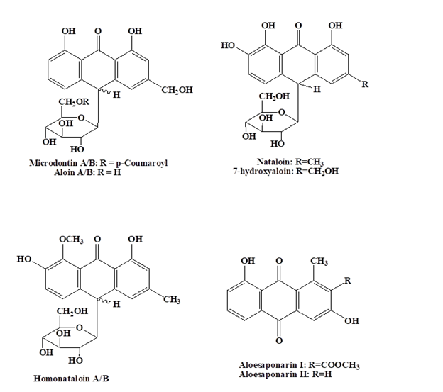
  Antimalarial Aloe Compounds Introduction Among the most prevalent diseases caused by protozoan parasites, malaria is caused by parasites o...