Wednesday, May 4, 2022

Current Research Progress on Pharmacological Activities and Clinical Application of Guhan Yangshengjing


Current Research Progress on Pharmacological Activities and Clinical Application of Guhan Yangshengjing

Introduction

Guhan Yangshengjing (GHYSJ) is based on the essence of the bamboo slips “Healthcare Prescriptions” and “Endo-meridians” from the ancient tombs of the Western Han Dynasty in Mawangdui, Li Congfu, Liu Bingfan, and Ouyang Qi of the Hunan Academy of Traditional Chinese Medicine, condensed a compound recipe with their decades of clinical experience. GHYSJ,, composed of a variety of Chinese herbal medicines, has the efficacy of invigorating the kidney for strengthening the spleen, tonifying qi for benefitting essence, which is used for dizziness, palpitations, dizziness, tinnitus, forgetfulness, insomnia, impotence and spermatorrhea and fatigue and weakness caused by deficiency of qi and yin and insufficient kidney essence [1]. GHYSJ has been put into production since 1986,of which two dosage forms, oral liquid and tablets are developed with multiple specifications and models, which can meet the purchasing needs of different groups. In the process of longterm clinical application, it has been found that GHYSJ also has a good effect on many other diseases, such as atrophic gastritis, coronary heart disease, alopecia areata, chloasma, hypotension, Meniere’s syndrome, senile habitual constipation and menopausal syndrome [2].
There are still some problems in the research of GHYSJ. Firstly, the pharmacological and clinical research of GHYSJ are mostly performed in the 90s, which is hard to keep pace with the times. With the advance of research methods, its depth and breadth are difficult to fully satisfy the current application requirements of clinical drugs. Secondly, the small sample size of the clinical research of GHYSJ is hard to guarantee the clinical efficacy, so it cannot provide reliable scientific foundation for its pharmacological activity research. Finally, the effective material basis and mechanism of GHYSJ have not been clarified. In the process of clinical application, it has been found that GHYSJ has certain effects on many diseases, but the active substances are not well studied and the response mechanism between the active substance and the target proteins has not been clarified. Hence, it is necessary to carry out the secondary development of GHYSJ and the research of new direction, broaden the scope of clinical application, and further study its pharmacodynamic substances and mechanism of action. This paper summarizes the pharmacological and clinical study of GHYSJ with intent to provide research ideas and scientific basis for its further development. So that GHYSJ can better adapt to the requirements of the booming health industry and prepare for health protection for the upcoming aging society.

Pharmacological Activity

Antioxidant Activity

Numerous studies have shown that anti-oxidation plays an important role in preventing aging, because free radicals or oxidants can break down cells and tissues, affect metabolic functions, and cause different health problems [3-5]. GHYSJ can eliminate excessive oxidative free radicals and has anti-oxidation and anti-aging effects. Wei, et al. [6] found that GHYSJ can repair and protect red blood cells damaged by aging, exhibiting an antiaging effect by improving the vitality of red blood cells. Zhang, et al. [7] found that GHYSJ could inhibit the content of malondialdehyde (MDA) in the heart, liver and brain mitochondria, microsomes and erythrocyte membranes of middle-aged and elderly Wistar rats and can resist lipid peroxidation. Yu [8] proved that GHYSJ could increase the content of catecholamine (CA) in the brain by increasing the activity of superoxide dismutase (SOD) in the blood of aged rats and regulate monoamine transmitters.
GUYSJ has also been found to improve the function of hypothalamus-pituitary-gonadal axis and promote the synthesis of testosterone in old male rats. Zhang, et al. [9] demonstrated that GHYSJ can improve the metabolism of free radicals in female rats, effectively increase the content of SOD in the blood, enhance the body’s ability to scavenge oxygen free radicals, and play an antiaging effect. The proper amount of oxygen free radicals is beneficial to the body’s various life activities, but the accumulation of excessive free radicals would cause damages to the body [10]. In conclusion, GHYSJ can effectively remove excessive oxygen free radicals in the body and has a good antioxidant effect.

Regulate Immune Function

As one of the body’s important defense mechanisms, the immune system plays a vital role in the treatment of infections, inflammatory diseases and cancer [11]. And GHYSJ just has a certain immune regulation effect. Zhang, et al. [12] determines the content of T cells and antibodies in the blood of rats fed by GHYSJ and researched the changes in peripheral blood red blood cell immune status in 32 cases of tumor patients, which explains that GHYSJ has the effect of enhancing humoral immunity now that it alleviates the immunosuppressive state of tumor patients and regulates the overall immune effect. As a component of natural immunity, red blood cells lay an important foundation for the body’s own immune balance and stability. Wen [13] finds that GHYSJ not only improves the immune adhesion function of red blood cells inhibited by exercise stress, but also enhances the immune adhesion activity of red blood cells (RBC) and regulates the body’s immunity. GHYSJ has a good regulatory effect on the immune function of the body and mainly acts on the body’s first line of immune defense with the increasing number and enhancing function of immune cells, and immune active substances.

Anti-Fatigue Effect

Fatigue, or sports fatigue, refers to the inability of the body’s physiological processes to continue its function at a certain level and/or the inability to maintain a predetermined exercise intensity [14]. If fatigue is not eliminated in time, it can cause the body’s endocrine dysfunction and decrease immunity, which will cause a series of physiological problems and seriously affect human health [15]. Wen, et al. [16] finds that GHYSJ can enhance the activity of SOD, reduce the content of MDA in the blood, reduce the damage of RBC membrane structure, and achieve anti-fatigue effect. Li, et al. [17] observes the influence of GHYSJ on the physical functions of competitive athletes, and the results show that: GHYSJ can not only promote protein synthesis, reduce blood urea nitrogen content, improve heart function, but also promote hematopoietic function, increase the hemoglobin concentration, improve the body’s circulation and aerobic energy supply, and have the functions of anti-fatigue and delaying the appearance of fatigue.
Wen, et al. [18] and Lin, et al. [19] suggest that good results obtained for GHYSJ increase the content of liver glycogen, muscle glycogen and blood sugar in mice, increase glycogen reserves, and enhance exercise ability. Also, GHYSJ has turned out to reduce exercise fatigue and lipid peroxidation mediated by free radical and have good anti-fatigue effects. Chen, et al. [20] finds that the anti-fatigue mechanism of GHYSJ may be related to the reduction of lactic acid production in the blood, the increase of SOD, activity and enhancement of the body’s scavenging of oxygen free radicals. GHYSJ has an anti-fatigue effect by enhancing the ability of red blood cells, improving blood oxygen supply, increasing glycogen reserves, promoting the synthesis of related proteins, and scavenging oxygen free radicals. GHYSJ is such an excellent anti-fatigue drug that it can significantly alleviate the body’s fatigue after exercise.

Hypolipidemic and Anti-Atherosclerosis

Atherosclerosis is a chronic vascular inflammatory disease that mainly affects cerebral arteries, coronary arteries and peripheral blood vessels [21]. Abnormal lipid metabolism is the pathological basis of atherosclerosis and controlling blood lipid concentration is vital to the treatment of atherosclerosis [22]. Studies have shown that GHYSJ can significantly reduce the levels of total cholesterol (TC) and triglyceride (TG) in the blood and has a good effect on lowering blood lipids and anti-atherosclerosis. Qin, et al. [23] establishes a hyperlipidemia rat model by feeding a high-fat diet to study the lipid-lowering effect of GHYSJ and finds that GHYSJ can reduce the platelet adhesion rate, the maximum platelet aggregation rate, and the rat serum TC and TG, has anti-platelet adhesion and aggregation effect and lipid-lowering effect. Dai, et al. [24] and Liu, et al. [25] show that the GHYSJ has a super effect on lowering blood lipids and anti-atherosclerosis, which can significantly reduce the content of TC and low density lipoprotein (LDL) in the serum. It can also block plasma lipid peroxidation and LDL oxidative modification and denaturation in the arterial wall, inhibit lipid deposition on the arterial wall, increase prostaglandin I2 (Prostaglandin, PGI2) levels, and adjust thromboxane A2 (Thromboxane, TXA2) and PGI2 balance.

Other Pharmacological Effects

GHYSJ also has some other effects besides the above-mentioned pharmacological activities. Xiao’s research [26] shows that GHYSJ can improve the inflammatory response in rats with kidneyyang deficiency and asthma. Its mechanism may be related to the inhibition of interleukin 6 (Interleukin, IL-6), tumor necrosis factor-α (TNF-α) and high mobility group protein B1 (HMGB1) expression. Zhang [27] found that GHYSJ can regulate the gene expression of P62 protein and transforming growth factor-β (TGF-β1) and inhibit airway remodeling in asthmatic rats with kidney-yang deficiency. In addition, Qin, et al. [28,29] found that GHYSJ can significantly reduce the content of aspartate aminotransferase, creatine kinase, and lactate dehydrogenase in serum through acute cerebral ischemia models and increase the content of SOD in serum and brain homogenate, at the same time increase cerebral blood flow, reduce cerebrovascular resistance, and protect brain tissue.
GHYSJ shows a good awareness of the effects of improving postoperative cognitive dysfunction (POCD), Hou [30] finds that GHYSJ can significantly reduce the levels of pro-inflammatory cytokines TNF-a and IL-6 in the hippocampus of aged rats after surgery and relieve the inflammatory response in the hippocampus of aged rats after surgery. Wang’s research [31] shows that GHYSJ can improve postoperative cognitive dysfunction in aged rats by inhibiting the activation of astrocytes and microglia in the CA1 region of the hippocampus. Deng [32] bring to light the mechanism of GHYSJ improving postoperative cognitive dysfunction in rats may be related to the up-regulation of hippocampal synaptic protein SYN-1 and PSD-95 expression.

Clinical Applications

Treatment of Insomnia, Senile Brain Atrophy and Vascular Dementia

GHYSJ has the effects of replenishing qi, nourishing the kidney, and replenishing essence, which can treat neurasthenia, insomnia, forgetfulness, fatigue and weakness caused by deficiency of spleen and kidney, insufficient qi and blood, and loss of mental energy. Liu [33] compared the therapeutic effects of GHYSJ and Guilu Erxian Ointment on senile brain atrophy and detected that the total effective rate of the GHYSJ Group was 96.4%, which was 68.4% higher than that of Guilu Erxian Ointment. Yin, et al. [34] observed the therapeutic effect of GHYSJ on patients with neurasthenia and found that GHYSJ has a certain effect on neurasthenia, because the healing rate of GHYSJ (62.50%) is better than that of the control group Zishen Jiannaoye (47.22%). Yu, et al. [35] outcropped that the GHYSJ combined with oryzanol has a good effect on insomnia, with a recovery rate of 72.55% and a total effective rate of 92.16%, which is equivalent to the suldia. Yu [36] treated vascular dementia with GHYSJ supplemented by cerebral vasodilators and brain cell metabolism activators. The total effective rate was 76.1%, which was better than the 60.8% of the control group.

Treatment of Male and Female Infertility, Sexual Dysfunction, and Prostate Inflammation

GHYSJ can nourish the kidney and nourish the essence, invigorate qi and nourish blood, and have a good therapeutic effect on reproductive system diseases. Xia, et al. [37] used zinc gluconate in combination with GHYSJ to treat male infertility and found that it can significantly improve sperm activity and sperm number and has a significant therapeutic effect on idiopathic oligoasthenospermia. Zhou, et al. [38] observes the clinical efficacy of the GHYSJ for sexual dysfunction, and the results show that it can improve the sexual dysfunction, it’s apparent efficiency and total effective rate are significantly higher than those of the Jinkui Shenqi Pill. Zhang, et al. [39] found that GHYSJ have a good effect on prostate hyperplasia caused by spleen and kidney deficiency, and its total effective rate is as high as 95%. The study conducted by Wu, et al. [40] showed that the total effective rate of the GHYSJ in treating prostatitis caused by insufficient kidney yang or spleen deficiency and dampness deficiency was 92%. Niu [41] find that the total effective rate of treating women’s leucorrhea with GHYSJ is 92%, which is better than that of leucorrhea pills.

Treatment of Leukopenia

White blood cells are an important part of the body’s defense system and play an irreplaceable role in resisting the invasion of the body by pathogens. GHYSJ can resist the reduction of white blood cells due to some drugs or treatments and restore the normal functions of the human body. Lin, et al. [42] used GHYSJ to treat the leukopenia caused by clozapine because it can increase the body’s immunity and enzyme activity, enhance the cell oxygen carrying capacity and the detoxification capacity, and improve the blood circulation, the oxygen carrying capacity of cells, and the vitality of SOD in the human body, and reduce the production of free radicals in the human body. Xu, et al. [43] compared the effects of GHYSJ with oral inosine, salanol, and aminopeptide on white blood cells after radiotherapy and chemotherapy in patients with intermediate and advanced tumors, pointed that GHYSJ can not only stimulate bone marrow stem cells to increase white blood cells, but also can alleviate the toxic and side effects during the chemotherapy and radiotherapy of tumor patients, and reduce the decline of white blood cells. Chen [44] revealed that the GHYSJ has a protective effect on white blood cell (WBC), RBC and hemoglobin (Hb), can reduce the bone marrow suppression caused by chemotherapy, and can enhance the patient’s resistance to chemotherapy. Zhang [45] think that GHYSJ is an ideal treatment for leukopenia after tumor chemotherapy drug, because it can significantly enhance the ability of WBC, promote its recovery, and play an important role in increasing Hb, platelet (Platelet, PLT) and enhancing immune function.

Treatment of Other Diseases

Li, et al. [46] show that GHYSJ can significantly improve Qi deficiency and fatigue symptoms of perimenopausal women with Qi deficiency. It also helps to maintain anti-Müllerian hormone (AMH) and bone mineral density in women with qi deficiency in perimenopausal period. Zhao, et al. [47,48] and Zhang, et al. [49] pointed that: GHYSJ has a better conditioning effect on subhealthy people with yang deficiency and qi deficiency, which can improve their heart function, bone density, AMH, total sperm motility, and other indicators to improve exercise endurance and prevent the sub-health state from turning into disease. Sun, et al. [50] found that the total effective rate of GHYSJ on osteoporosis is 91.2%, which is better than Erxian Decoction. Chen [51] showed that GHYSJ can reduce the serum cholesterol level of patients with hypercholesterolemia by 17.1% without adverse reactions.

Conclusion and Prospect

GHYSJ has broad application prospects in the medical field, and its secondary development is of great significance, because the antioxidation, anti-aging, immune function adjustment, anti-cerebral ischemia, anti-fatigue, hypolipidemic and anti-atherosclerosis effects of GHYSJ covers multiple human systems such as nervous system, blood system, immune system and reproductive system.. It can treat insomnia, senile brain atrophy, male and female infertility, sexual dysfunction, prostate inflammation, and hypercholesterolemia. In addition, it can assist in the treatment of osteoporosis, tumor patients with leukopenia, coronary heart disease, angina pectoris, and syringomyelia, also can regulate the body functions of subhealthy people with yang deficiency and qi deficiency. Sub-health is a kind of intermediate state between health and disease, which is considered a precursor to several cardiovascular diseases such as hypertension, inflammation, atherosclerosis, diabetes, myocardial infarction, and other cardiovascular diseases [52]. Studies have shown [53] that human beings in a sub-health state for a long time may develop into a sub-clinical state, appear pathological changes, and then develop into diseases.
This process is irreversible, and certain measures can be taken to restore the body to health in a sub-health state. Using the reversibility of the sub-health state to implement intervention is the key to disease prevention. This is consistent with the “zhi wei bing” (preventative treatment) of traditional Chinese medicine, and it reflects the advantages of traditional Chinese medicine in disease prevention and treatment. An increasing number of people are in sub-health status because of the high pressure and fast-paced social environment. The reason why the GHYSJ plays an important role in sub-healthy people is that it can fill the marrow, regulate the function of various organs, enhance the body’s resistance, and prevent diseases. The secondary development of GHYSJ can contribute to alleviating the pressure on the medical system. With the decline of fertility and the improvement of medical standards the aging population has become a serious social problem around the world, especially in China, the most populated nation, which puts a significant health, economic, and social burden on society.
Improving the fertility level of the young population, improving the physical condition of the elderly population, and alleviating the phenomenon of population aging are problems that need to be solved urgently. GHYSJ replenishes the innate essence, nourishes the acquired foundation, and has a good effect on male and female infertility and various reproductive system diseases, such as Epimedium and Dodder [54,55] can improve the vitality of male and female reproductive cells, reduce the natural aging of reproductive cells, repair damaged cells, and protect reproductive organs. In addition, GHYSJ has a good effect on some senile diseases such as senile brain atrophy, vascular dementia, etc. It also has the effects of regulating immunity and improving physical functions and has certain adjustments to various physical indicators of the elderly. To sum up, GHYSJ can make a certain contribution to these two social problems, because it can regulate the body functions of sub-healthy people and prevent the occurrence of diseases, which is in line with the “preventive treatment” of Chinese medicine, and it is also very suitable for solving the problem of population aging. So the follow-up development of the GHYSJ should be combined with current social problems to serve the health of the country and even the whole mankind.
Starting from the two levels of the whole prescription and the single medicine, multi-directional and multi-level representation of the chemical substances in the GHYSJ and illustrate the material basis of GHYSJ promoting gastrointestinal motility and explore the mechanism of action from an organic and inorganic combination point of view. Combining the seven emotions, four sexes, guijing, and monarch and adjudicator of traditional Chinese medicines, study the compatibility of the individual medicines in GHYSJ, explore the role targets of GHYSJ in the human immune system and reproductive system, and lay the foundation for follow-up research. At the same time, metabolomics and proteomics can be used to study the in vivo absorption, distribution and metabolic process of GHYSJ, and to clarify its dose-effect relationship. To better guide the application of the GHYSJ in the concept of “preventing disease” and the problem of population aging, and to serve the national health.

For more Articles on :https://biomedres01.blogspot.com/


Sustainability of Biologic Therapy is Less in Ulcerative Colitis than Crohn’s Disease Patients, Independent of Prior Biologic Experience

Sustainability of Biologic Therapy is Less in Ulcerative Colitis than Crohn’s Disease Patients, Independent of Prior Biologic Experience

Introduction

Inflammatory bowel disease (IBD) is a chronic inflammatory condition that predominantly affects the bowel and includes both Ulcerative Colitis (UC) and Crohn’s Disease (CD). The burden of IBD is rising globally with a marked increase in the prevalence of IBD worldwide over the last 30 years [1]. The therapeutic goal in the treatment of IBD has evolved from focusing on symptomatic relief to aiming to achieve intestinal mucosal healing. Emerging longitudinal data suggests that individuals who obtain mucosal healing, which is observed more frequently with biologic than conventional therapies, have a superior long‐term prognosis with lower risk of surgery and hospitalization [2]. While there has been an expansion in the number of medications available for treating IBD including anti-tumour necrosis factor (TNF) agents, selective anti-integrins, anti-interleukins (IL) and most recently small molecule inhibitors such as the janus kinase (JAK) inhibitors, there still remains a large unmet clinical need for further therapies. Studies have shown between one third to half of patients commenced on anti-TNFs do not maintain response and require a switch to a second biologic [3]. Reasons for discontinuation vary but are often secondary to nonsustained response to a particular agent (primary non-response and secondary loss of response) or development of intolerance to a particular biologic. Chen et al looked at over 8000 patients with IBD on biologics and found after 1 year less than 50% of patients were still on this biologic agent [4].

With the evolution of medical management of IBD and new treatment goals there is a need to identify factors that predict response to biologics. To date age at diagnosis, disease duration, disease severity, and elevated inflammatory markers are all factors that have been identified to influence response to biologics [5]. However, few studies have specifically looked at real world sustainability of response to biologics, factors which might determine this and whether there is a difference between UC and CD. Given the distinct biological differences between UC and CD we aimed to look at the difference in sustainability between biologics in biologic-naïve and biologic-experienced patients depending on whether they had a diagnosis of UC or CD.

Material and Methods

Participants

A prospectively maintained electronic database of 4200 patients with IBD attending four hospitals in a single healthcare network was interrogated from January 2008 to March 2018. All patients with an established diagnosis of CD or UC currently and previously on biologic therapy were included in this analysis.

Study Design

Basic demographic information was collected including age, gender, disease subtype, age at diagnosis, type of biologic therapy used and concurrent immunomodulator therapy. Date patients commenced on a particular biologic was collected along with the date of discontinuation of biologic therapy (or last follow-up date if patients were still on this agent) due to inadequate treatment response or adverse effects. Where treatment was discontinued for other reasons (patient choice, sustained remission, or nonadherence) these treatment episodes were excluded. Whether a patient was biologic-naïve or had prior treatment with a different biologic agent (biologic-experienced) was also recorded for analysis. Ethical approval for this study was granted by the Ethics Committee at St Vincent’s University Hospital.

Outcomes

The primary outcome of interest was time to discontinuation of biologics in biologic-naïve and biologic-experienced patients’ dependent on whether patients were diagnosed of UC (UC and IBD-U) or CD. Secondary outcomes of interest included the identification of disease characteristics that impact discontinuation of biologics including immunomodulator co-therapy, age at diagnosis, disease duration, gender and disease location for both UC and CD.

Statistical Methods

Descriptive statistics were provided to summarize demographic characteristics using mean [95% confidence interval (CI)] or median [interquartile range (IQR)] for continuous variables, and number and percentage for categorical variables. Cumulative survival rates were estimated using the Kaplan-Meier method and mean and median survival time used. The log-rank test was used to compare the survival curves across the subgroups. A multivariate (adjusted analysis) Cox proportional hazards model was utilized to identify risk factors relating to survival. A two-sided P-value ≤ 0.05 was considered statistically significant. All data analyses were performed using SPSS 25.0.

Results

Baseline Characteristics

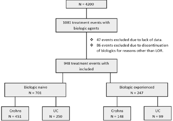
A total of 1081 treatment events with biologic therapy occurred at our centre at the time of this study. 47 events were excluded due to lack of data and 86 were excluded due to discontinuation of a biologic agent for reasons other than loss of response. In total 948 independent treatment events with biologic therapy were included in this study in 712 patients. In total 516 (54.4 %) patients were male. Median age at diagnosis was 33.8 years (IQR 24.6 – 44.8). 349 (36.8 %) patients had a diagnosis of UC. 353 (37.2 %) patients in total discontinued their biologic agents. 312 (32.9 %) patients were on co-therapy with immunomodulators. Mean follow-up was 2.3 years (IQR 0.64– 3.2) (Table 1). Patients were sub-divided depending on whether they were biologicnaïve (Group1) or biologic experienced (Group 2) (Figure 1).

Table 1: Baseline characteristic.

Figure 1: Flow diagram of patient selection.

Sustainability of Biologics Between UC and CD

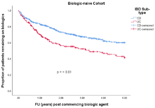
• Group 1: 701 (74 %) patients were biologic-naïve. 387 (55.2 %) were male. Median age at diagnosis was 33.3 years (IQR 23.7 – 44.5). 250 (35.7 %) had a diagnosis of UC and 451 (64.3 %) had CD. Median disease duration was 7.5 years (IQR 3.1 – 13.9). 256 (36.5 %) patients discontinued their biologics. 215 (30.7 %) patients were on co-therapy with immunomodulators (Table 1). 329 (46.9 %) patients received infliximab (IFX), 337 (48.1 %) adalimumab (ADA), 32 (4.6 %) golimumab (GLM) and 3 (0.4 %) received ustekinumab (UST). Mean time to discontinuation of biologics in the UC cohort was 2.9 years (95% CI 2.6 – 3.22 years) which was significantly shorter than in the CD cohort which was 3.6 years (95% CI 3.4 – 3.8 years) (p-value = < 0.001) (Table 2 & Figure 2).

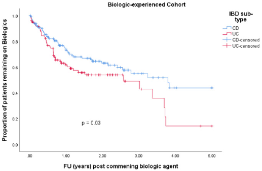
• Group 2: 247 (26.1 %) patients were biologic experienced. 129 (52.2 %) were male. Median age at diagnosis was 34.9 years (IQR 27.8 – 45.0). 99 (40.1 %) had a diagnosis of UC and 148 (59.9 %) had CD. Median disease duration was 10.5 years (IQR 5.2 – 15.9). 97 (39.3 %) patients discontinued their biologics. 97 (39.3 %) patients were on co-therapy with immunomodulators (Table 1). 83 (33.6 %) patients received IFX, 57 (23.1 %) ADA, 40 (16.2 %) vedolizumab, 26 (10.5 %) GLM and 41 (16.6%) received UST. Mean time to discontinuation of biologics in the CD cohort was 3.1 years (95% CI 2.7 – 3.5 years) compared with 2.3 years (95% CI 1.9 – 2.8 years) in the UC cohort (p-value = 0.030) (Table 3 & Figure 3).

Table 2: Survival analysis biologic-naïve cohort.

Table 3: Survival analysis biologic-experienced cohort.

Figure 2: Time to discontinuation of biologic agents in our biologic-naïve cohort.

Figure 3: Time to discontinuation of biologic agents in our biologic-experienced cohort.

Factors Affecting Discontinuation of Biologics

We looked at gender, age at diagnosis, co-therapy with immunomodulators, IBD subtype and disease duration as potential factors impacting biologic discontinuation.

• Group 1: Gender had no impact on discontinuation of biologic agents with a hazard ratio (HR) 0.8 (p-value 0.07, 95% CI 0.6-1.0). Older age at diagnosis was associated with a slight increased risk of discontinuing a biologic agent with a HR 1.009 (p-value 0.002, 95% CI 1.00-1.02). Shorter disease duration was protective against discontinuation of biologics with a HR of 0.97 (p-value <0.001, 95% CI 0.95 – 0.98). Co-therapy with immunomodulators increased the risk of discontinuing biologic therapy in a biologic-naïve cohort with a HR 1.6 (p-value 0.001, 95% CI 1.2 – 2.1). UC as seen in our survival analysis increased your risk of discontinuing biologic therapy in our biologic naïve cohort with a HR 1.5 (p-value 0.003, 95% CI 1.2 – 1.9) (Table 4).

• Group 2: Neither gender nor age at diagnosis showed any independent association with time to discontinuation of biologics in our biologic-experienced cohort. Shorter disease duration was again protective against discontinuation of biologics with a HR of 0.95 (p-value 0.002, 95% CI 0.92-0.98). Co-therapy with immunomodulator therapy had no significant impact on discontinuation of biologics in our biologic-experienced cohort with a HR 0.9 (p-value 0.8, 95% CI 0.9 – 1.5) and neither did a diagnosis of UC (p-value 0.2, 95% CI 0.9 – 2.1) (Table 5).

Table 4: Factors affecting discontinuation of biologics in biologic naïve cohort.

Table 5: Factors affecting discontinuation of biologics in biologic experienced cohort.

Discussion

Over the past two decades there has been significant advances in the medical management of IBD especially since the introduction of biologic agents. Unfortunately, despite current advances loss of response to biologics still affects up to 30-40% of patients in clinical practise [5]. Identifying factors that predict the efficacy of biologics is important to efficiently use these agents along with minimising side-effects and improving cost-effectiveness. To date certain disease related factors which impact response to anti-TNF agents include gender, age at diagnosis or the presence of fistulating disease [5,6] but overall data is limited. Interestingly, despite the anatomical, genetic, and immunological differences between CD and UC there is no evidence to date indicating biologics work more effectively in either sub-type of IBD. Our study is the first to investigate the impact of IBD subtype on sustainability of biologics and our findings show that in both biologic-naïve and biologic-experienced patients’ biologic therapy is more durable in CD than UC. In our biologic-naïve cohort patients with CD were significantly less likely to discontinue a biologic agent compared with UC and similar results were seen in patients who were biologicexperienced. More research is required to identify unique reasons why biologic therapy appears more sustainable in CD but one can hypothesis it is most likely multi-factorial. Certain disease related differences between CD and UC including genetic, immunogenic and differences in the gut microbiota can impact response to treatment.

Other modifiable factors such as differences in the inflammatory burden between CD and UC and how each physician manages and adjusts their patient’s treatment can certainly influence response to treatments. In CD, it is well known that surgery is not curative, and it is standard practice to favor bowel-sparing surgical approaches, and resection of the affected part of bowel only is preferable [7]. Interestingly after the introduction of anti-TNF therapy the risk for IBD related hospitalization have only decreased in CD but not in UC [8]. Unlike CD with UC surgery is often considered curative and this can impact both physician and patient decision making and treatment goals. From recent studies there is also a clear difference in the prescribing practise of biologics between UC and CD. One cross-sectional study analysing United States (US) health care utilization in 964,633 IBD patients between 2010 and 2012 showed that UC patients were much less frequently treated with biologics than CD patients (3.5% vs 16.8%) [9]. A second study in the US of over 400,000 IBD patients showed that the proportion of patients using biologics increased for both UC and CD between 2007 to 2015, but to a much higher extent in patients with CD (from 21.8% to 43.8%) than in patients with UC (from 5.1% to 16.2%) [10]. Numerous disease related factors impact response to biologic therapy other than the sub-type of IBD. In our study shorter disease durations and older age at diagnosis had a negative impact on sustainability of biologics.

Regarding disease duration studies are conflicting. Several studies have shown in CD shorter disease duration improves response to biologics [11-13] however the opposite has been in seen in two recent studies, one looking at anti-TNF therapy and one looking at vedolizumab response [14,15]. In our study, like Nguyen et al’s study patients with a shorter disease duration were more likely to discontinue biologic agents sooner. In our study older age at diagnosis was associated with a slightly increased risk of discontinuing biologic agents in our biologic-naïve cohort. Desai et al showed similar results in a small study looking where an age over 60 years correlated with an increased risk of discontinuation of biologics [16]. Co-therapy with immunomodulators is advised by ECCO guidelines for management of patients with moderate to severe IBD treated with IFX to help achieve and maintain remission however not for adalimumab [17]. Our study found in our biologic-naïve cohort co-therapy with immunomodulators had a negative impact on sustainability of biologics with a HR 1.6 which is expected given 48% of our biologic-naïve cohort were treated with adalimumab and 47% with IFX. In our study 37% of patients discontinued a particular biologic agent over the 5-year follow-up. The incidence of primary nonresponse to anti-TNFs varies from 10 to 30% in clinical trials and clinical practise [18-20].

The annual risk of secondary loss of response in one particular review for IFX was 13% per patient year [21] and 20.3% for adalimumab in two separate study [22]. This data highlights the need for a personalised approach to treatment for each patient with IBD. Promising targets for new therapeutic strategies are currently being developed and our study highlights the need to broaden treatment options available for IBD to ensure patients both achieve and sustain mucosal healing given the chronic nature of IBD. Limitations of our study include the retrospective and observational nature without randomization or blinding. Secondly data was not collected on disease severity at onset of biologics which can impact response to treatment. Another limitation is the variability in prescribing biologics and escalation of treatment between different gastroenterologists at our centre.

Conclusion

In conclusion our real word data indicates sustainability of biologic therapies is less in UC than CD and this is not strongly determined by prior biologic exposure. The reason biologics are more sustainable in CD is most likely multifactorial however this finding is important in determining how biologic agents are employed in both IBD subtypes. Our data also highlights the need for new biologics and small molecules inhibitors as there is currently an unmet medical need for treatments in UC and CD given the high rates of primary and secondary loss of response.

For more Articles on : https://biomedres01.blogspot.com/
Monday, May 2, 2022

Assessment of Body Composition of Adult Females among Weight Managing and not Managing Groups


Assessment of Body Composition of Adult Females among Weight Managing and not Managing Groups


Introduction

Human body can be understood in the form of a pyramid with 5 distinct levels. These are Chemicals, Cells, Tissues, Organs, Organ Systems [1,2]. The earliest assessment of body composition dates back to Cadaver Analysis, conducted by Clarys, Martin and Drinkwater between 1979 and 1980 [3]. Principal components such as fat, Total body water, bone minerals, non osseous minerals and proteins have also been obtained from Cadaver Analysis [4]. The different methods employed to assess body composition are Cadaver Analysis, Skin fold Measurements, Body Mass Index, Total Body Water, Total Body Counting and Neutron Activation, Body Density, Dual Energy X Ray Absorptiometry, Bioelectrical Impedance Analysis Bioimpedance or biological impedance is defined as the ability of biological tissue to impede electric current [5]. Body impedance (Z) is defined as the opposition of a conductor to the flow of an alternating current, and consists of two components: Resistance (R) and Reactance (Xc) (Figure 1). Resistance (R) is the major opposition of the conductor to the flow of electric current [6]. Reactance is an additional opposition or the storage of an electrical charge by a condenser for a short period of time; the lipid component of the membranes of the body cell mass behave as capacitors and reduce the flow of intracellular ions. In practice, Impedance is the amount of dropped voltage when a small constant current (800 micro ampere) with a fixed frequency (50 kHz) passes between electrodes spanning the body [7].

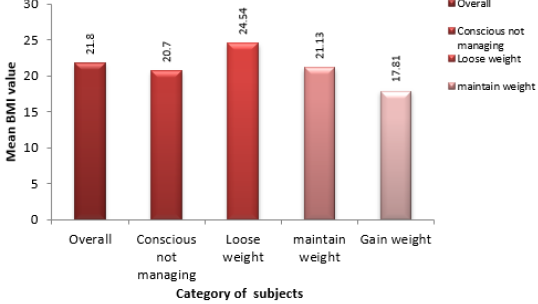
Figure 1: Mean body mass index in adult females of different categories.

Objectives

The study was framed keeping the following objectives in mind
1. To study the body composition of the adult females with reference to Body Mass Index (BMI), fat mass, fat mass per cent, fat free mass (Kg), fat free mass per cent, total body water per cent.
2. To compare the body composition parameters in different groups of adult females namely Conscious for weight but not managing weight (CNM), making attempts to loose weight (group I), making attempts to maintain weight (group II), making attempts to gain weight (group III) (Figure 2).

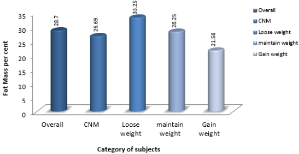
Figure 2: Mean fat Mass per cent in adult females of different categories.

Methodology

To fulfil the objectives of the study, a sample of 147 subjects (Young females in the age group of 18 to 30 years) were selected through convenience sampling from an academic institution in the city of Ajmer. The subjects on the basis of attempts for weight management were then classified into 4 groups namely, conscious for weight, but not managing weight (CNM), making attempts to loose weight (group I), making attempts to maintain weight (group II), making attempts to gain weight (group III). The body composition analysis of the subjects was done through an automatic body composition analyser namely “Tanita BC 420 MA”, operating on the principle of Bioelectrical Impedance Analysis (Figure 3). For each subject, parameters namely weight, height, Body mass index, fat mass (Kg), fat mass per cent, fat free mass (Kg), Fat free mass per cent, total body water (TBW) (Kg), and total body water per cent were measured and analysed. Subjects in different groups were expected to differ with respect to the above body composition parameters.

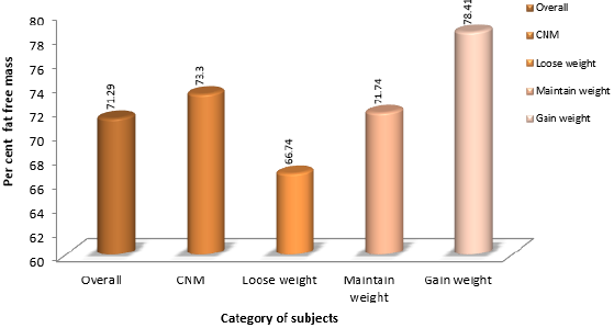
Figure 3: Mean fat Free Mass per cent in adult females of different categories.

Results

The results for the assessment of body composition parameters have been discussed below

Individuals Conscious for Weight but not Managing Weight (CNM)

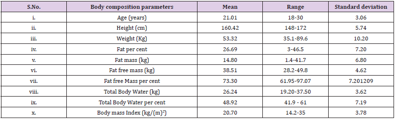
This group comprised of 66 subjects who were weight conscious, but due to limitation of resources, focus on Academics and profession, limitation of time, could not make any efforts to manage weight. The results for the body composition analysis of the group “Conscious for weight but not managing weight” have been presented below
i. Age (Yrs): The mean age of the subjects was 21.01 years.
ii. Height (cm): The mean value for height was 160.42 cm
iii. Weight (kg): The mean weight of the subjects was 53.32 Kg.
iv. Fat mass per cent: The mean value for fat mass per cent was found to be 26.69 per cent.
v. Fat Mass (kg): The mean value for fat mass was found to be 14.80 Kg.
vi. Fat Free Mass (kg): The mean value for fat free mass (kg) was 38.51.
vii. Fat Free mass Per cent: The mean value for Fat free mass per cent were found to be 73.30 per cent.
viii. TBW (kg): The mean value for total body water was 26.24 Kg.
ix. TBW Per cent: The mean value for total body water per cent was 48.92
x. Body Mass Index (kg/(m)2): The mean value of Body Mass Index was found to be 20.70 Kg /m2 (Table 1).

Table 1: Body composition parameters of subjects conscious for weight but not attempting to manage weight (CNM).

Body Composition Analysis of Subjects Conscious for Weight and Managing Weight (Loose Weight) (Group I)

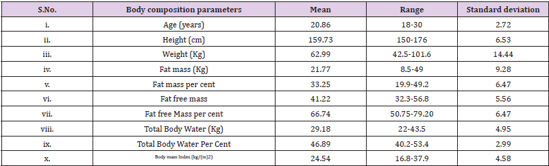
This group comprised of 46 subjects who were weight conscious, and made different dietary modifications and exercise patterns to manage weight (Loose weight). The results for the body composition analysis of this group have been presented below
i. Age (Yrs): The mean age of the subjects was 20.86 years.
ii. Height (cm): The mean value for height of the subjects was 159.73 cm
iii. Weight (kg): The mean weight of the subjects was 62.99 Kg.
iv. Fat mass per cent: The mean fat mass per cent value was 33.25 per cent.
v. Fat Mass (kg): The mean fat mass value was 21.77 kg
vi. Fat Free Mass (kg): The mean value of fat free mass was calculated to be 41.22 Kg
vii. Fat Free mass per cent: The fat free mass values obtained in kilogram when converted into percentage showed a mean of 66.74 per cent.
viii. TBW (kg): The mean value of the total body water in Kilogram was 29.18 Kg.
ix. TBW Per cent: The mean value for the total body water percentage was 46.89. This indicated that the subjects were normal with respect to body water percentage, but were on the lower margin, since a body water content of 45 to 60 % is considered normal for females.
x. Body Mass Index (kg/(m)2): The mean value for Body mass Index was found to be 24.54 Kg /m2 (Table 2).

Table 2: Body composition parameters of subjects conscious for weight and managing weight (loose weight).

Body Composition Analysis of Subjects Conscious for Weight and Managing Weight (Maintain Weight) (GROUP II)

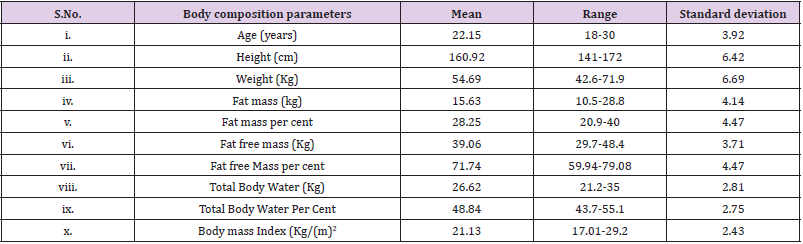
This group comprised of 26 subjects who were weight conscious, and made different dietary modifications and exercise patterns to manage weight (Maintain weight). The results for the body composition analysis of this group have been presented below
i. Age (Yrs): The mean age of the subjects was 22.15 years
ii. Height (cm): The average height of the subjects was 160.92 cm
iii. Weight (kg): The average value of weight for the subjects comprising this particular group was 54.69 kg
iv. Fat mass per cent: The values of fat mass when converted into percentage, demonstrated a mean value of 28.25 per cent.
v. Fat Mass (kg): Fat mass values exhibited an average value of 15.63 kg.
vi. Fat Free Mass (kg): The mean value of fat free mass was 39.06 Kg.
vii. Fat Free mass Per cent: The fat free mass values so obtained when converted to percentage, exhibited a mean value of 71.74.
viii. TBW (kg): The average value for the total body water content in kilogram was 26.62 Kg.
ix. TBW Per cent: A mean value of 48.84 was obtained for the total body water percentage values. The mean value indicated that the subjects could be categorised as normal with respect to body water percentage.
x. Body Mass Index (kg/(m)2): The mean value of BMI for the subjects was 21.13 Kg/ (m)2 (Table 3).

Table 3: Body composition parameters of subjects conscious for weight and managing weight (maintain weight).

Body Composition Analysis of Subjects Conscious for Weight and Managing Weight (Gain weight) (GROUP III)

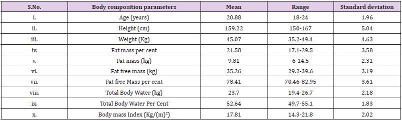
This group comprised of 9 subjects who were weight conscious, and made different dietary modifications and exercise patterns to manage weight (Gain weight). The results for the body composition analysis of this group have been presented below i. Age (Yrs): The mean age of the subjects was 20.88 years. ii. Height (cm): The mean value of height for the subjects was 159.22 cm. iii. Weight (kg): The mean weight of the subjects was 45.07 Kg. iv. Fat mass per cent: The obtained average value for fat mass per cent was 21.58. v. Fat Mass (kg): The calculated mean value for fat mass was 9.81 Kg vi. Fat Free Mass (kg): The mean value of fat free mass was 35.26 Kg. vii. Fat Free mass Per cent: The average fat free mass per cent value was calculated to be 78.41. viii. TBW (kg): The total body water values exhibited a mean of 23.7 Kg. ix. TBW per cent: The mean value for total body water per cent was found to be 52.64. Thus the subjects could be classified as normal with respect to the body water compartment. x. Body Mass Index (kg/(m)2): The mean value of BMI measurements taken for the subjects in the group was found to be 17.81 Kg/ (m)2 (Tables 4 & 5).

Table 4: Body composition of subjects conscious for weight and managing weight (gain weight).

Table 5: Mean ± Standard Deviation of body composition parameters in different groups of subjects (18-30 years).

BMI is the one of the most important and basic parameter that helps in the assessment of nutritional status. Therefore a study of the distribution of the subjects in various BMI categories was done, the results of which have been presented below: 1) Conscious for Weight, but not Managing Weight (CNM): Employing the BMI criteria, 21 subjects (31.82%) could be classified as Underweight, 32 (48.48%) as Normal, 5 (7.58%) as Overweight and 8 (12.12%) as Obese. Thus majority of the subjects fell in the “Normal” category. 2) Loose Weight (Group I): Employing the BMI criteria, 3 subjects (6.52%) could be classified as underweight, 16 (34.78%) as Normal, 12(26.08%) as Overweight and 15(32.60%) as Obese. Thus majority of the subjects fell either into the “Overweight or Obese category”. 3) Maintain Weight (Group II): Employing the BMI criteria, 2 (7.69%) subjects could be classified as Underweight, 19 (73.08%) as Normal, 3 (11.54%) as Overweight and 2 (7.69%) as Obese. Thus majority of the subjects were “Normal” with respect to BMI. 4) Gain Weight (Group III): With respect to BMI 3 (33.33%) subjects could be classified as “Normal”, while, majority that is 6 subjects (66.66%) were classified as “Underweight”. This clearly shows that majority of the subjects fell into the “Underweight” category. A statistical analysis using SPSS was done to assess significance difference in the body composition parameters of different groups. The difference in the BMI was highly significant (P<0.001) both within and between subjects of various categories Conscious not managing (CNM), loose weight (Group I), maintain weight (group II), gain weight (group III). The difference in fat mass per cent was highly significant (P<0.001) both within and between subjects of various categories (Conscious not managing (CNM), loose weight (Group I), maintain weight (group II), gain weight (group III). The difference in fat free mass per cent was highly significant (P<0.001) both within and between subjects of various categories (Conscious not managing (CNM), loose weight (Group I), maintain weight (group II), gain weight (group III). The difference in body water per cent was significant (P<0.05) both within and between subjects of various categories Conscious not managing (CNM), loose weight (Group I), maintain weight (group II), gain weight (group III).

Summary and Conclusion

In reference to BMI it was seen that majority of the subjects falling in group I (Loose weight) fell in the Overweight and Obese category. Alternately subjects falling in group III (gain weight) were found to be “Underweight”. Lastly majority of the subjects falling in the conscious not managing group (CNM) and in group II (maintain weight) fell in the Normal category. The fat mass per cent values were found to be highest in subjects of group I (loose weight), while the values were found to be lowest in subjects of group III (gain weight). The above results justify the attempts of the subjects falling in group I and group III to loose and gain weight respectively. The fat free mass per cent was found to be highest in subjects of group III (gain weight), while the values were found to be lowest among subjects of group I (Loose weight). Since subjects falling in group III (gain weight), had highest quantity of fat free mass they similarly possessed highest quantity of total body water per cent. The category wise BMI distribution of the subjects, along with the above parameters simultaneously justifies the weight management objective of the subjects. Many a times females may have an inappropriate body image perception which may make them frame incorrect objective for weight management. As such it can also be said that the subjects in the present study, had an appropriate body image perception. But the practices employed for weight management should be absolutely healthy so that along with weight management, an optimum Nutritional Status can be achieved.

For more Articles on : https://biomedres01.blogspot.com/  

Immediate and Long-Term Impacts of COVID-19 on Surgical Services and Patient


Immediate and Long-Term Impacts of COVID-19 on Surgical Services and Patient


Introduction

Corona virus comprises of a large family coronaviridae sub family coronavirinae of viruses that infect a large range of hosts including human beings as well animals (camels, cattle, cats, and bats).
There are four different strains of corona virus [1].
1. Alpha coronavirus- HCoV-22E and HCoV-NL63 (infect humans), Pig,Cat,dog CoVs.
2. Beta coronavirus- HCoV-OC43, HCoV-HKU1, MHV, a) HCoV-SARS (that causes severe acute respiratory syndrome, or SARS)
b) MERS-CoV(that causes Middle East Respiratory Syndrome, or MERS)
c) SARS CoV-2(novel coronavirus that causes the coronavirus disease 2019, or also known as COVID-19)
3. Gamma corona virus-Chicken and turkey CoVs
4. Delta corona virus - Bird CoVs

Sometimes corona virus from animals infects humans and spread further via human to human transmission such as with MERS-CoV, SARS-CoV, and now with this COVID 19 (Corona disease 2019). The virus that causes COVID-19 is designated as severe acute respiratory syndrome corona virus 2 (SARS-CoV-2) previously, referred to as nCoV-2019.In December of 2019, this novel corona virus was found to be a cause of upper and lower respiratory tract infections in Wuhan, a city in the Hubei Province of China. It rapidly spread throughout China and then gradually epidemic spreading to other parts of the world in pandemic proportions. It has spread almost every continent in this world. In February 2020, the World Health Organization designated the disease COVID-19, which stands for corona virus disease 2019 [2,3].

Mode of Transmission

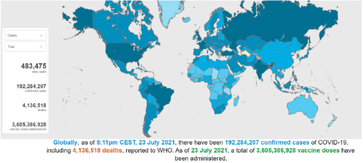
The virus is released along with the respiratory secretions when an infected person coughs, sneezes or talks. These droplets carrying the infection can spread infection to others if they make direct contact with the mucous membranes. Infection can also spread by touching an infected surface and followed by eyes, nose or mouth. Patients are thought to be most contagious when they show clinical symptoms of active disease. Some spread might be possible before symptoms appear, but this is not thought to be a common occurrence [4-6]. Asymptomatic carriers (also referred to as super spreaders) have been shown to spread the COVID-19. These are cases that have no symptoms and radiological manifestations but can transmit the virus to others [7]. Globally, as of 23rd july 2021, 17:11 GMT there have been 4,489,503 confirmed cases of COVID-19, 192,284,207 cases including 4,136,518 deaths, reported to WHO [8] Figure 1.

Figure 1: Countries, territories or areas with reported confirmed cases of COVID-19, 23 JULY 2021 [source -WHO].

Pathophysiology of COVID-19

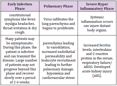
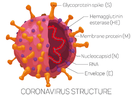
The Coronaviruses are a large family of single-stranded RNA viruses (+ssRNA) that can be isolated in different animal speciesSARS-CoV-2 belongs to the betaCoVs. It has a round or elliptic and often pleomorphic form, and a diameter measuring approximately 60–140 nm (Figure 2). According to recent research, this spike mutation, which probably occurred around late November 2019, triggered transmission to humans. The disease progression can be divided into below mentioned three distinct phases [9-11] (Table 1).
a) Early infection phase
b) Pulmonary phase
c) Severe hyper-inflammatory phase

Table 1: COVID-19, 3 phases of disease progression.

Figure 2: Structure of SARS CoV2 / COVID 19.

Clinical Features

a) Incubation period- approximately 4 to 14 days following exposure [12,13]
b) Common clinical features at the onset of illness were [13,14].
c) Fever in 88-99 %
d) Fatigue in 38-70 %
e) Dry cough in 59-68%
f) Anorexia in 40 %
g) Myalgia in 15-35 %
h) Dyspnea in 19-31 %
i) Sputum production in 27-34 %

Laboratory Findings

Total number of peripheral white blood cells is normal or decreased, the lymphocyte count is reduced, and some children have increased liver enzymes, lactate dehydrogenase (LDH), muscle enzymes, and myoglobin some critically ill patients have increased troponin, D-dimer and ferritin and the number of peripheral blood lymphocytes have progressively decreased.

Radiology

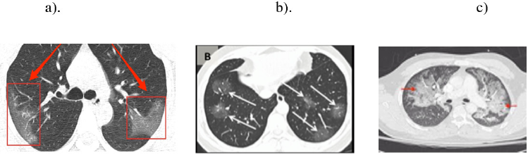
The most important finding in early stages is a single or multiple limited ground-glass opacity which is most commonly located under the pleura or near the blood vessel of the bronchus especially in the lower lobes. Severe period is very rare, manifested by diffuse unilateral or bilateral consolidation of lungs and a mixed presence of ground glass opacities [15]. Consolidation with surrounding halo seen in pediatric patients and was suggested as a typical sign in pediatric patients [16] (Figures 3 & 4).

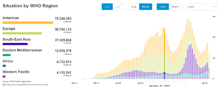
Figure 3: Comparison of confirmed case indifference countries ( source -WHO).

Figure 4: CT thorax finding of COVID patient showing ground glass opacity in basal segment (a) diffuse consolidation with surrounding holo (b) and ground glass opacity with overlap of organizing pneumonia with diffuse alveolar damage(c)

Impact of COVID 19 Pandemic on Delivery of Surgical Care

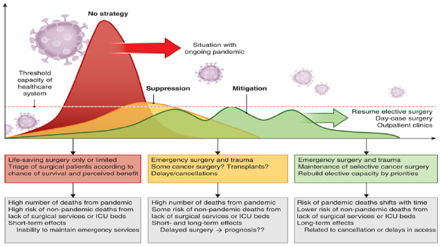
The pandemic of COVID-19 caused by the coronavirus SARSCoV- 2 is disrupting global health, social welfare and the economy in a proportion unparalleled in modern history [17]. In addition to the effects of the COVID 19 pandemic itself on public health, a collateral effect from panuniversal disruption and cancellation of operative interventions has emerged. The current pandemic has unprecedented implications for surgical services and patients with surgical conditions [18]. Surgical capacity may experience extreme challenges in pandemic zones [19,20]. The ability of surgical services to play a key role depends on the development of the peak of the pandemic, the spread of the infection and disease, the duration of societal regulations and implementation of societal laws, and the duration and temporal epidemic cycle repeats by which the disease burden will approach the threshold of maximum capacity of the critical care resources [21] (Figure 5).

Figure 5: Burden of pandemic and impact on surgical survices [21].

Number of surgeries being put on a pause and how this burden will be addressed in the fallout of the pandemic. Rough estimates suggest that approximately 330 million operations are done worldwide annually [22]. The vast majority in high-income countries now following a strong policy of deferring of all nonemergency surgeries (North America and European countries).
With a global average of about six million a per week, the total numbers of patients who will be affected over the coming months are growing rapidly .In the current pandemic, patients may prefer to have elective surgery deferred due to fear of contracting the disease during the hospital stay .However, this fear also leads to patients not seeking timely care for conditions that would otherwise have been correctable or curable by presenting at an earlier stage; loss of function, reduced life expectancy and chronic debility may be the result of delayed presentation and hence an untimely diagnosis. This burden will only increase with the duration and severity of the pandemic. When the destructive effects of natural or man-made forces overwhelm the ability of a given area or community to meet the demand for healthcare [23].
Trauma admissions still occur during a pandemic, although there are unconfirmed reports of a reduction in acute and trauma admissions due to social distancing and overall reduced activity in society. Although trauma admissions may drop, so may the recruitment of the regular bunch of blood donors, potentially leading to a shortage of blood products in many systems. In addition, coagulation may be affected in patients who are COVID- 19-positive, potentially adding to issues with bleeding injuries [24]. Patients who were admitted for an acute abdomen or any pathology requiring acute surgery may be co-infected with COVID-19, and hence represent a risk for infecting the healthcare personnel and other patients, even when asymptomatic or experiencing severe symptoms suggestive of COVID-19. One of the main concerns is the ability asymptomatic patients may have to infect during the incubation period [25].
Pandemic effects on organ transplantation is related to its impact on the donor pool and the risk of transmission of infection, risk to healthy living donors and overall access to scarce resources [26,27]. The scenario of exposing a healthy donor to a major surgical procedure in concordance with the risk of being infected with COVID-19 must be considered and calls for caution, even if cases of successful recovery exist [28]. As the overall strain of the pandemic intensifies, this might dynamically change organ allocation and prioritization of policies [26,27].

Material and Methods

The information and statistical data is collected from various international journals published worldwide and electronic media.

Results

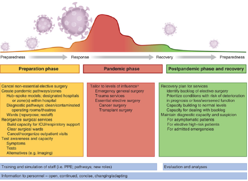
Impacts of COVID pandemic on surgical services are profound potentially long-lasting and extensive. Due to the cancellation of surgeries, there is drastic effect not only on wellbeing of the patient but also on emotional, psychological and physical health. Surgeries which was planned in pre COVID week are cancellation after lockdown because of that patients can experience feelings of sadness, disappointment, anger, frustration and stress [29]. This superimposes on the potential economic consequence (loss of jobs, sick leaves or reduction in work availability) and impact on family life. A complete neglect of certain surgical services can be considered unwelcome collateral damage, and unwittingly increase the number of deaths and life-years lost where the onus lies on the COVID-19 pandemic. This can create ethical dilemmas at a time of scarce resources and high pressure, burnout, guilt and unbearable psychological pressure on health care workers. As elective surgery has been cancelled on a scale never seen before in history of modern medicine, the collateral damage to health and well-being, maintenance of functionality, and risk of shortened lifespan is present for patients in countries of all income and development statuses. With a long-lasting lockdown and social distancing in effect, there will be millions of operations cancelled, postponed or simply not done at all over the times to come. The cancellation of all elective operations creates a huge backlog of patients who have been, or would be, seen for planned surgery (Figure 6).
The effects of COVID-19 on surgical outcomes (and vice versa) are not known. Pathological changes in blood coagulation [30], inflammatory response [31], and co-morbidities added to single or multiple organ failures, [32] may be considered as mechanisms for added risk for surgery. Blood and blood products reserves could be affected to a great extent as blood donations decrease due to decrease in frequent (regular) blood donors, potentially leading to as shortage of blood and blood products in some systems. In addition, coagulation system may be deranged in patients who are COVID-19-positive, potentially adding to issue in patients undergoing major surgeries requiring blood transfusion and traumatic bleeding injuries. Limiting access to transplantation is associated with a significant fall in prognosis for most patients on the transplant list, there is decreased donor pool, risk of transmission of COVID infection to the healthy donor. Lung transplant recipient have risk of getting infection via infected lungs. One of the main concerns is the ability asymptomatic patients may have to infect during the incubation period [33], and if large number of senior surgeons is exposed to infected patients, the possibility for them to become infected and requiring self-isolation is real and could potentially result in a dangerous shortage of senior expertise within surgical teams. The competition for scarce resources related to hospital, ICU beds, health care workers, PPE and blood transfusions mandates strict prioritization in several surgical disciplines, notably transplantation, cancer surgery and other highrisk procedures. As a result, some patients may unfortunately not receive the treatment they would otherwise have received. The post pandemic evaluation and future planning should involve strategies to tackle shortage and include surgical services as an essential part to maintain appropriate surgical care.

Abbreviations: PPE: Personal protective equipment; WHO: World Health Organization

Figure 6: Framework for surgical planning through pandemic phase.

Discussion

As elective surgery has been cancelled on a scale never before seen in modern history, the collateral damage to health and wellbeing, maintenance of function, and risk of shortened lifespan is present for patients in countries of all income designation, poor and marginalized will be affected most severely. In a situation where resources are scarce, one should not treat or prioritize on a first-come, first-served principle [34]. With a long-lasting lockdown and social distancing in effect, there will be millions of operations cancelled, postponed or simply not done at all over the times to come. Surgery is already recognized as a scarce resource in much of the world [35-36]. with a global challenge to provide safe surgery and anesthesia as a recognized area of priority [37,38].
A strong advocacy agenda is needed that includes investigation, planning research and communication for surgical services for future pandemics. Surgical delivery before, during and after a pandemic, owing to its cross-cutting nature and synergistic effects on health systems at large needs to be addressed into the WHO agenda for national health and planning. In this pandemic situation mindful utilization of financial and human resources is very important. Judicious utilization of resources and manpower is of utmost importance in healthcare. It is important to ensure the ability of surgeons, so we need to change the policies in the country like construct surgical systems in terms of infrastructure, workforce, care delivery, information management, financing and governance so that we can combat the damage caused by this pandemic. To inform COVID-19 surgical plans and address patients’ concerns effectively, there is an urgent need for high-quality multi center research. Data on the effects of surgical cancellation on patients’ complete well-being, including emotional and physical health, are much required. In addition, the effect of the stacking up of operations and the fallout of the pandemic will need to be analyzed. Guidance is urgently needed on how to deliver surgical services safely and effectively in the face of pressures placed by the COVID-19 pandemic [17-19].

Conclusion

Surgical services need a contingency plan for maintaining surgical care in an ongoing or post pandemic phase. The current situation is unparalleled in history of modern medicine, and so no readily available information exists to compare or extrapolate the effect of disruption of surgical services and operations on public health during the global pandemic. To mount an effective response to the COVID-19 pandemic, hospitals should prepare detailed context-specific pandemic preparedness protocols and teams for surgical services and key domains for the same includes prepare a pandemic response plan for surgical services ,ensure staff are trained to deliver surgery safely during pandemic, hospital response to COVID-19,agree a team-based approach for running emergency services .Important areas for future research may include finding the impact of COVID-19 infection on postoperative outcomes, identifying risk stratification strategies, and determining whether prophylaxis using drugs for COVID (example HCQ) reduces rates of hospital-acquired COVID-19 infection. There is also a need to determine whether COVID-19 is present in abdominal fluids (urine, bile and peritoneal fluid) or aerosols created during introduction of pneumoperitoneum, in order to inform recommendations around the benefits and risks of open and laparoscopic surgery during the COVID-19 pandemic.

For more Articles on : https://biomedres01.blogspot.com/ 

 

Pyometra in a Cat: A Clinical Case Report


Pyometra in a Cat: A Clinical Case Report


Introduction

Pyometra is an acute or chronic suppurative inflammation of the uterus. It is characterized by endometrial hyperplasia with cystic dilation of endometrial glands and accumulation of a neutrophil-rich exudate in the uterine lumen. The incidence of feline pyometra is still not well documented and probably underestimated because queens often don’t present with clinical signs [1]. No prevalence data for pyometra have so far been described in cats, but observations of most veterinarians are that the disease is observed less commonly than in dogs. The most common clinical finding in case of 75% of pyometra cases is mucopurrulent to hemorrhagic vaginal discharge [2]. The clinical presentation of pyometra is similar in cats and dogs. In ‘open-cervix pyometra’ a blood stained; purulent vaginal discharge may be the only clinical sign. Animals with ‘closed-cervix pyometra’ may not show any vaginal discharge and are more commonly systemically ill because resorption of bacterial toxins from the uterine lumen into the circulation can result in endotoxaemia. Bacteremia may also occur. Non-specific clinical signs such as anorexia, vomiting, lethargy, loss of weight and unkempt appearance can also be observed [3]. Polyuria and polydypsia do not occur as often as in dogs. They were reported only in 9% of the cases [4].
Abdominal ultrasound is the most important diagnostic tool in a pyometra case. The uterine horns typically appear distended with hypo-/ to hyperechoic fluid with or without flocculation. The uterine wall often appears thickened with irregular edges and small hypoechoic areas consistent with cystic changes of the endometrial glands. The pyometra can be diffuse or segmental. Cytology of the uterine or vaginal discharge is likely to reveal degenerative neutrophils and phagocytized bacteria. Leukopenia can be present in around 5% of the cases [4]. Treatment includes correction of fluid deficits, proper administration of antibiotics against bacterial organisms and removal of infected uterine contents. The other management includes surgical removal of ovary and uterus (ovariohysterectomy) or use of by PGF2α [3]. The decision to try medical or surgical therapy is based on the physical status and breeding capacity of the queen.
However, some complications may develop after ovariohysterectomy (OHE), such as ovarian remnant syndrome (ORS). This syndrome may develop because of the failure to totally remove both ovaries (most commonly the right ovary) at OHE, or the presence of a partial or complete separation of a portion of normal ovary (the fragment may be located near the ovary or in the broad ligament) that is not detected at OHE. In some cases, uterine stump pyometra may occur because of ovarian remnants and this situation may be fatal in affected queens. Worldwide, fatal complications occur as a result of surgical errors in routine OHE. In this article, we report and discuss the procedure and importance of ORS in a queen.

Materials and Methods

History and Clinical Examination

An eleven years old local breed cat was admitted to Teaching and Training Pet Hospital and Research Centre, Chittagong Veterinary and Animal Sciences University, Bangladesh, with history of anorexia, chronic emaciation. At first, general physical examination was done, then special examination was done. On physical examination, body temperature found 1010C, heart rate 174 beats per minute and respiratory rate 42 breaths per minute.

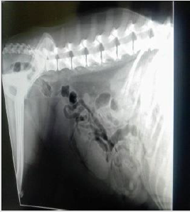
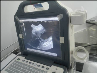
On abdominal ballottement the uterus felt harder and enlarged than normal. Lateral radiograph revealed multiple tubular, radioopaque fluid filled structures from caudal to mid abdomen (Figure 1). The structures appeared distinct and separate from the intestinal loops. Ultrasonography was performed using a B mode real-time 5MHz linear transducer. The finding of abdominal ultrasound was found multiple anechoic fluid filled area without foculation (Figure 2). Then blood sample was collected for doing routine examination and serum analysis.

Figure 1: Multiple tubular, radio-opaque fluid filled structures

Figure 2: Multiple anechoic fluid filled pocket.

Surgical Management

Restraining and Anesthesia: Firstly, the cat was being held on its side with its back against the handler, while the handler grasps the front and back legs, with a forearm across the cat neck. As premedication agent atropine sulphate was administered (Injection Atropine®, Techno drug, Bangladesh, 0.04mg/kg body weight intramuscularly) and as muscle relaxant xylazine hydrochloride (Injection xylazine®, Indian Immunologicals Ltd, India, 1mg/ kg BW intramuscularly) administered. Again, as a general anesthesia ketamine hydrochloride (G-ketamine®, Gonoshasthaya Pharmaceuticals Ltd., Bangladesh, 15 mg/Kg body weight intravenously) was administered. The maintenance anesthetic dose was given half of the initial dose during the surgery. Preparation of surgical area was carried out after shaving and removing hairs. 70% alcohol scrubbed onto the skin around the surgery area, the area is then covered until surgery, since nothing must touch it once it is cleaned.
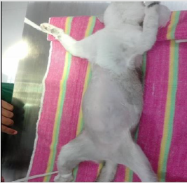
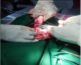
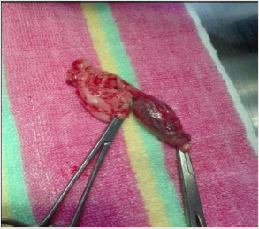
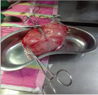
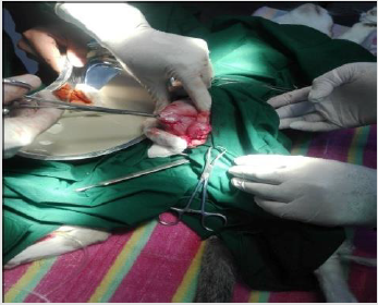
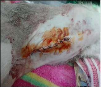
Surgical Procedure: The cat was being laid on her back (Figure 3) and a sterile drapper was placed over her. Close monitoring of temperature, blood pressure, heart rate, gum color, pulse strength and depth of anesthesia was done. An incision was made in the middle of the underside along the length of the abdomen. After exposing the abdomen by laparotomy, the uterine and ovarian blood vessels were properly secured and the ovaries, uterine horns and uterus were completely removed. The abdominal wall was closed with catgut (size: 1-0). The skin was then closed with cross-mattress suture pattern using silk. The sutured wound was covered with the benzoin seal. During the entire operative period, 5% dextrose saline was intravenously infused.

Figure 3: Lying the cat at dorsal recumbent position.

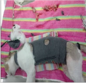
Post-Operative Care: After surgery, antibiotic ceftriaxone @20 mg/Kg body weight (Injection Triject vet 1gm®, SK+F Pharmaceuticals, Bangladesh) was administered intramuscularly daily for 7 days. Antihistaminic chlorpheneramine maleate @1mg/ Kg body weight (Injection Astavet®, Acme Laboratories Ltd., Bangladesh) was administered intramuscularly daily for 7 days. Analgesic (Injection meloxicam @40 mg/Kg body weight and Injection Melvet®, Acme Laboratories Ltd., Bangladesh) was administered subcutaneously daily for 5 days for pain management. The patient was kept in clean squeeze cage and observed for 7 days. No complication was noted, and the bitch recovered uneventfully. On the 14th day, the suture was removed, and it was noticed that the surgical site was healed completely (Figures 4-9).

Figure 4: Ligating the uterus at the base of the uterine body.

Figure 5: Removal of ovaries.

Figure 6: Removal of uterus.

Figure 7: Release of pus from uterus.

Figure 8: Application of benzoin seal.

Figure 9: Cat after surgery.

Results and Discussion

Pyometra is a uterine inflammatory disorder characterized by cystic endometrial hyperplasia [5]. Potter et al. Potter et al. [6] concluded that the prevalence of pyometra in cats increases with age in sexually intact female cats and mainly after parturition, while Agudelo, [7] suggested that the disease is common in queens older than three years and in other queens older than five years with no relationship to the number of parturitions, these close to findings were reported in this case. Hagman et al. [8] found comparatively higher prevalence of pyometra in Bengal cat which is almost similar to this study. Pyometra is a disease of the middle-aged or older animal which was also stated by Brady et al. It could be speculated whether this increase is related to degenerative changes in the uterus or other conditions such as ovarian pathologies or uterine neoplasia that more often affect older animals and may predispose for developing pyometra. But it has been described also in younger cats [9-11].

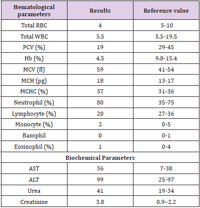
Present study before treatment the hemoglobin level of cat was decreased indicating anemia which agrees with the previous reports [12,13]. This might be due to loss of red blood cells by diapedesis into uterine lumen apart from depressed feed intake and impaired erythropoiesis under toxemic condition in severely affected cases [14]. The PCV level was decreased in the bitches indicating a mild normocytic, normochromic philia might be due to and regenerative type of anemia [15]. According to Greene et al. [16] total erythrocyte count before treatment was decreased in the bitches affected with pyometra indicating anemia which is similar to this study. It might be associated with the toxic depression of the bone marrow whereas severe non-regenerative, microcytic, hypochromic anemia accompanied by extremely high white blood cell levels might be indicative of a concurrent blood loss possibly by diapedesis into luminal pus and due to shortened life span of circulating erythrocytes associated with iron deficiency [17]. Different degree of leucocytosis was observed in bitches affected with pyometra which is consistent to this study. It might be due to severity of the inflammation varying between animals.

In the present study, absolute neutrophilia, lymphopenia, monocytosis with normal eosinophil count was the most consistent finding among the bitches affected with pyometra. Neutrophilia with regenerative shift to the left might be due to retention of purulent exudates in the uterus which exerts a chemotactic effect on neutrophils resulting into accelerated granulopoiesis and lymphopenia might be due to severe stress and elevated monocyte count might be due to chronic suppurative process [12]. Neutrophilia is a typical feature in hematology of bitches affected with pyometra [18] which might be due to influence of toxins in pyometra [19]. The ovariohysterectomy is the useful treatment of pyometra. Potter et al. [6] recorded that 61% of affected cats were spayed or died because of complications relating to reproductive tract disease. The case fatality for pyometra overall was 5.6%. In dogs it is reported to be 3% to 4% [20]. The reason for the higher fatality rate in cats is not known, but one theory could be that this species is less sensitive to endotoxin, or not as prone to show clinical signs unless they develop sepsis [21].

Pyometra can cause liver and kidney function changes (Nak et al. 2001). Occasionally, in this study ALT level moderately increased which support the finding of Nak [22]. Because of septicemia hepatocellular damage were happened resulting diminished hepatic circulation and cellular hypoxia in the dehydrated cats. In this study, decreased ALT result can explain by a process of inhibition of liver enzyme synthesis or possible hepatic membrane damage. Renal dysfunction may develop secondary related to bacterial endotoxin to pyometra. In this case, blood urea nitrogen and creatinine concentration increased it might be due to dehydration [23-27]. A high creatinine concentration was determined in 12% of a group of cats with pyometra Kenney et al. [28].

In this study, ovariohysterectomy was performed under general anesthesia using xylazine hydrochloride and ketamine hydrochloride which is almost similar to study of Deniz et al. [28]. The causes of postoperative wound dehiscence include rough handling and tearing of tissues during surgery, improper selection of the suture material, inefficient suturing, infection, hematoma or seroma formation, failure to obliterate dead space and training of the animal. In such cases, debriment and fresh coaptation of the wound is indicated [29].

Table 1: Hematological and biochemical analysis of pyometric cat.

For more Articles on : https://biomedres01.blogspot.com/ 
 

Phytoremediation of Organophosphorus Pesticides from Aqueous Media Using Azolla Filiculoides (Case study: Anzali Wetland)

  Phytoremediation of Organophosphorus Pesticides from Aqueous Media Using Azolla Filiculoides (Case study: Anzali Wetland) Introduction Org...