Monday, February 10, 2025

Complex PCL Avulsion and Hoffa Fractures with a Unique Posteromedial Approach: A Case Report


Complex PCL Avulsion and Hoffa Fractures with a Unique Posteromedial Approach: A Case Report

Introduction

Posterior Cruciate Ligament (PCL) avulsions are rare injuries of the knee joint. What makes this injury even more complex is its association with Hoffa fracture, which is an intra-articular distal femur fracture affecting one or both of the femoral condyles in the coronal plane Hoffa [1] and constitutes around 0.65% of all femur fractures [2]. The posteromedial knee approach has been suggested as an appropriate approach to posterior tibia plateau, and femoral condyle fragments fixation with a safe complication profile and good clinical outcomes [3,4] Yet, not many surgeons are familiar with this approach. A thorough literature review was conducted on PubMed, Web of Science and Google Scholar till July 2021. It is to the best of our knowledge that no such cases with this combination of injuries and this surgical approach have been reported in the literature before. This case report presents an unusual PCL avulsion injury associated with an ipsilateral lateral femoral condyle Hoffa fracture. Also, we highlight our surgical treatment and patient’s clinical, functional, and radiological outcomes 18 months following management of this unique injury pattern.

Case Report

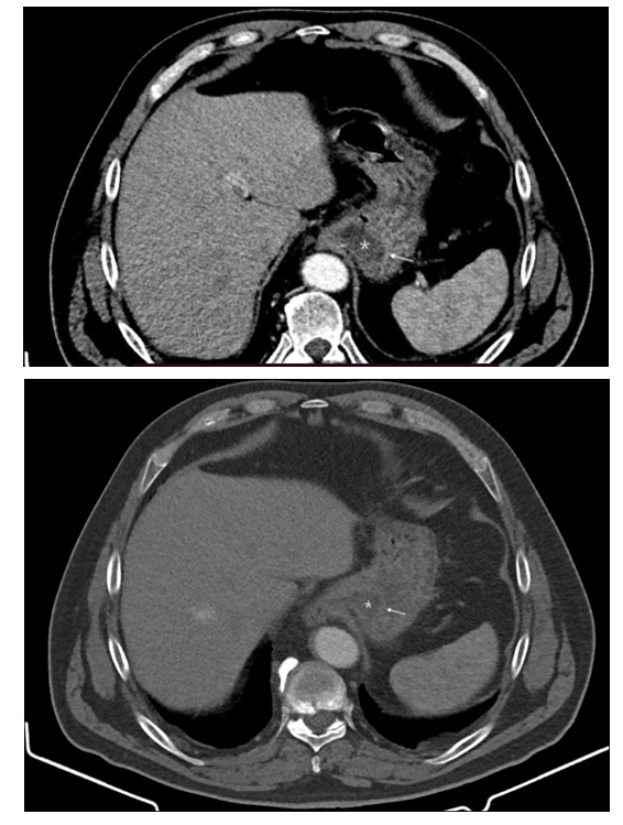
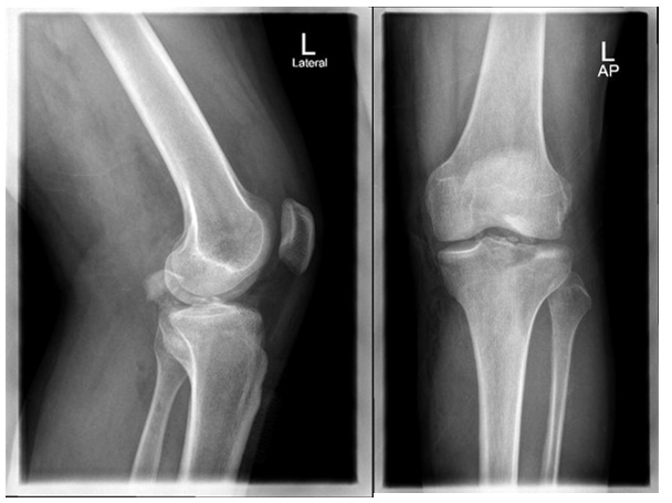
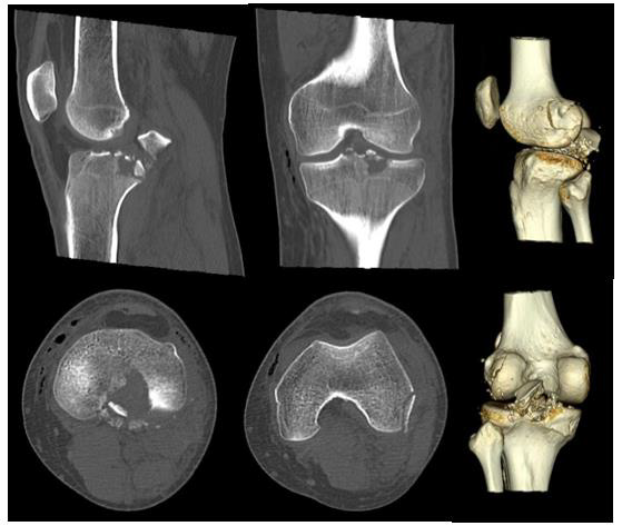
A 35-year-old gentleman sustained a road traffic accident after developing an epileptic generalized tonic-clonic seizure while driving his car. He complained of severe left knee pain and swelling. On physical examination, the patient was hemodynamically stable, conscious, and oriented. The left injured knee was moderately tender, swollen, with superficial abrasions anteriorly. The range of motion was limited to 0-30 degrees of extension-flexion. Also, he had tenderness over the lateral femoral condyle area. Varus stress test was suggestive of grade 2 injury of lateral collateral ligament of the left knee. The posterior drawer test could not be assessed at that time due to the severity of pain but tested positive intraoperatively. All compartments were soft with intact lower limbs neurovascular exam. Preoperative radiological evaluation with plain radiographs & CT scan of the left knee revealed a comminuted tibial plateau fracture extending posteriorly with multiple intra-articular fragments, comminuted Hoffa fracture of lateral condyle of the left femur (Figures 1 & 2).
biomedres-openaccess-journal-bjstr

Figure 1: Preoperative AP & Lateral X-rays.
biomedres-openaccess-journal-bjstr

Figure 2: Preoperative knee CT scan.

Open reduction and internal fixation of left PCL avulsion and lateral femoral condyle fractures was performed by a senior knee orthopaedic surgeon at a trauma 1 center. In prone position and after scrubbing, prepping and draping the left lower limb, a proximal thigh tourniquet was inflated. Then a posteromedial knee approach was taken as described by Burks and Schaffer [5]. First, the PCL avulsed fragment was reduced and fixed with one 3.5 millimeters lag screw and a washer. Next, another posterior lateral 1 x 3 cm fragment was fixed using a 3.5 mm lag screw. Finally, a backup fixation was done with No.5 non-absorbable braided suture in the PCL and tied around the screw and washer. Furthermore, a small posterolateral incision, as an extension of the posteromedial approach in a lazy “S” fashion, was made on the left lateral femoral condyle, and a shell of bone was fixed with two headless 3.5 mm screws. Special knee tests were negative at the end of the procedure. Fluoroscopy confirmed excellent reduction and hardware placement.

Outcome & Follow-up

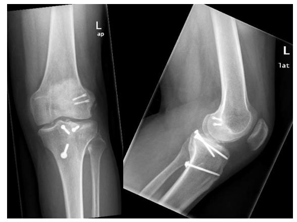
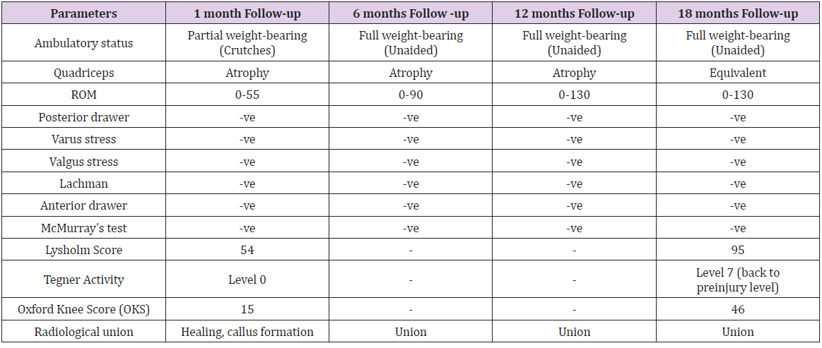
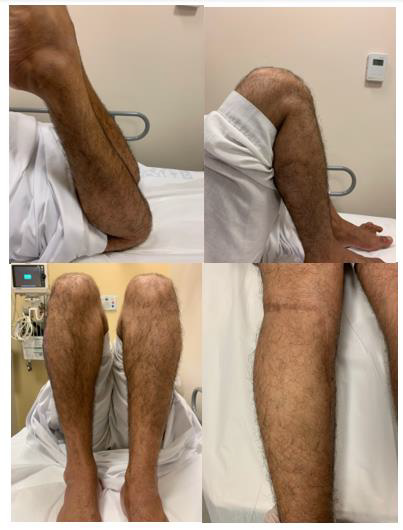
The patient was observed for a total follow-up period of 18 months following surgery and had frequent clinic visits at 1, 6, 12 and 18 months with the following findings (Table 1). Thorough clinical and radiological assessment was done in each visit, validated knee functional outcome scores including Tegner activity, Lysholm score and Oxford knee score (OKS) were utilized. Radiographic and bed side images at final follow up are shown in Figures 3 & 4, respectively.
biomedres-openaccess-journal-bjstr

Figure 3: Final AP & Lateral X-ray at 18 months follow-up.
biomedres-openaccess-journal-bjstr

Table 1: A summary of the postoperative follow up parameters. PWB: Partial weight bearing. FWB: Full weight bearing.
biomedres-openaccess-journal-bjstr

Figure 4: Clinical images at the final follow-up period of 18 months postoperatively.

Discussion

PCL avulsion fractures are rare and challenging injuries. While the best treatment options remain controversial, poorly treated PCL avulsions might lead to debilitating long term consequences of an unstable knee, malunion and nonunion [6,7]. These fractures can also be associated with other bony and ligamentous injuries around the knee joint, increasing surgical complexity. PCL avulsion fractures often have a similar mechanism of injury to PCL intrasubstance tears, such as dashboard injury, where an anteroposterior force applied to a flexed knee and sports traumatic injuries with a knee in hyperextension position [8]. Also, Hoffa and tibial plateau fractures are associated with high-energy trauma such as motor vehicle accidents, particularly in the younger population. While non-operative management could be considered in some PCL avulsion cases with minimal displacement of less than 5mm [9], most surgeons advocate for surgical fixation, taking into account various factors including fracture characteristics, displacement, comminution, and associated injuries [10,11]. The use of posteromedial knee approach has been reported in the literature with some technical variations, however, a recent paper describe the method used in our case and showed excellent radiological and functional outcomes [12] (Figure 5).

biomedres-openaccess-journal-bjstr

Figure 5: Intraoperative clinical image of the posteromedial knee approach used (from a previous case).

Patient Perspective

“After the horrific accident and how badly injured I was, I never thought I will be back on my legs walking. I’m really thankful to have reached this level of activity and getting back to my normal life”


For more Articles on: https://biomedres01.blogspot.com/

Thursday, February 6, 2025

ZSM-5-5-FU as a Drug Delivery Platform for 5-Fu


ZSM-5-5-FU as a Drug Delivery Platform for 5-Fu

Introduction

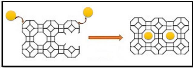
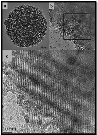
Cancer as the most prevalent diseases worldwide is one of the main public health concerns. (Figure 1) In spite of intensive efforts for treatment of cancer, the necessity of developing effective agents isn’t ignorable [1]. Designing an ideal drug delivery system for targeting cancer cell is considered as a hot topic in life science research. MOFs with crucial features including high drug loading capacity, high surface area, as well as tunable pore size is used for drug delivery intensively[2]. MOFs plays an important role as an carriers in drug delivery because they are non-toxic as well as the uptake of drugs and getting across the cell membrane has been facilitated via controlling the size of MOFs [3]. 5-florouracil (5-FU) is anticancer drugs which is able to induces cytotoxic and increase DNA damage [4]. Although, 5 FU frequently applied, developed drug resistance and severe side effects affected its clinical application [5]. Encapsulate of 5-FU using various DDS could be an effective idea [6]. In present work, the drug loading capacity of zsm-5 for 5-FU as an anticancer drug was evaluated. Upon exposure by zsm- 5-5Fu the in vitro cytotoxicity against cancer cells were assessed. Finally but contrary to the original goal of this project, which was to use a muff, because of the simpler and faster synthesis, we carried out this project with a zeolite. (Figure 2)

biomedres-openaccess-journal-bjstr

Figure 1: Placement of drug in the structure of MOF.

biomedres-openaccess-journal-bjstr

Figure 2: TEM images (a, b) of mesoporous ZSM-5 microsphere of different magnifications and the HR-TEM image (c) from the area marked by a black square in (b).

Nano Composite

Nanocomposite is a multiphase solid material where one of the phases has one, two or three dimensions of less than 100 nanometers (nm) or structures having nano-scale repeat distances between the different phases that make up the material. [7] The idea behind Nanocomposite is to use building blocks with dimensions in nanometre range to design and create new materials with unprecedented flexibility and improvement in their physical properties. In the broadest sense this definition can include porous media, colloids, gels and copolymers, but is more usually taken to mean the solid combination of a bulk matrix and nano-dimensional phase(s) differing in properties due to dissimilarities in structure and chemistry. [8] The mechanical, electrical, thermal, optical, electrochemical, catalytic properties of the nanocomposite will differ markedly from that of the component materials. Size limits for these effects have been proposed.

1. <5 nm for catalytic activity

2. <20 nm for making a hard magnetic material soft

3. <50 nm for refractive index changes

4. <100 nm for achieving superparamagnetism, mechanical strengthening or restricting matrix dislocation movement [9].

Nanocomposites are found in nature, for example in the structure of the abalone shell and bone. [10] The use of nanoparticle-rich materials long predates the understanding of the physical and chemical nature of these materials (Figure 3). Some researchers investigated the origin of the depth of color and the resistance to acids and bio-corrosion of Maya blue paint, attributing it to a nanoparticle mechanism. From the mid-1950s nanoscale organo-clays have been used to control flow of polymer solutions (e.g. as paint viscosifiers) or the constitution of gels (e.g. as a thickening substance in cosmetics, keeping the preparations in homogeneous form). By the 1970s polymer/clay composites were the topic of textbooks, although the term “nanocomposites” was not in common use. [11] In mechanical terms, nanocomposites differ from conventional composite materials due to the exceptionally high surface to volume ratio of the reinforcing phase and/or its exceptionally high aspect ratio. The reinforcing material can be made up of particles (e.g. minerals), sheets (e.g. exfoliated clay stacks) or fibers (e.g. carbon nanotubes or electrospun fibers). [12] The area of the interface between the matrix and reinforcement phase(s) is typically an order of magnitude greater than for conventional composite materials. [13] The matrix material properties are significantly affected in the vicinity of the reinforcement. Some scientists be aware that with polymer nanocomposites, properties related to local chemistry, degree of thermoset cure, polymer chain mobility, polymer chain conformation, degree of polymer chain ordering or crystallinity can all vary significantly and continuously from the interface with the reinforcement into the bulk of the matrix (Figure 4). This massive quantity of reinforcement surface area means that a relatively small amount of nanoscale reinforcement can have an observable effect on the macroscale properties of the composite [14].

biomedres-openaccess-journal-bjstr

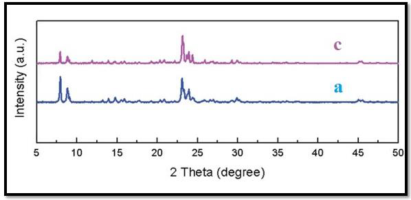
Figure 3: XRD patterns of ZSM-5 samples obtained 5-FU: (a) ZSM-5 (c) ZSM-5-5-FU.

biomedres-openaccess-journal-bjstr

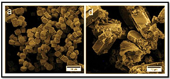
Figure 4: SEM images of ZSM-5 samples obtained 5-FU: (a) ZSM-5, (d) ZSM-5-5-FU.

Zeolite

Zeolites are a group of crystalline materials made up of evenly sized pores and tunnel systems. When purifying VOCs and hydrocarbons, we use a synthetic hydrophobic zeolite. When the contaminated air passes through the material, the hydrocarbons are adsorbed. The material can adsorb a certain amount of hydrocarbons before needing to be regenerated. [15,16] A smaller flow of hot air is then directed through the material so that the hydrocarbons release from the zeolite in a higher concentration. This enables more cost-effective incineration. One of its strengths is that it is non-combustible–meaning it can withstand very high temperatures. [17] This means that we are also able to purify volatile hydrocarbons such as fumes emitted from vulcanization, plastic smoke and styrene, all of which require very high temperatures during regeneration. The resistance to high temperatures and the structure of the material also allows the zeolite to be completely regenerated – meaning that the VOCs completely release from the zeolite when heated. This means that the system maintains its high purification rate year after year and that the material does not have to be replaced, which gives it a long lifespan and a minimal need for maintenance. [18] Our systems have an availability of over 99% and a lifespan exceeding 25 years. Combining the benefits of zeolite with our 30 years of experience in working with air purification gives our customers a supremely sustainable and customized system with low operating costs and high availability.

Reversible Hydration and Dehydration

During drying it comes to the removal of free and bound water from the crystal grid, which is then counterbalanced back in contact with materials such as stored grain and feed, pet litter, in flue gas to prevent condensation and the like. [19] Clinoptilolite stabilize moisture at a low dose of volume and avoid the adverse effects of water. [20]

Results and Discussion

Characterization

The chemical structure of the zsm-5-5Fu was characterized with different analytical methods such as XRD, SEM & TEM.

Drug Loadings and Release

The MOF- with the proper size and the accessible porosity could be used for loading and release of 5-Fu. The loading capacity of zsm-5 under physiological condition (pH 7.4) was investigated. the results showed high drug loading capacity (DLC) (90%) and drug loading efficiency (DLE) 70% by UV–Vis spectroscopy. The results of release profiles of zsm-5-5Fu revealed sustained for 72 h despite with an initial rapid release.

Cytotoxicity Assay

In order to determine the in vitro cytotoxicity of the zsm-5, 5-FU drug, and zsm-5-5Fu HT-29 cell lines, MTT assay was conducted [21]. The obtained results of the cell viability assay showed that zsm-5-5Fu and 5-FU drug inhibited cell growth in a time and dosedependent manner while the zsm-5- showed less growth inhibition after 48 h compared to drug loaded zsm-5 and free drug 5-FU. Based on this results, one may conclude that MOFs with low toxicity could be used effectively for biological applications in the future [3].

Conclusion

In this study, zsm-5 was applied for delivery of 5-Fu. The obtained nanostructure poses spherical morphology with an average diameter of 39-52 nm. Results showed the high loading capacity (90%) and sustained drug release behavior. Moreover, upon exposure by zsm-5-5Fu, higher cytotoxicity than those for zsm-5 and 5-Fu drug against PC3 cells was determined indicating zsm-5-5Fu may could be a promising anticancer drug delivery system in the future.


For more Articles on: https://biomedres01.blogspot.com/
Tuesday, February 4, 2025

Recent Methods in Cancer Treatment and Diagnosis


Recent Methods in Cancer Treatment and Diagnosis

Introduction

The large number of cancer patients and the resulting deaths made cancer the center of attention. Ac-cording to the Center for Cancer Statistics in America in 2022, every minute four cases of cancer are diagnosed, and one patient dies [1]. Cancer is defined as a rapid random proliferation of cells. Its resistance to programmed cell death is what distinguishes it from normal cells which undergo programmed death when they malfunction [2,3]. The cancer cells may undergo metabolic changes that support their rapid growth and division [4]. The most important causes of cancer are aging, tobacco, cancer family history, alcohol, exposure to sunlight and to radiation [5]. The type of tumor, its spread and the age of the patient play the major role in conventional cancer treatment and diagnosis, such as biopsy, surgery, chemotherapy and radiotherapy [3]. An excisional biopsy could be used for conventional diagnosis and treatment, but it could not be per-formed in many types of cancers such as leukemia. In addition, the patient’s age can be a contraindication for surgery [5,6]. One of the traditional treatments for leukemia and lymphoma is chemotherapy, but some tumors do not respond to chemotherapy such as colon and pancreas cancers, in addition to its significant side effects such as hair loss, gastro-intestinal disturbance and lack of selectivity [7,8]. Skin and pancreatic cancers don’t respond well to radiotherapy, which has significant side effects, such as infertility [8]. Recently, novel methods of treatment and diagnosis of cancer have emerged, including nanotechnology that depends on the very small size of drugs (1- 100 nanometer), which helps in their penetration through different cellular barriers without causing damage to healthy cells, and consequently leading to have the desired therapeutic effect at the tumor site [9]. The aim of this work is to shed light on some recent and promising nanotechnologies that are used in the treatment and diagnosis of cancer.

Nanotechnology in Cancer Treatment

Nanotechnology has been used in medicine, especially in the targeted treatment of cancer [10,11]. The advantages consist of the easily cross of cells barriers, control size and shape, increasing efficacy and reducing toxicity of anticancer drugs [12,13]. All these features, and more, have made nanotechnology the focus of attention [13].

Sodium Chloride Nanoparticles and Cancer (SCNPs)

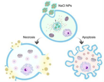
The use of salt nanoparticles is one of the most recent methods for cancer treatment that combines nano-science and immunotherapy NaCl nanoparticles were studied and found to have the ability to eliminate tumor by 66% in the 16th day of starting treatment in experimental mice and 20% of mice became completely free of tumor without causing serious side effects, such as the toxicity to major organs, hair loss or weight loss in experimental mice which lived 8 months after the treatment. The mechanism by which these particles act was causing a dramatic increase in the osmolality of the cancer cell and this leads to apoptosis and necrosis. The high selectivity of NaCl nanoparticles is because the cancer cell contains more [Na+] than the normal cell and so it is exposed to more shock than the normal cell. In addition, the cancer cells have greater affinity for nanoparticles, and the dose needed to kill the tumor is much lower than the dose that affects the normal cell (Figure 1) [13,14]. NaCl nanoparticles are exploited as a novel type of cancer treatment which is taken up by cancer cells through endocytosis and release large amounts of ions inside them. This causes a drastic increase of os-molarity, leading to cell apoptosis and necrosis. This process is highly immunogenic, stimulating an anti-cancer immunity that improves local and systemic tumor control [14].

biomedres-openaccess-journal-bjstr

Figure 1: Sodium Chloride Nanoparticles (SCNPs). The mechanism of Action [14].

Doxil® (DOX) and Nanotechnology

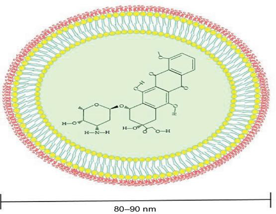
Doxil® is the first FDA-approved nano drug used to treat breast cancer, bladder cancer, Kaposi’s sarcoma, lymphoma, and acute lymphocytic leukemia [15,16]. The PEG component of Doxil® liposome causes in-creasing in blood circulation time decreasing side effects especially on heart cells [17-19], whereas Doxil® liposome is surrounded by a phospholipid bilayer and coated with methoxy polyethylene glycol. The en-capsulating Doxil® in liposomes help decreasing systemic side effects while PEGylation protects the lipo-some from recognition by the mononuclear phagocyte system and increasing its circulation time (Figure 2) [20- 22]. Doxil® Carbon Nanotubes (CNTs) can be described as graphite sheets that are rolled up into cylindrical shapes. The length of CNTs is in the form of micrometers with a diameter of about 100 nm [23]. The reasons for using carbon nanotubes are making drugs more biocompatible, fast electron transfer kinetics, ultra-light weight, chemical inertness, high tensile strength, and can be used in wide number of antibacterial and antifungal drugs, although it can act as protein carriers [24,25]. Although Doxil® liposome can be accumulated at tumors based on the enhanced permeability and retention effect, it stays around periphery of the blood vessels with limited internalization and intracellular release due to the hindrance of the PEG component [22,25,26]. Therefore, it was necessary to study other methods.

biomedres-openaccess-journal-bjstr

Figure 2: Doxil-conjugated liposome structure [19].

Cancer Diagnosis and Detecting

Detecting cancer at very early stages is associated with good patient prognosis as it allows immediate in-terventions to prevent cancer progression, so it is important to ensure earlier detection for effective treat-ment of cancers to reduce cancer disease spread and mortalities [27]. Among the novel strategies in cancer detecting and diagnosis is nanotechnology which has led to several promising results in the diagnosis cancer disease and detecting cancer cells, including drug delivery, gene therapy, drug carriage, biomarker mapping and targeted cancer therapy [28,29].

Magnetic Nanoparticles (MNPs)

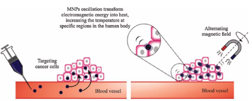
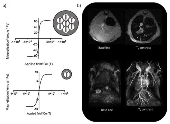
Engineered Magnetic Nanoparticles (MNPs) represent a cutting-edge tool in medicine because they can be simultaneously functionalized and guided by a magnetic field (Figure 3). Use of MNPs has advanced magnetic resonance imaging (MRI), guided drug and gene delivery, magnetic hyperthermia cancer thera-py, cancer diagnosis, tissue engineering, cell tracking and bio separation [30]. Synthesis of iron oxide magnetic nanoparticles has been achieved via physical and chemical methods [31,32]. MRI has been used as a contrast technique in cancer imaging soft tissues; nevertheless, the continuous development of magnetic nanoparticles as contrast agents has made possible the improvement of the qual-ity of the images [33]. Due to the great usefulness of the MRI technique in clinical diagnosis and the tremendous potential of iron oxide nanoparticles (IONPs), the proper functionalization of the latter (e.g., with epithelial growth factor receptor, antibodies, and short peptides sequences), as an advanced contrast agent in MRImediated molecular imaging has enabled the targeted diagnosis of various types of cancer, including breast, stomach, colon, kidney, liver, and brain cancer (Figure 4) [30,33].

biomedres-openaccess-journal-bjstr

Figure 3: Magnetic nanoparticles accumulate in tumors, targeting the cancer cells [32].

biomedres-openaccess-journal-bjstr

Figure 4:

(a) Change in the magnetic behavior of iron oxide nanoparticles with the decrease of the core size, from superparamagnetic (top) to paramagnetic (bottom),

(b) Mouse liver T2-weighted MRI using iron oxide nanoparticles with bigger core size (top), and T1-weighted MR angiography using iron oxide nanoparticles with smaller core sizes (bottom) [30].

Consequently, a series of promising applications have emerged in these recent years, for example, an at-tractive study described the design of a short peptide and ligand library to functionalize IONPs which were able to choose the ligand that provided IONPs with the most advantageous features for in vivo ap-plications and obtaining a considerable enhancement in contrast between the liver tumor and the healthy liver tissue in comparison with a commercial MRI contrast agent [33]. In another interesting study, researchers developed fine-tuned IONPs coated with polyethylene glycol and useful in MRI cancer imaging. In vitro characterization, the results revealed high biocompatibility and relativity values greater than the commercial alternative Ferumoxytol®. The in vivo characterization with breast cancer mouse models showed the capability of these PEG-coated IONPs to be totally preserved at the tumor site for up to 24 h, thus indicating their strong potential as MRI contrast agents for real-time long-lasting monitoring of tumor progression [30,33].

Quantum Dots (QDs)

Quantum Dots (QDs) are semiconductor nanocrystals that emit fluorescence on excitation with a light source. They have excellent optical properties, including high brightness, resistance to photobleaching and tunable wavelength. Their unique optical properties, such as high brightness, long-term stability, simultaneous detection of multiple signals and tunable emission spectra, make them appealing as potential diagnostic and therapeutic systems [34,35]. The lack of ability to penetrate objects limits the use of visible spectral imaging, quantum dots that emit fluorescence in the near-infrared spectrum (700-1000 nanometers) have been designed to overcome this problem, making imaging colorectal cancer, liver cancer, pancreatic cancer, and lymphoma [36,37]. A second near-infrared (NIR) window (900-1700 nm) with higher tissue penetration depth, higher spatial and temporal resolution has also been developed to aid cancer imaging. Also, the development of silver rich quantum dots containing a sulfur source has been reported to allow visualization of better spatial resolution images over a wide infrared range. Despite all the advantages, the applications of these nanoparticles are limited for the use in human system, and this is mainly because of the toxicity associated with heavy metals [38,39].

Colloidal Gold Nanoparticles (AuNPs)

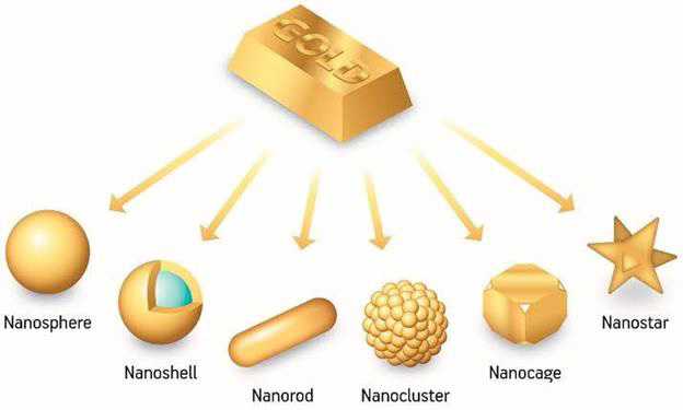
Colloidal gold nanoparticles have been prepared from HAuCl4 using aqueous solution, X-ray irradiation, and chemical reduction method. Gold nanorods were synthesized according to the seedmediated growth method. The colloidal gold nanoparticles were characterized by using transmission electron microscopy, X-ray diffraction, and UV-VIS absorption spectroscopy [40]. The literature is usually associated with controlled methods of synthesis, allowing for the acquisition of AuNPs with defined shapes and sizes (Figure 5) [41]. Gold nanoparticle is a good contrast agent because of its small size, good biocompatibility, and high atomic number. The research showed that AuNPs work by both active and passive ways to target cells. The principle of passive targeting is governed by gathering the gold nanoparticles to enhance imaging because of the permeability tension effect in tumor tissues. Mixing AuNPs with liver cancer cells and using X-ray imaging showed that the clusters of liver cancer cells in the gold nanocomposite group were significantly stronger than those in the liver cancer cells alone. These findings have important implications for early diagnosis allowing tumors as small as a few millimeters in diameter to be detected in the body [40,41].

biomedres-openaccess-journal-bjstr

Figure 5: Various shapes and sizes of Gold Nanoparticles [41].

Conclusion

Many researches have conducted to investigate nanotechnology and introduced its significant role in treating and diagnosing cancer, and that because nanotechnology has a remarkable fingerprint in the medical field and it is becoming famous due to its superiority over other technologies. The efficiency of nanotechnology in the treatment and diagnosis of cancer is proven due to the ability of controlling the particles size and shape, its selectivity towards cancer cells, the improvement of the bio-compatibility and nanoparticles do not make toxicity to the normal cells as conventional cancer treatments.


For more Articles on: https://biomedres01.blogspot.com/
Monday, February 3, 2025

Evaluation of Target Definition by Multimodality Imaging for Management of Low-Lying Rectal Cancer with Short Course Radiation Therapy (SCRT)


Evaluation of Target Definition by Multimodality Imaging for Management of Low-Lying Rectal Cancer with Short Course Radiation Therapy (SCRT)

Introduction

Colorectal cancers are among the most frequent cancers worldwide [1,2]. Rectal cancer composes a significant proportion of colorectal cancers, and low-lying rectal cancers are an important subgroup possessing different characteristics. Patients with rectal cancer may have a pplethora of symptoms which may profoundly deteriorate quality of life. Also, morbidity and mortality due to rectal cancer is not uncommon. Within this context, rigorous management of rectal cancer is warranted to achieve optimal patient outcomes. Currently, surgical resection, systemic therapies, and Radiation Therapy (RT) may be utilized alone or in combination with respect to disease stage, patient, tumor, and treatment characteristics. Total Mesorectal Excision (TME) has been introduced as a viable modality of surgical management leading to improved treatment results and is widely accepted as a prominent surgical approach [3]. In the context of irradiation, both short course RT (SCRT) and long course RT may be feasible and may be utilized after thorough patient evaluation and selection [4]. The recent coronavirus disease 2019 (COVID-19) pandemic came up with some administrative measures which led to modifications in treatment facility practice patterns to a large extent.

SCRT may allow for minimizing treatment visits which may improve patient and treatment facility convenience under the special circumstances of COVID-19 pandemic. Nevertheless, larger fraction size and high fractional doses should be used vigilantly to refrain from adverse radiation effects. Any deviation from accuracy in target definition may result in treatment failure or untowards toxicity. Given the importance of precise target definition in the context of SCRT, we herein assess multimodality imaging for target definition of low-lying rectal cancers.

Materials and Methods

Patients referred for SCRT for low-lying rectal cancer following thorough multidisciplinary assessment were included with consideration of suggested therapeutic strategies, patient, tumor, and treatment related features. Lesion size, localization and association with surrounding critical structures was taken into account along with symptomatology and expected outcomes of SCRT. Contribution of multimodality imaging on target definition of low-lying rectal cancer was assessed in terms of interobserver and intraobserver variations. All included patients underwent Computed Tomography (CT)-simulation, and target definition was performed by use of CT-simulation images only or by incorporation of Magnetic Resonance Imaging (MRI). A prominent group of experts have defined the ground truth target volume to be utilized for actual treatment and comparative evaluation. In this context, thorough patient assessment was performed with colleague peer review and consensus. Following treatment simulation at the CTsimulator (GE Lightspeed RT, GE Healthcare, Chalfont St. Giles, UK), acquired planning images have been transferred to the contouring workstation (SimMD, GE, UK) via the network for delineation of treatment volumes and critical structures.

We have used either CT-simulation images only or registered CT and MR images for target definition of low-lying rectal cancer. Comparative analysis has been performed to assess target definition by CT only and with incorporation of CT-MR registration. Individualized treatment dose calculation has been performed in the Treatment Planning System (TPS) unit by accounting for electron density, CT number and HU values in CT images with consideration of tissue heterogeneities. Synergy (Elekta, UK) linear accelerator (LINAC) was used for SCRT with daily incorporation of image guidance by electronic digital portal imaging and kilovoltage cone beam CT.

Results

Critical organ dose limitations were considered on an individual basis and requirements of optimal treatment planning were taken into account in light of recent guidelines along with reports of American Association of Physicists in Medicine (AAPM) and International Commission on Radiation Units and Measurements (ICRU). Treatment dose calculation has been carried out by considering the tissue heterogeneity, electron density, CT number and HU values in CT images. Coverage of treatment volumes has been a priority in treatment planning by expert radiation physicists while maintaining optimal normal tissue sparing. Determination of ground truth target volume has been performed after meticulous evaluation, detailed collaborative assessment, colleague peer review, and consensus by board certified radiation oncologists. We used the graound truth target volume for actual treatment and also for comparative evaluation. Treatment delivery has been accomplished by use of Synergy (Elekta, UK) LINAC with daily integration of image guidance by kilovoltage cone beam CT and electronic digital portal imaging. Our study with the primary focus on target definition by CT-only imaging and by CT-MR registration based imaging has revealed that target definition by CT-MR registration based imaging was identical with the ground truth target volume for low-lying rectal cancer.

Discussion

Colorectal cancer comprises a major public health concern as a leading cause of cancer related morbidity and mortality around the globe [1,2]. Among the group of colorectal cancers, low-lying rectal tumors require utmost attention with their characteristics. Prognosis for low-lying rectal cancers may be relatively poorer. This may be in part due to difficulties in surgical management, absence of peritoneum as a barrirer to tumor spread, and possible tendency for systemic metastases. Within this context, optimal management of low-lying rectal cancer poses a formidable challenge to the treating physicians. Surgical techniques, systemic therapy strategies, and RT have evolved over time and significant advances have occurred in recent years. COVID-19 pandemic has resulted in critical modifications in treatment practice, and the utility of SCRT has been revisited in the meantime. Improving patient and treatment facility convenience under the special circumstances of COVID-19 pandemic has been a critical endpoint in decision making for patient management. In the context of rectal cancer, SCRT has been well recognized within the scientific community as a viable mode of radiotherapeutic management.

Using condensed treatment schedules has been more popular during the COVID-19 pandemic with shorter overall treatment time. This may clearly have implications for improved utilization of available sources, reduced contact and treatment visits under the special circumstances of COVID-19 pandemic. Nevertheless, the utility of SCRT has been assessed for rectal cancer for a long time, and its safety and efficacy as a viable alternative to long course RT has been supported by high level evidence [4]. Clearly, the use of large fraction size and high fractional doses should be performed vigilantly. Image guidance and image registration methods may aid in improving radiotherapeutic management. Target definition composes an integral part of optimal radiotherapeutic management. While determination of larger than actual treatment volumes may profoundly increase toxicity, definition of smaller than actual treatment volumes may lead to geographic misses and resultant treatment failures. In the awareness of this critical situation, there is need for exploiting the advantage of multimodality imaging. CT has the inherent limitation of low contrast resolution which renders detailed assessment of rectal wall layers and the sphincter rather complex.

MRI with appropriate slice thickness may significantly improve rectal cancer imaging for RT target definition [5,6]. In clinical practice, CT-simulation is typically performed for radiation treatment planning in majority of cancer therapy centers. MRI may provide additional information which may assist is precise and accurate target definition. The superior contrast resolution of MRI may offer several clinical implications regarding SCRT for low-lying rectal cancer. Currently, threre has been an increasing trend towards utilization of multimodality imaging for improved target definition of several tumors throughout the human body [7-41]. From this perspective, this study may add to the growing body of evidence regarding the incorporation of multimodality imaging based target definition for SCRT of low-lying rectal cancer. Recent years have witnessed several improvements in radiation oncology discipline with incorporation of contemporary therapeutic equipment and adaptive irradiation strategies, Image Guided RT (IGRT), Adaptive Radiation Therapy (ART), Intensity Modulated RT (IMRT), Breathing Adapted Radiation Therapy (BART), molecular imaging methods, automatic segmentation techniques, and stereotactic RT [42-78]. For the meantime, introduction of sophisticated RT approaches comprises an evolving area of active investigation and there may still be room for future advances.

In conclusion, utilization of multimodality imaging data for improved visualization and use of sophisticated image registration and fusion techniques may assist in accurate target definition for SCRT of low-lying rectal cancer despite the need for further supporting evidence.


For more Articles on: https://biomedres01.blogspot.com/
Atypical Appearance of Cameron Lesion Casting Diagnostic Doubt

  Atypical Appearance of Cameron Lesion Casting Diagnostic Doubt Introduction Hiatal hernia (HH) is a common endoscopic finding noted in up ...