Saturday, August 8, 2020

Molecular Docking, Synthesis, in Silico and in vitro Screening of Substituted Aryl Ureido Analogues as BACE1 Inhibitors to target Alzheimer’s Disease

Molecular Docking, Synthesis, in Silico and in vitro Screening of Substituted Aryl Ureido Analogues as BACE1 Inhibitors to target Alzheimer’s Disease

Introduction

In Alzheimer’s disease, one of the defining crucial hallmark in brain is the formation of extracellular amyloid-beta (Aβ) peptide plaques and the other being intracellular aggregation of hyper phosphorylated tau to form neurofibrillary tangles. Literature directs, cerebral Aβ accumulation begins at an earlier stage i.e. 10-20 years before the inception of dementia, which indicates that targeting Aβ deposition in brain will be apt for AD pathogenesis [1]. BACE1 (β-site APP cleaving enzyme, β-secretase) is the first protease of the amyloid cascade which initiates cleavage of the amyloid precursor protein (APP) to generate two fragments viz. N terminal fragment and a Aβ-containing C-terminal fragment. Subsequently, the C-terminal fragment is cleaved by γ-secretase to liberate Aβ peptides (Aβ40 or Aβ42) and an APP intracellular domain (AICD). In AD, Aβ42 is over produced due to genetic mutations of either the APP gene or other genes. The Aβ42 peptide is more hydrophobic and stickier than Aβ40 so it readily clumps resulting in formation of amyloid plaques [2,3].

As BACE1 is the key rate limiting enzyme of the amyloidogenic pathway so, inhibition of β-secretase can be considered as a prominent therapeutic target for treatment of AD as the disease still lacks an effective treatment [4-6]. BACE1 is highly expressed in brain and belongs to aspartyl protease family [7]. Literature survey indicates that the active site of BACE1 has two catalytic aspartic residues (Asp32 and Asp228),[8] optimum acidic pH and the correct sequence specificity for enzymatic activity [5]. The reported crystal structure of BACE1 complexed with OM00-3, an octapeptide inhibitor (PDB: 1m4h); depicts eight sub sites for ligand-enzyme interaction [9]. Literature reports discovery of many peptidomimetic as well as non-peptide BACE1 inhibitors [10,11]. Although, peptidomimetic inhibitors displayed potent activity against BACE1, their relatively large molecular size, low metabolic stability and poor bioavailability rendered their development into therapeutic drug candidates difficult [12]. Hence, now the research interest are directed more towards development of small non-peptide organic moieties as lead compounds [13-16].

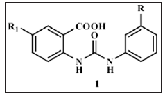
Some notable small-molecule nonpeptide BACE1 inhibitor classes are Vertex biarylnaphthalenes. acylcarbinamine, acylguanidine, aminothiazines, and aminoquinazoline-based scaffolds [6]. Huang and co-workers have reported their work on cell permeable diphenyl urea analogues [17]. Zhu et al revealed unique interaction pattern of CNS penetrant amidine containing heterocylic analogues with BACE1 catalytic dyad [18]. Prompted by above findings we tried to explore the possibility of substituted aryl groups on the two amidine nitrogen’s and replacement of one of the nitrogen with oxygen to simulate ureido functionality. By this approach we designed, synthesized and screened substituted aryl ureido analogues (1) derived from substituted anthranilic acid as potential non-peptide BACE1 inhibitors. In silico ADME studies were carried out to evaluate drug like properties of the candidates (Figure 1).

Figure 1: Substituted phenyl ureido benzoic acid scaffold.
biomedres-openaccess-journal-bjstr

Results and Discussion

Docking approach was employed to explore the BACE1 active site and to estimate binding interactions of these analogues with the receptor. For docking studies, the protein data bank (PDB: 1m4h) crystal structure of BACE1 complexed with OM00-3 inhibitor possessing good potency (Ki: 0.3 nM) was considered. This inhibitor exhibits significant binding to the sub sites and has been extensively studied [19]. The docking protocol was validated by reproduction of binding pose of OM00-3 inhibitor in the 1m4h active site (rmsd 1.51). The octapeptide inhibitor OM00-3 of PDB 1m4h occupies S1, S2, S1ꞌ and S2ꞌ sub sites. To judge further, some reported non-peptide BACE1 inhibitors were docked in the BACE1 active site. Many exhibited interactions with the crucial catalytic dyad involved in drug-receptor binding and enzymatic catalysis. They occupied S1, S2 and S1ꞌ sub sites in the active site [20]. As per the Hong et al amino acid residues present in various sub sites were: S1- Leu30, Asp32, Tyr71, Gln73, Phe108, Asp228, Gly230; S2- of Tyr71, Thr72, Gln73, Gly230, Thr231, Arg235 residues and S1ꞌ- Gly34, Tyr71, Thr232 and Arg307 [9]. On the same guidelines, series of 2-(3-substitutedphenylureido)-5-nitrobenzoic acids (Type I) was docked in the BACE1 active site (Table 1).

These analogues were found to be oriented in the centre of BACE1 binding pocket, occupying S1, S2 and S1ꞌ sub sites analogous to the reported docked inhibitors. In Type I series, the 5-nitro group in all analogues exhibited bidentate interactions (H-bond and electrostatic) with Gln73 residue through water molecule except 4c wherein the nitro group interacted with Thr72. Interestingly, in 4c both the ureido nitrogen’s formed H-bonds with the catalytic residue Asp32 of BACE1. In the other Type I analogues, apart from the nitro group, ureido as well the carboxylic acid group depicted interactions with BACE1. Like reported inhibitors, they were associated with Asp32, Gly34, Thr72 and Gly230 residues of BACE1 active site. These findings were similar to the reported motifs so it can be envisaged that these 2-(3ꞌ-(3ꞌꞌ-substitutedphenyl)ureido)-5-nitrobenzoic acids successfully bind to the BACE1 catalytic site and should therefore turn out to be fruitful candidates for BACE1 inhibition (Figure 2).

Further, to study the effect of introduction of basic group on interaction pattern of Type I series within BACE1 active site, the acidic nitro group was replaced by amino group to obtain a new series of 5-(3ꞌ-(3ꞌꞌ-substitutedphenyl)ureido)-2-aminobenzoic acids (Type II) (Table 2). These analogues occupied S1 and S1ꞌ sub sites. Analogous to 4c, nitrogens of the ureido group of all analogues of this new series depicted bidentate interaction with Asp32 via H-bond and electrostatic interactions except 5f whose ureido group interactions where similar to other Type I members. The newly introduced 2-amino group formed H-bonds either directly or through water molecule with Lys107 and Phe108 residues, which was different from Type I series ligands. On superimposing these Type II analogues with the Type I ligands, orientation of both the series was found to be different which may account for change in the interaction pattern within BACE1 active site (Figure 3).

In Silico ADME Studies

The in silico ADME (Absorption, Distribution, Metabolism and Excretion) properties of designed test set analogues and reported ones were determined to gain an understanding of their pharmacokinetic behavior invivo. The 2-(3ꞌ-(3ꞌꞌ-substitutedphenyl)ureido)-5- nitrobenzoic acids (Table 1) possessed descriptor values within the recommended range of QikProp software such as low molecular weight (MW < 350 g/mol), hydrogen bond donor (HBD) < 3 and QP logPo/w values in the range of 1.0-2.4 [21]. Bioavailability can be gauged by estimating the QP logP values, literature states that for therapeutic use, compounds with QP logP values lower than 5 may have improved drug properties compared to more hydrophobic analogues [22]. The QP logP values for these analogues appear to be favourable. Ghose et al has mentioned some guidelines to design good quality CNS drugs: favorable QikProp CNS parameter, total solvent accessible surface area (SASA) of 460−580 Å2 and HBD less than 3 [23]. For permeability through the blood brain barrier (BBB), QikProp predicts QP logBB; the brain-blood partition coefficient. The QP logBB values were obtained in range of -1.9 to -2.7 (Expected range: -3-1.2) except 4f analogue.

Similarly, the SASA values were found to be in the range of 572- 610, very much favourable to be the likely CNS candidates. Series of 5-(3ꞌ-(3ꞌꞌ-substitutedphenyl)ureido)-2-aminobenzoic acids (Table 2) exhibited some values similar to Type I series such as low molecular weight (MW < 320 g/mol). Comparison of QP logPo/w values (0.5-2.0) with Type I analogues indicated that they are less hydrophobic and HBD value was 4, well within QikProp limit but a digit more than the limit specified by Ghose et al. SASA values were observed to be in between 574-600, seems to be favourable. QP logBB values were observed in the expected range to be-1.6 to -2.9. The percent Human oral absorption for both the series were found to be moderate i.e. 40-60%. None of them violated the Lipinski’s rule of five.

Synthesis

The Type I series docking results were analogous to reported ones so the analogues were synthesized and the structures were confirmed by use of spectral techniques (Scheme 1). In addition, the in silico ADME results were more favourable for the Type I analogues. Further, they were subjected to screening for activity against BACE1.

Pharmacological screening

The in vitro BACE1 FRET (Fluorescence resonance energy transfer) inhibition study results were in agreement to the molecular docking outcome. All analogues of the series 2-(3ꞌ-(3ꞌꞌ-substitutedphenyl) ureido)-5-nitrobenzoic acids showed BACE1 enzyme inhibition at 50μM concentration (Table 1). The percent inhibition at 50μM concentration was found to be in the range of 20-40 %.

Conclusion

The intention to design this scaffold was to analyse the suitability of carboxylic acid group possessing compounds as prospective CNS candidates; as literature indicates that carboxylic acid group is not a very common functionality in the design of CNS drugs. It has been observed that many non-CNS drugs especially orally administered ones have carboxylic acid group while very few CNS drugs possess it. Only 3−4% of CNS drugs had a carboxylic acid group, whereas 25% of non-CNS oral drugs possessed a carboxylic acid group. The docking and in vitro screening studies comply with our assumptions that the substituted aryl ureido analogues can be targeted for potential CNS use. Comparison between the two serie projects the 2-(3ꞌ-(3ꞌ substitutedphenyl)ureido)-5-nitrobenzoic acids (Type I) to be more suitable candidates than Type II series. Amongst them the 4c and 4d analogues exhibit equivalent BACE1 enzyme inhibition. These analogues can be further optimized by suitable structural modifications to attain a more targeted concentration at the site for higher favourable response as BACE1 inhibitors.

Scheme 1: [a] Reagents: (a) Acetone, 40oC.

biomedres-openaccess-journal-bjstr

Experimental Section

Molecular Docking

Materials: On Red Hat Enterprise Linux (RHEL) workstation various software’s viz. Sybyl version 8.1.1 (Tripos International, Portugal) [24] and GOLD version 4.1.1 (CCDC Ltd., UK) [25] were utilized for ligand as well as protein preparation and docking studies. Designing of ligands and protein preparation. The method already reported in our publication was followed for ligand and protein preparation. The same protein data bank crystal structure (PDB code 1m4h) in which BACE1 is complexed with an octapeptide OM00-3 was prepared and further considered for docking studies [26].

Designing of ligands and protein preparation: The method already reported in our publication was followed for ligand and protein preparation. The same protein data bank crystal structure (PDB code 1m4h) in which BACE1 is complexed with an octapeptide OM00-3 was prepared and further considered for docking studies [26].

Docking protocol: The earlier published protocol was followed for docking studies of these analogues [26].

In silico ADME studies

Materials: For the studies Qik Prop version 3.0 (Schrodinger LLC, New York, USA) software on RHEL workstation was employed [21].

Method: Same method as followed in our earlier work was utilized to determine various descriptor values for the in silico ADME studies [26].

Synthesis

Materials: As per the requirement solvent used were distilled for purification purpose. Merck

F254 TLC plates were used to monitor progress of reaction. The title compounds were purified using recrystallization and/ or column chromatographic technique. Infrared (IR) spectra were recorded on Shimadzu IR Affinity-1 FTIR spectrophotometer using DRS-8000 (Diffuse Reflectance attachment). 1HNMR spectra and Mass spectra have been recorded on Mercury Plus 300 MHz (Varian, USA) NMR spectrometer and 410 Prostar Binary LC with 500 MS IT PDA detectors (Varian Inc, USA) mass spectrometer respectively.

For 1HNMR spectra tetramethylsilane (TMS) has been used as an internal standard and DMSO-d6 (Dimethyl sulfoxide) as solvent. 2-(3ꞌ-(3ꞌꞌ-Substitutedphenyl)Ureido)-5-Nitrobenzoic Acids (4 (a-f)): The 2-(3ꞌ-(3ꞌꞌ-substitutedphenyl)ureido)-5-nitrobenzoic acids were synthesized as per Scheme 1.

General Method for Analogues (4 (a-f)): To the solution of 2-amino-5-nitrobenzoic acid (2, 0.2 g, 1.0989 mmol) in 2 ml dry acetone, equimolar solution of respective 3-substitutedphenyl isocyanate (3 (a-f)) in 2 ml dry acetone was added gradually at 40oC in 15-20 minutes with stirring. The reaction mixture was refluxed at 40oC for 16 hours with addition of minimum amount of dry acetone if needed. Further, mixture was poured in 50 ml water for precipitation of crude product which was recrystallized from ethanol resulting in pure analogues as product (5 (a-f)) [27].

2-(3ꞌ-(3ꞌꞌ-acetylphenyl)ureido)-5-nitrobenzoic acid (5a): Yield: 64%; 206-08oC. IR (KBr) ʋ: 1708 (ureido), 3089 (C-H), 1695 (>C=O), 1593, 1357 (NO2), 1681 (COCH3) cm-1. 1HNMR (DMSO-d6): δ 10.60 (s, 1H), 10.28 (s, 1H), 9.33 (s, 1H), 8.19 (d, 1H, J = 8.8 Hz), 8.12 (t, 1H, J = 1.6 Hz), 7.90 (d, 1H, J = 8.8 Hz), 7.85 (d, 1H, J = 2.6 Hz), 7.82 (td, 1H, J = 6.6, 2.3 Hz), 7.63 (dd, 1H, J = 5.3, 2.4 Hz), 7.47 (t, 1H, J = 7.9 Hz), 2.57 (s, 3H). MS (acetone) [M]: 342.

2-(3ꞌ-(3ꞌꞌ-Chlorophenyl)Ureido)-5-Nitrobenzoic Acid (5b): Yield: 73%; 218oC. IR (KBr) ʋ: 1720 (ureido), 3035 (C-H), 1691 (>C=O), 1597, 1355 (NO2) cm-1. 1HNMR (DMSO-d6): δ 10.60 (s, 1H), 10.25 (s, 1H), 9.31 (s, 1H), 8.19 (d, 1H, J =8.8 Hz), 7.86 (d, 1H, J = 2.2 Hz), 7.83 (d, 1H, J = 2.2 Hz), 7.75 (t, 1H, J = 2.0 Hz), 7.40 (td,1H, J = 8.2, 1.6 Hz), 7.33 (t, 1H, J = 7.9 Hz), 7.08 (td, 1H, J = 5.7, 1.7 Hz). MS (acetone) [M]: 334.

2-(3ꞌ-(3ꞌꞌ-Cyanophenyl)Ureido)-5-Nitrobenzoic Acid (5c): Yield: 36%; 208oC. IR (KBr) ʋ: 1708 (ureido), 3055 (C-H), 1697 (>C=O), 1591, 1346 (NO2), 2249 (CN) cm-1. 1HNMR (DMSO-d6): δ 10.65 (s, 1H), 10.40 (s, 1H), 9.30 (s, 1H), 8.19 (d, 1H, J = 8.8 Hz), 8.00 (t, 1H, J = 6.6 Hz), 7.91 (d, 1H, J = 8.8 Hz), 7.85 (d, 1H, J = 2.2Hz), 7.75 (d, 1H, J = 8.1 Hz), 7.63 (d, 1H, J = 2.2 Hz), 7.48 (t, 1H, J = 6.2 Hz). MS (acetone) [M]: 325.

2-(3ꞌ-(3ꞌꞌ-Methoxyphenyl)Ureido)-5-Nitrobenzoic Acid (5d): Yield: 63%; 198oC. IR (KBr) ʋ: 1724 (ureido), 3034 (C-H), 1697 (>C=O), 1598, 1359 (NO2), 2843 (OCH3) cm-1. 1HNMR (DMSO- d6): δ 10.51 (s, 1H), 10.04 (s, 1H), 9.31 (s, 1H), 8.18 (d, 1H, J = 8.4 Hz), 7.83 (dd, 1H, J = 8.8, 2.6 Hz), 7.22 (t, 1H, J = 2.2 Hz), 7.21 (d, 1H, J= 1.1 Hz), 7.17 (d, 1H, J = 6.2 Hz), 7.08 (td, 1H, J = 8.1, 1.1 Hz), 6.61 (td, 1H, J = 8.1, 2.7 Hz), 3.74 (s, 3H). MS (acetone) [M]: 330.

2-(3ꞌ-(3ꞌꞌ-Methylthiophenyl)Ureido)-5-Nitrobenzoic Acid (5e): Yield: 63%; 184-186oC. IR (KBr) ʋ: 1720 (ureido), 3082 (CH), 1697 (>C=O), 1585, 1357 (NO2), 678 (SCH3) cm-1. 1HNMR (DMSO-d6): δ 10.54 (s, 1H), 10.07 (s, 1H), 9.31 (s, 1H), 8.18 (d, 1H, J = 8.8 Hz), 7.83 (dd, 1H, J = 8.6, 2.4 Hz), 7.50 (t, 1H, J = 1.8 Hz), 7.30 (d,1H, J = 1.8 Hz), 7.28 (t, 1H, J = 5.1 Hz), 7.25 (dd, 1H, J = 8.2, 3.5 Hz), 6.91 (td, 1H, J = 7.3, 1.5 Hz), 2.47 (s, 3H). MS (acetone) [M]: 346.

2-(3ꞌ-(3ꞌꞌ-Nitrophenyl)Ureido)-5-Nitrobenzoic Acid (5f): Yield: 74%; 202oC. IR (KBr) ʋ: 1720 (ureido), 3088 (C-H), 1703 (>C=O), 1602, 1354 (NO2) cm-1. 1HNMR (DMSO-d6): δ 10.70 (s, 1H), 10.57 (s, 1H), 9.32 (s, 1H), 8.56 (t, 1H, J =2.0 Hz), 8.19 (d, 1H, J = 8.8 Hz), 7.88 (d, 1H, J = 2.9 Hz), 7.85 (dd, 1H, J = 6.0, 2.7 Hz), 7.62 (t, 1H, J = 3.1 Hz), 7.59 (d, 1H, J = 7.3 Hz), 7.56 (d, 1H, J = 3.7 Hz). MS (acetone) [M]: 345.

Pharmacological Screening

Materials: BACE1 FRET (Fluorescence resonance energy transfer) inhibition study was performed by Department of Pharmaceutical Sciences, University of Bologna, Italy, by the M-2420 method [7,28].

Preliminary Screening: Similar procedure as reported in our earlier published work was followed for the BACE1 inhibition study using the same M-2420 method [26].

Lower Trapezius Muscle Transfer For Elbow Extension Reconstruction After Failed Nerve Transfer for Tetraplegia-https://biomedres01.blogspot.com/2020/08/lower-trapezius-muscle-transfer-for.html

More BJSTR Articles : https://biomedres01.blogspot.com

Friday, August 7, 2020

Lower Trapezius Muscle Transfer For Elbow Extension Reconstruction After Failed Nerve Transfer for Tetraplegia

Lower Trapezius Muscle Transfer For Elbow Extension Reconstruction After Failed Nerve Transfer for Tetraplegia

Introduction

For the last several years, it has been emphasized that restoring upper limb function takes precedence over any other objective during the rehabilitation of traumatic tetraplegia patients [1] According to an IFSSH (International Federation of Societies for Surgery of the Hand) scientific committee update [2] on hand surgery in tetraplegic patients, one of the main goals of reconstruction is elbow extension [2] Traditionally, elbow extension has been reconstructed by transferring the posterior deltoid [3] or biceps muscle [4]. Recently, transferring the posterior division of the axillary nerve or the motor branch to the brachialis to triceps branches has demonstrated consistent recovery of M4 elbow extension strength; thereby supplanting muscle transfers [5]. Failed nerve transfer reconstructions exist, however, often related to poor donor nerves, a prolonged interval between the injury and surgery, and more advanced patient age. In this situation, salvage surgery is complicated. On one hand, if surgeons have used the posterior division of the axillary nerve as the donor, the posterior deltoid muscle is unavailable for transfer. The biceps muscle would still be available.

However, concomitant to triceps reconstruction, thumb and finger extension are typically reconstructed by transferring the nerve to supinator to the PIN [5] In the case of a biceps transfer, supination will be lost. In our previous cases, when we used the biceps to triceps muscle transfer, some of our patients complained about lost elbow flexion strength. They particularly noticed this when they hooked their flexed arm around their thigh to change lower limb position (unpublished observations), a complication that others have also recently reported [6]. In fact, a roughly 50% decrease in elbow flexion strength is routine after a biceps- to triceps transfer [7] which makes us reluctant to propose this surgery in the first place. Bertelli et al. [8] has successfully transferred the lower trapezius muscle to reconstruct elbow extension in patients with brachial plexus injuries. We hypothesised that this technique could [3] also be useful to reconstruct elbow extension among tetraplegics when a nerve transfer fails to achieve adequate results. In this paper, we present two patients with traumatic tetraplegia who had improved inadequately after a nerve transfer procedure and subsequently underwent a lower trapezius transfer to reconstruct elbow extension.

Case Reports

In advance of any data collection, the protocol of the present study was approved by our local ethics committee. Each patient provided his verbal informed consent before participating, in accordance with the Declaration of Helsinki guiding biomedical research involving human subjects (http://www.cirp.org/library/ ethics/helsinki/).

Case 1

A 17-year-old gentleman presented with tetraplegia following an injury that he sustained diving into shallow water. On examination, nine months after the accident, he was found to be Group 1 on the right and Group 2 on the left, as per the International Classification (IC) for Surgery of the Hand in Tetraplegia. He had bilateral elbow flexion contractures. We planned nerve transfer surgeries for elbow extension, along with other nerve transfers, to enhance function in both of his hands. Bilaterally, the posterior division of the axillary nerve was transferred to the nerve innervating the long and upper medial heads of the triceps via axillary access, as reported elsewhere [5]. At 18-month follow up, elbow extension scored M4 on the right side, as rated using the British Medical Research Council (BMRC) scale grading muscle strength. However, left elbow extension strength only scored M2, which had not improved even when he was reassessed at 24 months post operatively. At that time, we performed a lower trapezius transfer to the triceps to reconstruct left elbow extension. Sixteen months after this second surgery, elbow extension scored M4 in the horizontal position. The patient was able to extend his elbow with his shoulder at 1000C of abduction, but 4 not in full abduction. However, he was able to keep his elbow extended with his shoulder fully abducted, which had been impossible for him prior to the trapezius transfer. Video 1 shows pre- and postoperative results for Case 1 post lower trapezius transfer.

Case 2

A 21-year-old man presented one year following a road traffic accident in which he sustained a spinal cord injury and was left tetraplegic. He scored as IC Group 2 for tetraplegia on the right and Group 3 on the left. Wrist extension scored M2 and M4 on the right and left, respectively. He exhibited no recovery of right-sided triceps function even 36 months after a posterior division of the axillary nerve transfer to the triceps long head motor branch. By then, left triceps strength had improved to M4. At this time, a trapezius-totriceps transfer was performed to restore right elbow extension. Twenty-four months after this surgery, the patient was able to extend his elbow with his shoulder at 90 degrees of abduction, scoring M3. Video 2 documents the pre- and postoperative views after lower trapezius transfer in Case 2.

Surgical Technique

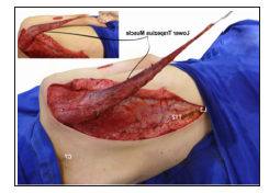
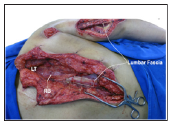
Under general Anaesthesia without relaxants, each patient was placed in a lateral decubitus position. An incision was made from the spine of the scapula to the T12 vertebra. The trapezius muscle was harvested from the T12 vertebra, in continuity with a 1cmwide strip of fascia over the spinous processes of T12 to L3. The trapezius was detached from the spine up to C7. Care was taken not to injure the rhomboid muscles (Figure 1). The XI spinal accessory nerve was identified and protected. The medial side of the lower trapezius, which was inserted into the spine, now was sutured to the medial border and spine of the scapula. A 10cm-long incision was created over the posterior surface of the shoulder and upper arm, and 5 another one, again 10cm long, over the triceps tendon. These incisions were made to permit tunnelling of the lower trapezius to the triceps tendon. With the arm adducted, shoulder extended, and elbow fully extended, the lower trapezius was sutured to the triceps tendon under maximal tension. In Case 2, a portion of lumbar fascia was removed to reinforce the lower trapezius tendon (Figure 2). After skin closure, the upper extremity was immobilised in a plaster cast, with the arm completely adducted and extended 30o, and the elbow flexed 20o. The shoulder was placed in 30o of extension to allow for some relaxation of the transplanted muscle. The cast was maintained for four weeks. Over this period, patients were asked to individualise their lower trapezius and contract it 10 times per cycle, three cycles per day. To instruct patients how to individualise lower trapezius muscle function, the surgeon gently squeezed the transferred muscle and asked the patient to contract where they felt pressure.

Figure 1: Intraoperative view of the dissected lower trapezius muscle and its transposition over the triceps.

biomedres-openaccess-journal-bjstr

Figure 2: Intraoperative view of the dissected lower trapezius muscle and its transposition over the triceps. A segment of lumbar fascia was harvested to reinforce the lower trapezius muscle’s attachment to the triceps tendon. (LT) lower trapezius sutured to the medial margin of the scapula, (RB) rhomboid muscles.

biomedres-openaccess-journal-bjstr

Discussion

Even though not strong enough to permit patients to raise their body up for transfer, the restoration of elbow extension was useful for improving arm positioning and, thereby, increasing its functional range. Before attempting this surgical technique, we were concerned that removing the lower trapezius might impair trunk control. This did not happen. In fact, both patients reported better trunk control after surgery: while sitting, they now used their restored elbow extension to control their balance, using the arm rest of the wheel-chair as support. We propose using the lower trapezius as an alternative for triceps reanimation for various reasons. First, trapezius transfers have already been employed successfully to reconstruct elbow extension in patients with brachial plexus injuries [8,9]. Second, the trapezius is innervated by the spinal accessory nerve, which has a cranial origin in the medulla and a spinal origin from the upper five cervical roots. Since both these components should be uninjured in surviving tetraplegics, the availability of the trapezius for muscle transfer should never be an issue.

Third, postoperative re-education was straight forward: squeezing the transferred belly of the trapezius muscle and asking patients to contract where they felt pressure helped them learn how to selectively contract their lower trapezius. Holtermann et al. [10] observed that all his volunteers were capable of independent control of the lower trapezius when examined by EMG. They believed that this resulted from the selective contribution of the cervical plexus to lower trapezius innerve [10]. The lower trapezius muscle also can be used, not only as a fall-back option, as in our patients, but in patients in whom the deltoid and biceps are unavailable for transfer. A final advantage of the proposed surgery is that the lower trapezius muscle’s insertion can be extended by harvesting fascia from the T12 to L3 spinal processes, thereby increasing the tendon’s length and eliminating any need for a tendon graft. In tetraplegic patients, our first choice for reconstructing elbow extension is to perform a nerve transfer. However, whenever transferring a nerve fails, transferring the lower trapezius muscle to the triceps is a safe and effective alternative.

Organ-Sparing Surgery in Stage I Testicular Sex Cord-Stromal Tumours: Results of a Small Series-https://biomedres01.blogspot.com/2020/08/organ-sparing-surgery-in-stage-i.html

More BJSTR Articles : https://biomedres01.blogspot.com
Organ-Sparing Surgery in Stage I Testicular Sex Cord-Stromal Tumours: Results of a Small Series

Organ-Sparing Surgery in Stage I Testicular Sex Cord-Stromal Tumours: Results of a Small Series

Introduction

Testicular sex-cord stromal tumours (TSCST) arise from non-germinal cell-lines of the male testis: Leydig cell tumours are the most common subtype (75-80%) but this group of tumours includes also Sertoli and granulosa cell tumours, mixed and undifferentiated tumours, thecomas and fibromas. They can occur at any age with a range from 12 to 76 years [1]. The most common clinical presentation is a palpable testicular mass but one third of patients presents gynecomastia, infertility, erectile disfunction or decreased libido. While most cases of TSCST are benign, a malignant behaviour with metastasizing potential is reported in approximately 10% of these tumours [2-6]. The identification of the favourable cases, performing frozen section analysis and using some histological findings, which can predict malignancy, can avoid an unnecessary orchiectomy [1-5]. We present a small series of patients with stage I TSCST, treated with organ sparing surgery, which confirm its feasibility and safety in selected patients.

Materials and Methods

Between 2005 and 2016 a TSCST was diagnosed in 11 patients at our department. The age ranged from 14 to 83 years (mean 46.8). The blood screening with alpha-fetoprotein (alfa-FP), human chorionic-gonadotropin (HCG) and lactate-dehydrogenase (LDH) was assessed preoperatively in all patients. No patient had hormonal disorders except one, who showed low testosterone levels and no patient presented gynecomastia. All patients underwent inguinal access to the testis. During the operation frozen sections were requested, as usual in our department, in order to choose the definitive surgical strategy. A testis-sparing surgery (TSS) was performed always in case of TSCST, waiting for the definitive histology, instead to perform a primary radical orchiectomy. A thoracic and abdominal computed tomography (CT) was performed after the surgery in all patients. The follow-up was scheduled according with the European Association of Urology guide-lines for testicular tumours.

Results

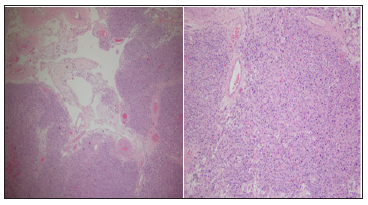
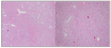
All lesions were intraparenchymal and detected in ultrasound. Alpha-FP, HCG and LDH were negative in all patients. The intraoperative histology showed a TSCST in 10 patients, which were treated with TSS. In 1 patient was performed an orchiectomy, because the frozen section suspected a seminoma, but the definitive histology showed a TSCST. The tumour-size ranged from 7 to 40 mm (mean 11.3 mm). There was no evidence of angioinvasion, margin infiltration or necrosis in the definitive histology. The mitosis-index was < 1% in all patients (Figures 1 & 2). All postoperative courses were uneventful, and the patients were discharged one day after the operation without complications. After discharge one patient developed an intratesticular hematoma, recovered within 4 weeks without any treatment. The CT showed absence of lymph node enlargement or distant metastases in all patients. Follow-up ranged from 18 to 108 months (mean 46.3 months), every 6 months for 5 years with ultrasound of the testis and ultrasound of the abdomen alternated with CT and then every year with ultrasound of the testis and ultrasound of the abdomen. All patient is free from disease and alive except one, who died after 108 months for other reasons (Table 1).

Figure 1: On the right side hematoxylin and eosin staining of a Leydig cell tumor. In the upper left corner vessels and atrophic seminiferous tubules are visible; all around the neoplastic cells. On the left side 10 x magnification of the bottom right corner.

biomedres-openaccess-journal-bjstr

Figure 2: On the right side hematoxylin and eosin staining of a Sertoli cell tumor: the seminiferous tubules surrounded by neoplastic cells are visible. On the left side 10 x magnification of the upper right corner.

biomedres-openaccess-journal-bjstr

Discussion

TSCST are very rare tumours of the male testis, accounting from 3 to 5% of all testicular neoplasms. From a review of the literature the median age of presentation is 35 years with a range from 12 to 76 years [1]. The most common clinical presentation is a palpable testicular mass, even if the incidental finding of asymptomatic and non-palpable lesions is now more frequently detected, due to the increased use of the ultrasonography [2]. Due to their endocrinological activity, erectile dysfunction, infertility or decreased libido could be present in one third of patients. A. gynecomastia is also present in about 15% of the patients [3-7]. The great majority of these tumours shows a good clinical outcome, but a malignant behaviour has been reported in about 10% of all cases [1,8,9]. The retroperitoneal lymph nodes are the most common metastatic regions, but liver, lung and bone metastases have also been described. [4,5,10]. In these cases, the progression of the disease is rapid and the prognosis poor, due to scarce response to CHT or RT [2].

Because of his low incidence and the lack of data with prospective studies, their management remains controversial: the EAU guide-lines recommend an organ-sparing procedure in every small ultrasound-detected, non-palpable intraparenchymal lesion to obtain a histological diagnosis and, in case of malignancy, to perform a secondary orchiectomy, even if they don’t give a cut-off for the tumour size. But the potential malignancy and the difficulty to individuate the unfavourable cases can be a reason for most urologists to perform an orchiectomy and the TSS remains only an option. The identification of some parameters to discriminate, which tumour can have a malignant behaviour, allows to decide, if a secondary surgery need to be performed and can avoid an unnecessary orchiectomy in patients with lesions, which have a good prognosis. Previous investigators have demonstrated that some histopathological findings can predict malignancy for Leydig cell tumours and that they can be applied to all stromal tumours; tumour size >5 cm, positive margins, lymph vascular invasion, necrosis, cellular atypia and an increased number of mitoses per high-powered field (>3) [1,5,11].

Furthermore, an age greater than 50 years old seems to be a predictor of increasing risk of malignancy. Rove et al. observed a significant difference, comparing the 5-year occult metastatic disease-free survival in patients with 0 or 1 risk factor to those with 2 or more: 98.3% vs. 48.1%. Age and tumour size >5 cm are the risk factors, that correlate better with the risk to develop disease recurrence or metastases [1]. In our small series of patients with clinical stage I TSCST no pathological risk factor was found, and no patient developed recurrence or metastases. Five patients were over 50 years old. Our experience confirms the previous findings, that TSS could be safely performed, in presence of a TSCST in frozen sections, when the tumour size doesn’t exceed 4 cm and the lesion is easy to resect, leaving sufficient testicular parenchyma. In presence of one or more pathological risk factors in the definitive histology and age over 50 years old a radical surgery with retroperitoneal lymphadenectomy should always be considered [1,3,4]. Although the good prognosis of these tumours we point out the need of a regular follow up.

Conclusion

Most of the patients with TSCST have a good prognosis. The reported cases show, that testis sparing surgery is safe in selected cases, if frozen sections are available and if the definitive histology shows 0 or 1 risk factors in patients under 50 years. By presence of 2 or more risk factors, more aggressive personalized treatments with surgery and afterwards eventually chemotherapy and radiotherapy should be considered.


More BJSTR Articles : https://biomedres01.blogspot.com
Incidence of Injuries in Athletes of Olympic Wrestling

Incidence of Injuries in Athletes of Olympic Wrestling

Introduction

The Olympic wrestling is contact sport and high intensity, with a pause between these thirty second periods and the maximum time to recover from any injury during combat is two minutes [1]. Because it is a contact modality its practice is related to a high incidence of orthopedic injuries. Injuries often occur at important points in the career, leading to withdrawal from competitions, participation of selections, and may even lead to career withdrawal. In the Olympic Wrestling, according to Garnés Roz, Gomez & Ignoto [1], the knee is one of the most injured joints during combat, followed by the shoulder joint. The same author, in another study, says that musculoskeletal injuries are especially common in the lower limbs, specifically the knee. These results are also found in the studies conducted by elite Brazilian athletes [2], where 85% of athletes suffered at least one injury, mostly in the lower limb, mainly knee, followed by shoulder injuries, thigh and ankle. The knowledge of the main injuries that occur in the Olympic Wrestling is necessary so that the professionals involved with this sport can elaborate a suitable training program and adopt measures that prevent the injuries without this diminishing the performance of the athletes.

Methodology

This research is a review of the literature, of the descriptive type, carried out through bibliographical research, obtained through the Lilacs and Scielo databases. The descriptors used to search the articles were: Olympic wrestling, sports injuries, and injuries in the Olympic wrestling. Regarding the recovery of the lesions presented by the athletes in this study, those who were treated conservatively had a period of remission of the practices of 1.6 months, while the lesions that required surgical treatment had a period of 7 months. These injuries are also associated to the practice time of the modality, as pointed out in this study where the athletes who had less than 24 months of practice had not suffered injuries and that the athletes with the longest practice had a higher number of injuries. And with regard to the period, in which these injuries occur, most injuries occur during training and the incidence of injury from exposure is about four times higher during competitions. In other words, the Olympic fight presents high injury rates, being considered the second sport with the highest number of injuries, behind only American football, according to Jarrett [3]. In addition to the injuries presented above, another study showed the occurrence of low back pain, spinal curvatures and shortening of the major muscles that control pelvic tilt in Olympic wrestling athletes. According to this study of Dezan [4], during the practice of the Olympic Wrestling, the requirement of a great strength and power of the hip flexors muscles of the athletes is visible, since many hip flexion movements are performed, with the intention to increase stability during defense, since the athlete has his center of mass lowered. However, such frequent movements become detrimental to the spine, as an overload occurs in the functional units of the spine. In the study by Dezan [4], with 12 elite male athletes, aged 23 years, stature between 1.71m and the weight in the 74kg range, all participants of the First Brazilian Olympic Wrestling Championship, held in Curitiba in 2001, as a training routine, approximately 10.5hours / week; it was verified that 58.3% of the athletes presented low back pain, where 71.4% reported chronic symptoms of low back pain and 28.6% acute pain. In addition, it was verified that athletes with chronic low back pain had a shorter shortening of the one and bi-articular flexors, of the hip and greater flexibility of the ischial-tibial muscles. It can be seen that reports of lumbar pain in athletes are frequent, and possibly related to postural alterations such as lumbar lordosis, because Dezan [4] confirm that athletes with low back pain also presented a greater angle of lumbar curvature, that is, lordosis. In addition, it is concluded that these back pains would also be related to the great muscular imbalance between the hip flexor musculature and the ischio-tibial musculature [5].

Final Considerations

From this reflection we can conclude that the majority of the athletes of Olympic fight have already presented some injury due to the practice of the modality and that the athletes with greater time of practice are more susceptible to these injuries. In addition, we can characterize Olympic wrestling injuries as having a higher incidence in lower limbs, more frequent involvement of the knee, shoulder, thigh and ankle anatomical regions, with the most frequent diagnoses being sprains, muscle injuries and bruises.

That is, the Olympic fight, as a contact sport and high intensity, requires a good physical training of the athlete to avoid subsequent injuries. This physical preparation should involve muscular strength, flexibility and a good development of the joint. In addition, there is a need to balance these valences between muscle groups, especially between the extensor muscle of the hip and the ischiostibial muscles, in order to reduce the incidence of back pain and postural changes in these athletes. That is, it is necessary for both muscles to be strong and flexible.

A Cross-Sectional Study of the Attitudes and Perceptions towards BiologicallyBased Therapies in the United Arab Emirates-https://biomedres01.blogspot.com/2020/08/a-cross-sectional-study-of-attitudes.html

More BJSTR Articles : https://biomedres01.blogspot.com
Wednesday, August 5, 2020

A Cross-Sectional Study of the Attitudes and Perceptions towards BiologicallyBased Therapies in the United Arab Emirates

A Cross-Sectional Study of the Attitudes and Perceptions towards BiologicallyBased Therapies in the United Arab Emirates


Introduction

Complementary and Alternative Medicine (CAM) is “a group of diverse medical and healthcare systems, practices, and products that are not presently considered to be part of conventional medicine” [1]. According to the National Center for Complementary and Alternative Medicine (NCCAM), CAM can be organized into the following categories: alternative medical systems, energy therapies manipulative and body based methods, mind-body interventions, and Biologically-Based therapies [2]. Due to the widespread use of CAM therapies, healthcare professionals are provided with continued medical education on the topic in addition to appropriate counseling techniques [3]. Biologically-Based Therapies (BBTs) are often used by patients in conjunction with conventional medicine and not simply as an alternative. BBTs consist of 4 types of therapies, namely: Diet, Herbal, Orthomolecular and Chelation Therapies [4-5].

Diet therapy is the practical application of nutrition for the prevention or treatment of a disease. This includes inclusion of certain beneficial foods into one’s diet in order to promote wellness, while eliminating other foods to reduce harm. Herbal therapy involves the use of herbs for the purpose of treating common ailments and promoting general well being and is considered to be one of the earliest forms of healthcare. Orthomolecular therapy includes the use of vitamins, minerals, trace elements, electrolytes, fatty acids and amino acids for the purpose of treating specific conditions and/or preventing diseases. Chelation therapy involves the use of a binding agent that removes toxic metals or minerals, such as, lead, copper, iron, or calcium from the bloodstream [6]. In the past few decades, there has been an exponential growth in the use of diets, dietary supplements, and herbal compounds due to the assumption that their use is considered to be safe. It is assumed that these products promote general well-being through the prevention, correction and/or treatment of certain health conditions [7].

Many BBTs are available for purchase over the counter in the UAE. Communication with healthcare professionals and BBT consumers vital because prescription drug interaction with herbal medicine is high [8]. It is essential for healthcare professionals to counsel patients on their use of BBTs and accurately assess their Dietary Reference Intake (DRI) for vitamins and minerals [9]. Despite a lack of clinical research on BBT’s, most consumers use supplements after having self-diagnosed a suspected health condition [10]. The general risks of utilizing herbal/dietary supplements are adverse reactions and drug-supplement interactions. In some instances, substances may interact with prescription drugs and lead to potentially dangerous outcomes such as hemorrhaging [11]. Safety is an important aspect when considering alternative therapies. Regarding the risk of CAM treatments themselves, some are empirically deemed as safe, such as using ginger to treat nausea [12].

The US Food and Drug Administration (FDA) does not regulate Herbal/dietary supplements as regular drugs, because safety and efficacy data is not available for them [13]. The FDA typically intervenes if adverse reactions have been reported after the supplements have appeared on the market [14]. Dietary supplements may contain dangerous contaminants, toxic ingredients, or commercial drugs. Contaminated dietary supplements by microbes, pesticides, and heavy metals, led to adverse effects and resulted in removal of the supplements from the market by the FDA. Other reasons for recall include an absence or excess of the advertised active dietary ingredient [14]. Healthcare providers can more confidently provide adequate healthcare if they are familiar with the biochemical action of each component of the supplement on the body. It is necessary to understand how the individual supplement components interact with one another, how diseased state alters nutritional status, and how the prescription medication can affect nutrient deficiency [15]. The objective of this study was to determine the level of usage of BBTs among residents of the United Arab Emirates (U.A.E) and to assess their knowledge on the subject. Additionally, the study attempts to determine if healthcare providers are taking into consideration their patient’s use of BBTs before prescribing or dispensing medications.

Materials and Methods

A cross-sectional survey was administered in the U.A.E focusing on the following variables: the number of people using BBTs in any form, public perception of BBTs, and if healthcare providers would consider any natural products or dietary supplements before prescribing/dispensing medications to patients. A crosssectional survey was chosen because it is the most thorough method of collecting a large quantity of information regarding behavior surrounding BBT consumption. This study did not aim in evaluating any temporal relationship with BBT consumption and perception of BBTs. A questionnaire comprising of 16 items, each with multiple selectable options was administered online using ‘Google Docs’. Questionnaires were personally handed to students at Ajman University, located in the Emirate of Ajman. Additionally, the questionnaire was also available online. The collection of responses started from the 4th of March 2011 until the 30th April 2011. The survey questionnaire and results are included as supplementary files and are available upon request.

A total of 145 responses were obtained during that period of time. Random sampling was used to select the population in order to provide the most unbiased representative sample of the relatively large overall population. This study size was determined by the number of people that agreed to fill out the questionnaire and did so completely. The data analysis and descriptive statistics were found using Microsoft Excel 10. There was no exclusion criterion applied with participants. They represented all age groups, males and females, and it was inclusive of all ethnicities. All respondents consisted of U.A.E residents. The data collection occurred only at the aforementioned time, with no follow-up. The exposure to BBTs and the perceptions relating to their consumption was measured. The reasons for consumption, familiarity with BBT safety, and healthcare provider awareness of BBT consumption were outcomes measured in respondents that consumed BBTs.

The only quantitative variable measured in this study was age, and it was only analyzed using descriptive statistics with no focus on stratification by gender or BBT consumption. All 145 participants responded to each question on the survey. There may be some potential confounding variables in the data collection. Respondents may not be fully educated or aware of what exactly qualifies as a BBT. The data was self-responded, so there may be a degree of reporting bias as well, as a result of respondents not wanting to provide complete or true information. Attempts to alleviate sources of bias were done by informing participants of the definition of BBTs. Randomization of the sample was used as the primary method to counter confounders. Additionally, stratification of demographic variables, such as age and gender, was used to control for confounding.

Results

Out of the 145 respondents, 33% did not think it was important to mention it to their pharmacist that they were using a dietary supplement and 31% thought it was safe to use supplements at any time. Another 31% answered that their pharmacist did not ask them about herbal or dietary supplements. It is the duty of the pharmacist to check with the patient if he is using any supplements before dispensing OTC drugs or filling a prescription and provide proper counseling. Out of the 110 respondents who use supplements, 72% of the respondents have spent more than United Arab Emirates Dirham 50 on supplements in the past year. To ensure that individuals are buying the right products at reasonable prices, especially in the case of herbal supplements, they need to have the right information about supplements. The amount of money spent on BBTs by respondents is also displayed in Table 2.

Out of all respondents, 110 used or currently use BBT’s, 45 (41%) did so on occasion, 32 (29%) used them regularly, 25 (23%) experimented only once, and only 8 (7%) consumed them diligently. Eighty-two percent of respondents were below the age of 30. It was determined that 30% acquired health information about herbal and/or dietary supplements from primary care providers, 15% from pharmacists, whereas 55% of people learned about supplements from other sources (such as friends, relatives, magazines, internet, nonmedical provider or other sources). Of this sample, 48% failed to inform their primary care providers about the use of supplements with only 12% having experienced an adverse reaction when asked about allergies. Most respondents did not believe it was necessary to inform their primary care providers on their use of BBT’s. This subjective omission during a patient encounter may potentially result in various drug- interactions that may become problematic if not elucidated.

Out of the total sample size (145), 70% were unaware of any possible food or drug interactions with herbal or dietary supplements. Concerning efficacy, 41% of all respondents used herbal/dietary supplements based on the assumption that it was as effective as conventional medicines, whereas 25% felt BBT’s were superior. It is interesting to note that the most respondents bought their herbal/dietary supplements from pharmacies. Many felt that herbal/dietary supplements had fewer side effects than conventional medications. It may be inferred that this segment of the U.A.E population preferred herbal or nutritional supplements to conventional ones if given a choice. It is unclear, however, if respondents received appropriate counseling on the usage of supplements and whether those who bought their supplements from other sources had accurate information on BBT’s.

Discussion

Optimal nutrition can be considered as a form of preventive medicine whereby BBT’s are consumed for the promotion of good health such as in the developmental growth, the prevention of chronic health conditions, and treatment of immunodeficiency [16]. It can be argued that there is an increased interest in herbal products based on the ease of availability. Pharmacists can play a key role in educating the public on healthy dietary regimens, accurate health information, and on the safe use of BBT’s. It is therefore essential that they educate consumers on the possible adverse interactions between herbal supplements and prescription medications in addition to certain foods [17]. Our study concluded that 76% of all respondents used or currently use herbal products or other dietary supplements for the purpose of maintaining good health. More significantly, 48% of respondents who admitted to using herbal remedies did not disclose this information with their primary care physicians or pharmacists, which poses a problem for healthcare providers when it comes to discussing adverse drug interactions. Healthcare providers should take the appropriate steps during patient encounters and especially when prescribing or dispensing medications. Furthermore, many people are unaware of what symptoms they need to report to healthcare providers due to the overlap of side effects experienced using conventional medications.

The study has some inherent weaknesses. Despite Arabic being the official language of the U.A.E, the survey was conducted in English. This may be the source of some selection bias. Pharmacists are required to be proficient in Arabic but the resident population of the U.A.E is diverse and other languages were not surveyed, which may underrepresent the population. The resident population of the U.A.E is fluid as most visas have a two-year validity and results may vary if conducted on a regular basis. Future cross-sectional studies with multilingual options might provide a deeper insight on the beliefs of the population if conducted with fair regularity as able. Table 1 and Figure 1 show that there is a high amount of BBT consumption with other medication while respondents were not fully confident on BBT safety. This is significant because it shows that there is a degree of unfamiliarity that surround BBTs, yet there is still consumption. Currently, dietary or herbal supplements carry warning labels, nutritional information, and recommendations for consumers to consult with healthcare providers for further information on potential adverse reactions.

Pharmacists provide a crucial service in the delivery of care whereby they regularly counsel patients about the proper use of their prescription medications and based on this accessibility, they may be a patient’s only immediate contact for healthcare information, especially as patient-physician interaction times are decreasing [18]. While the use of nutritional supplements is intended to enhance health, some BBT’s have been found to interfere with prescribed medications [19]. This study can be used for a basis for more specifically focused studies regarding BBTs and consumption behavior. Similar studies can be conducted in regions with diverse populations in other Emirates or Gulf countries in order to determine the amount of familiarity consumers have with BBTs. From a public health perspective, pharmacists have the added responsibility of delivering health information. Preventive campaigns should not be limited to smoking and obesity, but also to the use of BBT’s and potential harms that may result if not used properly. There is a plethora of BBT’s in the U.A.E product market and the number of new products increases annually.

Despite the intent to promote good health, information regarding BBT’s remains highly limited and relatively unclear for many consumers. Further research concerning the efficacy of BBTs needs to be conducted so that sound regulations are established regarding their consumption and distribution. Overall, the use of CAM, which includes BBT’s, has increased among patients that suffer from chronic medical illnesses and acute health conditions. Despite the wide array of BBTs, the prevalence and the scope of potential adverse effects with prescription drugs is unknown. Rapid changes in nutritional science and societal nutritional demands have led to challenges in the delivery of adequate healthcare. To compensate for these challenges, healthcare providers (such as dieticians, nurses and physicians) who are in direct contact with patients and consumers need to be actively involved in the dissemination of reliable information concerning BBT’s and CAM. Pharmacists should routinely provide updates to other healthcare providers on the commonly encountered BBTs as to raise awareness on the potential health complications with their use.

Continuing Medical Education requirements should have a CAM component so that healthcare providers are up to date on health information for established and new products. Though there are several BBT’s that are well studied and widely used, their efficacies are variable due to poor regulation and even contamination during the manufacturing process such as lead or mercury. Stricter regulatory mechanisms need to be adhered to by all CAM and BBT’s practitioners as this may, over time, lead to a wider acceptance by allopathic and osteopathic physicians alike [20]. Pharmacists need to educate patients regarding the efficacy and potential adverse effects of BBTs in a manner that is consistent with the principles of evidence-based medicine. It is therefore important that pharmacists take an active role in the nutritional assessment and counseling of patients to ensure a healthy lifestyle.

Pharmacists in the U.A.E should educate themselves on the local remedies that are commonly used and should remain vigilant for any symptoms that are not reported to their primary care physicians, as they may be a result of potential drug interactions. Visual assessment of the patient in addition to medication history during a consultation may elucidate potential drug-nutrient or drug-herbal interactions and therefore should be done routinely. Reporting adverse reactions to the local health authority in the U.A.E would result in the safe use of herbal and dietary supplements, raise awareness on serious side effects on the use of a form of BBT, and would solidify the vital role pharmacists have in health education.

Cerebral Metastases as First Clinical Manifestation of Ovarian/Fallopian Tube Carcinoma-https://biomedres01.blogspot.com/2020/08/cerebral-metastases-as-first-clinical.html

More BJSTR Articles : https://biomedres01.blogspot.com
Antimalarial Aloe Compounds

  Antimalarial Aloe Compounds Introduction Among the most prevalent diseases caused by protozoan parasites, malaria is caused by parasites o...