Thursday, September 22, 2022

Squamous Cell Carcinoma of the Breast: A Case Report and Review of Literature

Squamous Cell Carcinoma of the Breast: A Case Report and Review of Literature

Introduction

Globally, breast cancer is the most common malignancy seen in women, responsible for 25% of all cancers seen [1]. It is surpassed by lung cancer alone in terms of incidence and cancer related mortality and it was reported to be responsible for 6.6% of all cancer related death in women in 2018 [1]. The overall incidence and cancer related mortality is worse in developing countries. The yearly incidence in West Africa is estimated to be about 33% of all new cancer cases and it is said to be responsible for 24% of cancer deaths in 2018 [2]. There is no significant difference in the overall incidence and breast cancer related fatality rate in the different parts of Nigeria. In Ibadan, south-western Nigeria, it is the most prevalent female malignancy; accounting for 40.8% of all cancers seen at the University College Hospital [3,4]. Reports from Jos, North-central of Nigeria and Kano, North-western Nigeria placed breast cancer as the most common malignancy in women [5,6]. A report from Enugu, South-Eastern Nigeria also indicated an incidence of 16.9% and reports from North-Eastern Nigeria is of the same trend [7,8]. The most consistent factor in all the reporting is the noticeable rising trend in Nigeria. This has been attributed to an actual increase in incidence, increased awareness of breast cancer in the populace, and an improvement in access to health care and diagnostic tools [9,10]. Also, an upward trend in more aggressive histological types, younger age of occurrence at presentation and the presence of more biologically aggressive histological subtypes and triple negative molecular profile are said to be on the rise in Nigeria [10].

All over the world the most reported histological subtype is Invasive Ductal Carcinoma, constituting about 60-80%, although; a rare report has placed it to be about 50% [11]. This is similar to reports from Ibadan, south-west of Nigeria. The main histological subtype reported is invasive ductal carcinoma responsible for 89.2% of the cases [11]. This is the consistent finding in the south western part of the country [12,13]. The North-Eastern part of Nigeria where this report is coming from also has similar reporting [11,14]. The special types of breast cancer are said to occur at a very infrequent rate than that of the invasive ductal carcinoma. These include invasive lobular carcinoma, mucinous carcinoma, apocrine carcinoma, tubular carcinoma, papillary carcinoma, squamous cell carcinoma and lymphoma. The reported incidence from North- Central of Nigeria ranged from 3.9% to 5% [15]. The relative rarity of these subtypes is also reported in the South-Eastern [16], North- Eastern and South-Western parts of Nigeria [17,18]. Other parts of West Africa and the rest of the world also reported same pattern [19,20].

The squamous Cell Carcinoma of the breast is reported to be less than 0.1% of all the breast malignancies [21-23]. Its clinical presentation and radiological appearance are said to be unremarkable [23,24,26]. The complete metaplasia of ductal epithelium is the most widely reported mode of pathogenesis. [22,25,27]. It may arise as a sequelae of squamous metaplasia from chronic irritation, as seen in a chronic breast abscess and in the presence of non-biological breast implants [4]. It may arise from squamous metaplasia of a pre-existing adenocarcinoma of the breast [23,24,26,28]. It has been reported to arise from squamous metaplasia in benign lesions such as Epidermoid Cyst and Fibroadenoma [29,30]. It is important to differentiate from histopathological assessment, a primary Squamous Cell Carcinoma (SCC) of the breast from secondaries, either from direct extension from overlying skin or from extra mammary origin. This can be achieved through the exclusion of SCC of the overlying skin of the breast or nipple, secondaries from extra mammary origin, presence of unequivocal dominance (more than 90%) of areas with SCC, and the lack of other neoplastic cells of ductal or mesenchymal origin [31]. Immunohistochemical study of the cytokeratin profile of any tumour helps in making this distinction. The presence of diffuse CK7, CK8, CK19 staining and focal Carcino-Embryonic-Antigen positivity favors ductal origin [32-35]. The presence of membranous E-cadherin staining is diagnostic of ductal origin also [36,37]. A positive staining for HMW CK, histologic appearance and lamellar keratin formation are typical of squamous cell origin [36,37].

We present a case of a 43-year old married woman on long term steroid therapy, who developed malignant features in an 11-month old left breast lump 2 months prior to presentation. An initial diagnostic breast USS and Fine Needle Aspiration study were equivocal. A wide local excision was done and the postoperative pathological analysis revealed a primary SCC of the breast. We report the case because of its rarity and for being the first reported case from North-Eastern Nigeria.

Case Report

A 43-year-old Kanuri woman who resides in Maiduguri-Nigeria. She noticed a painless lump in the retroareolar region of the left breast 11-months ago. It was initially the size of a peanut, grew slowly over 9 months and started growing rapidly in the last two months. It developed a continuous dull aching pain, intermittently relieved with non-opiate analgesics and is currently the size of her fist. There is no ulceration, nodules or orange peel appearance of the overlying skin and the nipple-areolar complex is said to be preserved. There was no preceding history of nipple eczematous rashes, bloody discharge, deviation or retraction. A month prior to presentation, she noticed a painless left axillary mass with no ulceration of the overlying skin or ipsilateral arm swelling. The contralateral breast, axilla, neck and arm are said to be free of masses or swelling. She noticed progressive weight loss, anorexia and easy fatigue 2 months before presentation. There was no history suggestive of visceral, brain or bony metastasis. She attained menarche at the age of 14 years, married at 18 years and had her first child at 30 years. She gave birth to 7 children, breast fed all for an average of 18 months and has never used contraceptive drugs. There is no history of a first or second degree relative with breast malignancy. She was diagnosed with Scleroderma 12 years ago, has been on intermittent steroid, immunosuppressive and cytotoxic drugs for 5 years now.

An anxious young woman was seen with no evidence of physiologic derangement on general physical examination. A 10*10 cm mass was found in the retroareolar area, non-tender, hard, irregular with well-defined margins. The overlying skin showed peau d’orange and a solitary malignant nodule. There is an obvious nipple retraction and a bloody discharge was notice on expressing all the quadrants of the breast. There is a solitary, 2*2 cm, discreet, anterior axillary lymph node. It was not warm or tender, hard and irregular with free intrinsic mobility. The contralateral infra and supraclavicular spaces, contralateral breast and axilla were free. No clinical evidence of distant metastasis was noticed. A provisional clinical diagnosis of left breast mitotic lesion (T3N1Mx) to rule out chronic breast abscess was made.

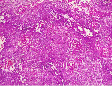
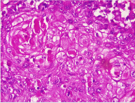
A diagnostic breast USS with a 5 MHz probe was done. A thickwalled 12 cm cystic lesion with multiple septations was seen. It has a 3 cm solid component, with no calcification or increased flow on Doppler interrogation. A fine needle aspiration was done, about 60 mls of straw-coloured fluid was drained and cytological analysis of the aspirate showed suspicious ductal cells. A wide local excision with axillary dissection and immediate breast reconstruction was done. Histopathological analysis showed malignant squamous epithelial cells that are disposed in sheets and nests with focal areas of keratin pearls (Figure 1A). The cells are polygonal with intercellular bridges (Figure 2B). Immunohistochemical staining with HMW Cytokeratin Antibody was positive. Immunohistochemical staining for focal Carcino-embryonic-antigen and E-cadherin was negative. The resection and deep margins are free of tumour and only the enlarged lymph node showed metastatic deposit. No adjuvant chemotherapy or radiotherapy given. Immunohistochemical study of molecular profile showed positivity for oestrogen only, progesterone receptor was negative and there is no over expression of the Her2-Neu receptor. Patient is currently on adjuvant selective oestrogen receptor modulator (Tamoxifen) at 20 mg daily for a two and a half years and will be subsequently switched to an Aromatase inhibitor {Anastrazole) for the same duration. Patient is to continue with follow-up visits to the surgical-out-patient-department. An informed consent was obtained from the patient for publication of the case report.
biomedres-openaccess-journal-bjstr

Figure 1: Photomicrographs of malignant squamous epithelial neoplasm that are disposed in sheets with focal areas of keratin pearls (A), A=X100.
biomedres-openaccess-journal-bjstr

Figure 2: The cells are polygonal with intercellular bridges (B); H and E stained, B=X200.

Discussion

There is no consensus on the origin of squamous cell carcinoma, whether it is a pure form of squamous cell carcinoma or it represents an extreme squamous metaplasia of an existing adenocarcinoma [39]. It is thus classified in to primary pure SCC, like our reported case and a mixed type (Figures 1 and 2). The primary pure SCC is characterised by the following features: The predominance (>90%) of malignant cells being of the squamous cell type, having no direct relation with the overlying skin of the breast, nipple and the areola, and the absence of evidence for a primary in an extra mammary site [40,41]. The mixed type is seen following a squamous metaplasia of an existing adenocarcinoma or a metastasis from an extra mammary origin [42]. The primary Squamous cell metaplasia is known to occur in benign epidermoid, dermoid and sebaceous cysts of the breast [43], as seen in our patient. It may arise within a long standing Fibroadenoma [44]. Primary squamous cell carcinoma of the breast has been observed to arise from squamous metaplasia of ductal epithelium in benign disorders of the breast, such as chronic abscess, after implantation of breast prosthesis and after radiation therapy [45-49]. Although there was USS evidence of a cystic mass in our patient, there was no history suggestive of chronic inflammation, use of implant for breast reconstruction or administration of therapeutic ionizing radiation.

The reported age of high incidence was the 5th -6th decades of life, with an average age of 50 years [50]. Our patient presented at the age of 43 years, almost a decade younger than the age in developed countries. The average age at presentation is 37.3 years in Enugu, south-eastern Nigeria [50]. Like the frequently seen adenocarcinoma, squamous cell carcinoma of the breast is also predominantly seen in post-menopausal women worldwide [4], but, some pockets of studies have reported seeing SCC in women within their gestational or lactating periods [23,32,51]. The decade age gap in the age of occurrence of SCC between women of Africa and those in Europe and North America has been observed by several authors in Nigeria and the sub Saharan Africa. The mean age of the patients at Calabar, South-South of Nigeria was 45.06 years, with an age range of 23 to 76 years [52]. This is similar to reports of the common age of 31-40 years in Lagos, South Western Nigeria [53], 40-49 years in Maiduguri, North Eastern Nigeria [54], and 40-49 years in Ghana, West Africa [55]. The reported ages in developed countries are 40-50 years in Hong Kong, 75-85 years in Maryland-USA and a median age of 62 years for all the histological subtypes [56].

Majority of patients with SCC present with a breast lump, usually a solid mass [25,57], but a significant proportion present with a fluctuant cystic mass, mostly a malignant epidermoid or sebaceous cyst or even a chronic breast abscess with a solid malignant component [58-61]. Our patient presented with a complex mass consisting of both solid and cystic components, probably a benign breast cyst with subsequent squamous metaplasia. Majority of SCC of the breast are said to be biologically aggressive, characterized by rapid growth over short duration and subsequent cystic and necrotic degeneration [28,60,61]. The index patient noticed such rapid growth with associated nodule formation just 2 months prior to presentation. Like mucinous adenocarcinoma, Squamous cell carcinoma of the breast is reported to be bulky, most often measuring greater than 5 cm (T3) in its widest dimension [59,60,61]. Our index mass measured 10 cm in its widest diameter. Many reports have indicated the preponderance of the left breast as the site of squamous carcinoma more than the right [43]. Our patient also presented with a left breast mass. Wynder et al. have reported the tendency for left-sided preponderance of the carcinoma of the breast of all types.

No identifiable risk factors were noted in the index patient, except for the age, presence of a breast cystic mass and long term steroid therapy. It has been noted above that most SCC in the African women are seen in the 4th decade of life [52-55], just like the age of 43 years at presentation for our patient. The presence of a cystic mass also reinforced the suspicion of a squamous metaplasia of the epithelium on the cyst wall, as reported by many authors [58,59]. The use of steroid and immunosuppressive drugs for more than 10 years could have impaired the body’s ability for immunologic surveillance, to arrest progression from dysplastic state and to halt lymphatic metastasis. Our patient presented with a clinically palpable ipsilateral axillary lymph node that showed positive metastasis on histology. Surprisingly, most reports indicated that Squamous cell carcinomas do not readily metastasize through the lymphatics like the adenocarcinomas. In fact, in only 10-30% of cases is lymph node involvement encountered intra-operatively [26,42,45]. However, up to 30% of the patients may harbour distant metastases at the time of presentation [26,42].

A diagnostic breast USS with a 5 MHz probe was done. A thickwalled 12 cm cystic lesion with multiple septations was seen. It has a 3 cm solid component, with no calcification or increased flow on Doppler interrogation. Other authors have reported also that there are no typical findings on the mammogram and a breast USS may only reveal a complex cystic mass with solid component or an inflammatory process [42]. Calcification and Cystic changes are often the frequent radiological finding [24-26,58]. A fine needle aspiration was done, about 60 mls of straw-coloured fluid was drained and cytological analysis of the aspirate showed suspicious ductal cells. A Tru-cut biopsy is the preferred method of obtaining a pathological diagnosis, as fine needle aspiration study is often not helpful [42]. In a study done by Gupta RK et al, very few patients have the histological diagnosis confirmed via a fine needle aspiration [62,63]. A wide local excision with axillary dissection and immediate breast reconstruction was done. Histopathological analysis showed malignant squamous epithelial neoplasm that are disposed in sheets and nests with focal areas of keratin pearls (Figure 1A). The cells are polygonal with intercellular bridges (Figure 2B). The reported histological evidence of a primary SCC of the breast is the microscopic appearance of malignant cells composed of infiltrative nests of atypical epithelial cells with irregular and hyperchromatic nuclei. There may be variable amounts of keratinous lamellar or a pseudo-sarcomatoid stroma [64-68]. It may have associated Ductal Carcinoma In-Situ or squamous metaplasia of ductal epithelium at the periphery of the invasive carcinoma [23,25,26,59,69]. Immunohistochemical staining with HMW Cytokeratin was positive. immunohistochemical staining for focal Carcinoembryonic- antigen and E-cadherin was negative. For adequate treatment, it is imperative to separate cutaneous SCC from primary SCC of the breast. Apart from the requirement of the predominance (>90%) of the squamous malignant cells in all the arrears of the tumour, the tumour should have no continuity with overlying epidermis or dermal appendages [31]. Immunohistochemical staining has emerged as an important diagnostic tool in making such distinction. Both the pure primary SCC of the breast and the SCC extending in to breast parenchyma from overlying skin stain positive with HMW-CK antibody. But, only primary pure SCC shows diffuse positive staining for CK 7, CK 8 and CK19 Antibodies [33- 36]. The distinction of pure primary SCC from secondary SCC from extra mammary sites such as the lungs is also difficult, because SCC differentiation demonstrate identical immunohistochemical staining in every organ in the body [21]. The demonstration of MAP2K4 gene mutation in a biopsied specimen should confirm an extra mammary origin, especially a lung SCC [21,70].

The resection and deep margins are free of tumour and only the enlarged lymph node showed metastatic deposit. No adjuvant chemotherapy or radiotherapy given. Literature review has shown that surgery is the mainstay of the treatment and a definitive role for adjuvant therapy has not been determined [24-26, 57- 59]. Both adjuvant radiotherapy and chemotherapy have been used with variable outcomes [24,26,47,57]. Majority of reports did not demonstrate a superior oncologic outcome with the use of adjuvant radiotherapy in primary SCC of the breast in stark contrast to extra mammary primary SCC [28,71]. Some studies have demonstrated an objective response and subsequent down staging of locally advanced pure primary SCC of the breast with the use of combined Neoadjuvant chemotherapy and radiotherapy [57,58]. Immunohistochemical study for molecular profile showed positivity for oestrogen only and progesterone receptor was negative, with no over expression of the Her2-Neu receptor. Patient is currently on adjuvant selective oestrogen receptor modulator (Tamoxifen) at 20 mg daily for two and a half years and will be subsequently switched to an Aromatase inhibitor for the same duration. The pure primary Squamous cell carcinomas are known to be mostly hormone receptor negative [40,41,46]. The over expression of c-erbB-2 is also usually not seen, but, some few reports have indicated an overexpression [69,72]. Patient is to continue with follow-up visits to the surgical-out-patient-department.

Because of its rarity, no much data has been published concerning the disease free survival and overall survival after curative treatment. A single-centre, small sample-size, retrospective study reported a 5-year survival of 67% in [26]. Menville has reported that prognosis is dependent upon the tumour biology and grade and may vary between deaths within 4 months in metastatic disease, to 16 years in early disease with curative resection [29]. Although the clinical course and prognosis of the SCC of the breast have remained obscure, many reports have considered a young age below 40 years, large tumour size and a histologically proven nodal metastasis to be markers of poor prognosis [25,26,57,71].

Conclusion

Despite its rarity, a primary squamous cell carcinoma of the breast is of clinical significance, because it can masquerade as a benign mass or arise within an existing adenocarcinoma. It’s often high histological grade, lack of hormone receptor positivity and often poor response to adjuvant care make its treatment herculean. Every benign breast mass removed must be subjected to pathological analysis, especially chronic cysts and abscesses.

For more Articles on: https://biomedres01.blogspot.com/

Wednesday, September 21, 2022

Parenteral Vs Enteral Nutrition as an Improvement in Wound Healing in the Severely Burned Patient in the ICU

Parenteral Vs Enteral Nutrition as an Improvement in Wound Healing in the Severely Burned Patient in the ICU

Introduction

Burns are defined as injuries produced in living tissues, due to the action of various physical agents (flames, hot liquids and objects, radiation, electric current, cold), chemical (caustic) and biological, which cause alterations ranging from a simple erythema until the total destruction of the structures [1]. Severe burn induces the patient to severe oxidative stress, a systemic inflammatory response, increased and persistent hypermetabolism and hyper catabolism with secondary sarcopenia, as well as organic dysfunction, causing sepsis, increasing mortality and energy deficit, the negative balance of proteins, antioxidant micronutrient deficiency during thermal stress giving poor clinical results [2]. Those with relevant criteria such as a severity index greater than seventy points, or with second or third degree burns as a whole, with greater than twenty percent of the total burned body surface (STCQ), patients under two years of age, or adults over sixty-five years of age with ten percent or more second or third degree burns, as well as all patients with respiratory or smoke inhalation burns, high voltage electrical burns, burns associated with multiple trauma and burns with associated serious diseases [3]. Therefore, the metabolic consequences of burns (especially those that exceed 20%- 30% of the STCQ), constitute a permanent challenge for those involved in the treatment of these, in their desire to achieve a prolonged survival, the successful rooting of the grafts, and adequate rates of tissue repair and healing [4].

The hypermetabolism that occurs due to the burn is immediate: increased susceptibility to infection, development of sepsis, multiple organ dysfunction, poor wound healing, loss of placed grafts, and eventual death; adding insufficient intake of energy, nitrogen, and micronutrients [4]. Caloric demands in critical condition are very high, assuming that it is accompanied by a large increase in basal metabolic output, that is, that the critically ill patient is strongly hypermetabolic. Therefore, when patients in a state of serious stress due to trauma, sepsis, burns or critical illness, they exhibit an accelerated catabolism of body proteins, and an increase in the degradation and trans nomination of branched chain amino acids in the skeletal muscle, with the consequent increase in the generation of lactate, alanine and glutamine, and a large flow of these substrates between the muscle (periphery) and the liver (central organ). The metabolic consequence is a marked elevation of glucose production in the liver, a process called gluconeogenesis. Gluconeogenesis prevents the accumulation of endogenous substrates from catabolism, which have no other way of purification. It also makes glucose available to those organs that depend on it for energy, such as the brain or bone marrow. Patients with severe sepsis, multiple trauma, or extensive burns require a higher protein intake of the order of 2.5 to 3.0 g / kg per day. So, when a burn occurs, gastric function is inhibited due to catabolism, since as mentioned above, it is necessary to have proteins and energy for the different organs of the patient’s system, and the absorption necessary to maintain an energy balance is deficient.

In the same way, during this process, they present hyper catabolism and hypermetabolism to compensate the hemodynamic state due to the increase in cardiac output, it is carried out by intestinal ischemia due to the burn, which affects the nutritional status of the patient and the healing of the wounds [1]. Also, the metabolism of PGQ can increase more than twice that of an unstressed subject and cause a significant depletion of lean body mass in the weeks following injury. Survivors may exhibit sequelae that will require prolonged specialized surgical treatments, therefore, the nutritional support in the PGQ acts by consuming the energy of the diet itself, avoiding hypermetabolism, ensuring the best response of the host to aggression, reducing the risk of complications, of skin grafts, the support of tissue repair and healing [4]; together with the shortening of the hospital stay and its impact on the Adult Intensive Care Unit (ICU). Consequently, the hypermetabolism observed after the burn is the result of the hormonal shift that under basal conditions would support the tissue deposition of substrates stimulated by the action of insulin, towards another that favors the mobilization and use of the stored substrates; and where anti-insulin hormones such as glucagon, cortisol and adrenaline participate. In this way, glycolysis and glucogenesis, fatty acid synthesis and deposition, and protein synthesis are inhibited; while glycogenolysis is activated, and gluconeogenesis in relation to muscle proteolysis [3].

On the other hand, it is important to mention that it has been shown that an accumulated caloric-protein debt throughout the stay in the ICU contributes to an increase in morbidity and mortality with a higher rate of infections, days of mechanical ventilation, as well as hospital stay. It has also been shown that one of the main consequences of poor nutrition is difficulty in healing due to the hypermetabolism that the critical patient presents at that time [4]. From the foregoing, it is concluded that nutritional disorders compromise wound healing and PGQ survival since there are decreased concentrations of minerals and trace elements in the blood of the burned patient, so nutritional therapy should be aimed at the replacement of the amounts lost as several weeks may elapse before serum concentrations of a particular micronutrient are restored to normal [4]. Therefore, the nutritional status of the patient has an important influence on the repair process, poor nutrition causes delays in healing. If the weight loss is above 30% of the appropriate weight, the deficiency of amino acids such as cystine and lysine will slow down the neovascularization process, the collagen synthesis process, and the final remodeling process [5]. The higher the percentage of total body surface area (SCT) affected, the greater the risk of developing systemic complications. In the case of patients who have complications due to an infectious process, they are exposed to the risk of sepsis and mortality, and it also causes local complications. Altered host defenses and devitalized tissue enhance bacterial invasion and growth.

The most frequent pathogens are streptococci and staphylococci during the first days and gram-negative bacteria after 5-7 days, although the flora is always mixed [5]. Among the main activities of the nursing professional in intensive care, feeding is a primary care. This is demonstrated in the mnemonic FAST HUG BID encompasses seven minimum aspects in critical patient care (diet, analgesia, sedation, thrombus prophylaxis, elevation of the head, prevention of stress ulcers and glucose control) [6]. Its compliance shows the improvement in the patient’s prognosis [6]. One of the nutritional therapies used in critically ill patients is enteral nutrition (EN), which is a nutritional support technique that consists of administering nutrients directly into the gastrointestinal tract through a tube; and parenteral nutrition (PN), which consists of administering nutrients to the body by venous and extradigestive routes [6], through specific catheters, to meet metabolic requirements [7]. EN is indicated when the patient in all cases in which the patient requires individualized nutritional support and does not ingest the necessary nutrients to meet their requirements [7]. On the other hand, PN is indicated when the needs are greatly increased and the patient is not able to cover them with the intake, when the patient does not tolerate the intake due to hemodynamic alterations such as heart or respiratory diseases such as bronchial dysplasia. Similarly, in patients who are unable to swallow due to oropharyngeal disorders, to take special foods with a bad taste and essential such as amino acid diseases or who cannot have prolonged fasting times: glycogenosis, alterations in the oxidation of fatty acids [7].

Having a contraindication in patients with intestinal obstruction [7]. The latter is used in patients with alterations in the gastrointestinal tract, being in many cases the only way to provide nutritional support, it is indicated in patients whose gastrointestinal tract is not usable for the administration, digestion, or absorption of nutrients, for a period greater than five days or when the gastrointestinal tract is usable, but it is desired to remain at rest for therapeutic reasons [8]. However, it is also associated with metabolic, infectious, and mechanical complications, which increases mortality and deterioration in the quality of life of the PGQ [8]. Within its content, PN requires water (30 to 40 ml / kg / day), energy (30 to 35 kcal / kg / day, depending on energy expenditure: up to 45 kcal / kg / day in critically ill patients), amino acids (1 to 2 g / kg / day, depending on the degree of catabolism), essential fatty acids, vitamins, and minerals [9]. Usually 2 L / day of the standard solution is needed. Solutions can be modified based on laboratory results, by underlying disorders, hypermetabolism, or other factors [9].

Justification

Burns are considered a public health problem worldwide, causing approximately 180,000 deaths per year, mostly in lowand middle-income countries [10]. According to the World Health Organization (WHO), patients with the highest risk of exposure are usually women, however, the injury rate is relatively higher in men than in women [10]. In the United States in 2016, an estimated 500,000 people suffer from unintentional burns annually, 80% of them (40,000 people) require hospitalization [11]. According to data from the National Fire Protection Association during 2005, in the United States, 600,000 people were burned, of which 25,000 required hospital handling and 4,000 of them died [12]. At the national level, the National Epidemiological Surveillance System reported in 2011 that burns ranked 17th in frequency of new cases of disease with a total of 129,779 patients with burns, which generated an overall national incidence of 118.82 (113.25 in women and 124.61 in men). By age group, new cases were more frequent (in decreasing order): from 25 to 44 years (43,321 cases), from 1 to 4 years (13,864 cases) and from 20 to 24 years (13,816 cases) [12], while in 2013, 126,786 new burn cases were reported, and from January to June 2014, 65,182 cases. Of these burns, 56% occurred in adults between 20 and 50 years of age and 32% in children between 0 and 19 years of age. Similarly, 85% of burns in adults occurred while carrying out work activities, while burns in children occurred in 90% of cases, within their homes, of which 80% were due to hot water (eleven).

PGQ medical care is expensive due to pre-hospital and hospital expenses (including the costs of consumable biotechnology, paraclinical studies, drugs, nutrition, etc.), estimating costs per patient from 30 thousand to 499 999 pesos [12]. It has been observed that both types of nutrition are used for severely burned patients, however, it is necessary to determine the nutrition that contributes to healing, as well as the Knowledge Generation and Application Line (LGAC) that supports the Program. Educational “Care for the person in critical condition” which belongs to the Quality and Nursing Care Research Group [13]. The healing process is complex and multiple factors influence it, such as nutrition. Nutrition and healing are closely related, in this way specific nutritional deficiencies may cause a delay in the progression of healing [13]. When mentioning healing, it is very important to identify up to which layer of the skin is burned or damaged, in order to identify the healing time according to the nutrition administered. In the same way, it can be determined that according to the characteristics of the wounds, the time is determined, however the healing ranges from 4 to 12 weeks for their complete healing. The previous analysis determines the importance of basing the care of the PGQ in relation to parenteral vs enteral feeding in order to base the care provided by the nursing staff specialized in intensive care, to achieve wound healing in the shortest time possible, so as to avoid the morbidity and mortality of PGQ in the Adult Intensive Care Unit [14].

Objective

To compare whether parenteral vs enteral nutrition improves wound healing in the severely burned patient in the Adult Intensive Care Unit.

PICO

Description of the Problem

Due to the hemodynamic instability and the high demand for care it requires, the PGQ merits admission to the ICU. These patients present hyper catabolism and hypermetabolism to compensate for the hemodynamic status due to the increase in cardiac output due to intestinal ischemia due to the burn, which affects the nutritional status of the patient and the healing of the wounds. However, burns care is now being provided to an increasing number of older adults who may have nutritional deficiencies, such as wasting and sarcopenia. These nutritional disorders could compromise the healing of the wounds and the survival of the patient and delay the evolution and have a poor prognosis. On the other hand, there are different nutrients that are very important in the evolution of the burn patient, for example, glucose, is the most important source for the healing process, among other nutrients, which in the same way help the physiological system. Due to burns, there is a significant loss of body weight, mainly of lean mass, which is transferred to immune compromise, delayed wound healing, infection, sepsis, organ failure, and death. The healing process is complex and multiple factors influence it, such as nutrition. Nutrition and healing are closely linked, in this way specific nutritional deficiencies may cause a delay in the progression of healing.

Addressing wounds in a comprehensive manner is the role of nursing, which is why it is important to know the current approach to nutrition in the PGQ [13]. It is important to mention that in the PGQ the small intestine can present affectations due to the systemic response due to the burn, and therefore inhibits the digestion and absorption of the necessary nutrients for the patient and in turn decreases wound healing [11]. The extent of injuries, pain, involvement of other organs and systems in burn injuries (such as the airways and lungs, which in many cases would involve intubation and mechanical ventilation) could make the use of nutrition impossible enteral and require the placement of venous enteral accesses to supply the estimated amounts of nutrients improving wound healing Field code changed [15]. Therefore, the decision of what nutrition to have in the PGQ will be decisive for your improvement in the ICU.

Questions that can be Answered

1. What is the best adequate nutrition for the critically burned patient?

2. What is the incidence of the population to be big burned?

3. Why does parenteral nutrition best benefit a severe burn patient?

4. After how many hours, is it necessary to administer parenteral nutrition to the severely burned patient?

5. What impact does parenteral nutrition have on a severe burn patient?

6. What is the most important nursing activity in the nutrition of the great burn patient?

7. What benefits can parenteral nutrition bring to the severely burned patient?

8. What nutrients are the most important in a large burn patient on parenteral nutrition?

9. How is the epidemiology in the great burn patient and what is its incidence?

10. How important is parenteral nutrition in severe burn patients?

Analysis of The Question with Its Components

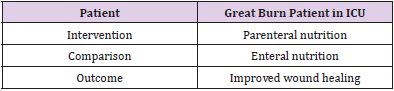
After analyzing the questions that could be answered, the one that best described the elements of interest of the present study was selected, which is presented in the following (Table 1).

biomedres-openaccess-journal-bjstr

Table 1: Elements of the PICO.

Wording of the Question

Does parenteral nutrition compared to enteral nutrition improve wound healing in severe burn patients in ICU?

Search Methodology

Search Strategy

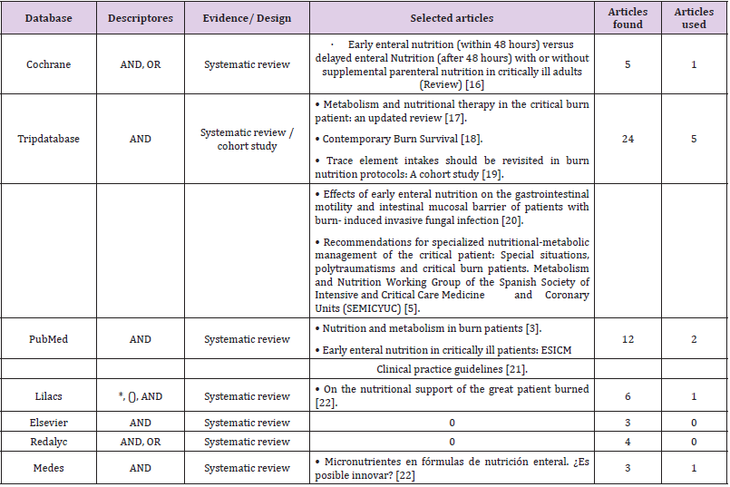
Regarding the search strategy, after having raised the problem and taking into consideration the elements of the PICO (Patient, Intervention, Comparison, Outcome), the terms were identified and translated into a documentary language, adapting to the Health Sciences Descriptor (DeCS), the Medical Subject (MESH) as well as free terms, to have a controlled language for effective search (Table 2). These terms were raised in both English and Spanish and Portuguese. It is worth mentioning that the information search was carried out in two stages: the first, through databases of secondary sources (Cochrane and Tripdatabase) and the second stage in databases of primary sources such as the Virtual Health Library (VHL) portal, United States National Library of Medicine (PubMed), Elsevier, Redalyc, Lilacs and Medicine in Spanish (Medes). In the same way, the AND and NOT and limiters * and () were used as Boléan operators to obtain more specific results. Studies related to adults with severe burn injuries and nutritional therapy in patients with severe burn injuries, which have a preferential time of less than five years of publication, and in other options up to ten years old were considered according to their level of evidence and grade. recommendation.

biomedres-openaccess-journal-bjstr

Table 2: Translation of the question into documentary language.

Note: *DeCS; **Mesh; ***Free term

Databases Queried

A query of information in databases was analyzed in the period from November 4, 2020 to February 22, 2021, 63 articles of interest were found in the databases, Cochrane, Tripdatabase PubMed, Redalyc, Lilacs, BVS, Elsevier and Medes. These articles were selected through critical reading to determine the level of evidence and grade of recommendation. With this, discarding all those that are not related to nutrition therapy in patients with great burns, obtaining a total of ten articles used [16-22]. To synthesize the evidence found, the Scottish Intercollegiate Guidelines Network (SIGN) scale was used to assess and establish the grades of recommendation according to the level of evidence (Table 3).

biomedres-openaccess-journal-bjstr

Table 3: Translation of the question into documentary language.

Note: Evidence resulting from the query made in databases

Results

Relevant Studies

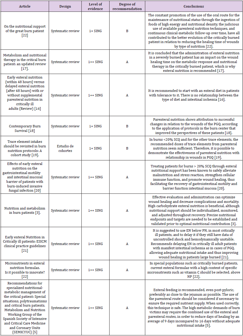
Of the 63 articles located with the aforementioned strategies, ten were used when meeting the desired criteria with the use of templates for the analysis of clinical trials and systematic reviews with the use of the “Critical Appraisal Skills Program” (CASPe) and with the Scottish Intercollegiate Guidelines Network (SIGN) scale to assess and establish the degrees of recommendation according to the level of evidence. Taking articles from systematic reviews and cohort studies, there are no experimental studies that verify the usefulness of both nutritional’s and the greater usefulness of either of these. The analysis of two out of ten articles determined that nutritional support is necessary as soon as resuscitation and resuscitation of the PGQ are completed, and hemodynamic stability and tissue perfusion are ensured [5]. The oral route should be preferred to feed the patient, the placement of naso enteral tubes could be necessary in many of them to avoid gaps in the provision of nutrients [17]. However, the dietary prescription could also be supplemented with energy-dense enteral nutrients in order to satisfy the high nutritional requirements found in the burn. Immunomodulation diets incorporating antioxidants, glutamine, and nucleotides have been described for use in nutritional support of burn injuries, but the results obtained with their administration have been mixed.

According to current knowledge, enteral nutrition is of choice; Likewise, during the resuscitation phase, the PGQ should receive high doses of vitamin C, as well as parenteral antioxidant cocktail supplements, which should be administered for variable periods of time according to the SCTQ [5]. Likewise, enteral glutamine intake seems to be a safe strategy capable of optimizing therapy, although more robust evidence is needed to support its use, thereby reducing healing times from nine days to 16 days on average [5].

Synthesis of the Evidence Found

Once the evidence was obtained from the articles, the conclusions about nutrition in the PGQ were compared.

On the other hand, it is based in a specific way on the methodological and design aspects, but not on the dimension of the perspective of suffering a disease or considering the economic implications of the recommended measures; situation that puts at risk the feasibility of its use in Latin American medical practice. Therefore, the articles are compared, as well as the conclusions obtained in each one of them (Table 4).

biomedres-openaccess-journal-bjstr

Table 4: Items with a grade of recommendation sing scale.

Implementation Plan

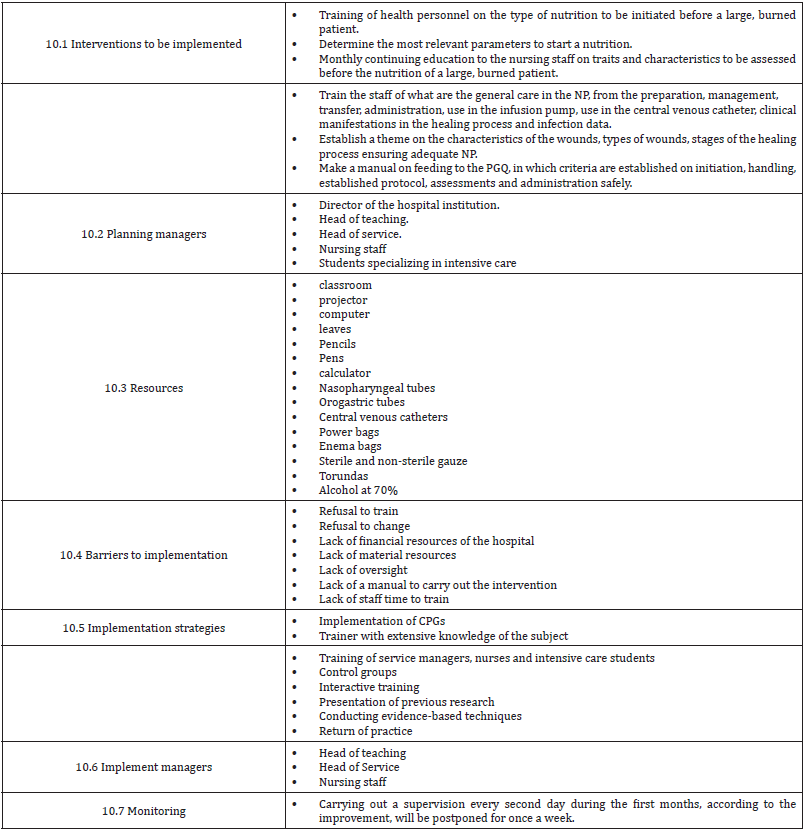
In this paragraph, note that it is a proposal by the authors for a possible implementation: The implementation of the research results is a long and complex process that requires effort on the part of all the agents involved, both patients, families, health professionals and the health system. Therefore, this section describes the necessary interventions to carry out (Table 5).

biomedres-openaccess-journal-bjstr

Table 5:

biomedres-openaccess-journal-bjstr

Table 6.

biomedres-openaccess-journal-bjstr

Table 7.

Schedule

Evaluation Plan

Conclusion

Once the topic was finished, it has been possible to reach the conclusion that the nursing staff carries out, being fundamental in the clinical part about the type of nutrition applied to the great burn patient. The evidence shows that the type of diet to be applied to a PGQ is preferably NE over PN. Making a high caloric intake, supplementing with Vitamin C, Glutamine among other proteins. Parenteral nutrition is an option for severe burn patients with intestinal ischemia, although it is recommended to start with a bowel rest to prioritize EN. In case of not being able to provide enough caloric intake, supplement it with a parenteral diet, and in this way you would be having an adequate combination of both nutritional’s. Similarly, go to total parenteral nutrition, if and only if, the enteral option is not possible, as in the case of patients with burns in the face area or inhalation burns. Going to show that NE is effective in wound healing in PGQ, since it is dependent on the nutrients that can be provided, and not on the site or route that nutrition is administered.

Therefore, the following recommendations are analyzed:

• Identification of the PGQ with the characteristics according to the GPC.

• Monthly training of staff on the characteristics of wounds, stages, and processes of healing, and identify what type of nutrition is the most appropriate according to the framework established for staff.

• Design and / or implementation of research by nursing staff in relation to the nutrition of PGQ.

• Detection of intestinal ischemia.

• Nutritional rest of the patient until hemodynamically stable.

• Use of first choice NE.

• Use of complementary PN according to the characteristics of the patient.

• Use of total PN, only in cases of not being able to provide firstline enteral nutrition.

• Maintain the patient’s nutrition with high levels of glutamine in NE and PN, as well as other nutrients to be required to provide a positive metabolic effect in relation to wound healing. Therefore, it is suggested to carry out more studies in the field of nutrition in the burn patient, as well as more systematic reviews.

For more Articles on: https://biomedres01.blogspot.com/

Tuesday, September 20, 2022

Inflamyar™ Possesses Anti-Inflammatory Effect on Human Immune Cells and Cytokine Expression In Vitro

Inflamyar™ Possesses Anti-Inflammatory Effect on Human Immune Cells and Cytokine Expression In Vitro

Introduction

Since virtually all organisms are constantly exposed to the influences of the living environment, the immune system has a great importance for the physical integrity of humans. It protects against threatening external influences such as infestation by microorganisms and parasites, but also against threats from the inside of the body, like e.g. necrotic and apoptotic cell material as well as functionally degenerated cells [1,2]. The human immune system is made up of several components. A distinction is made between a cellular and a humoral part. The cellular immune system comprises highly specialized immune cells that are either mobile (e.g. in the blood) or located in various tissues, e.g. Monocytes, Granulocytes, B cells, T cells, NK cells. The humoral immune system is the part of the immune system based on plasma proteins (antibodies, complement factors and cytokines) [2,3]. Upon induction of an immune response, humoral components of the immune system are initially released by the cells located in the affected tissue. By secreting these factors, other immune cells are lured to the focus of infection [2,3]. The inflammatory response in the body is important for the resolution of the cause of the inflammation but is also the cause of the symptoms of the disease [4]. The immune response is not entirely specific to its cause; even healthy tissue is always damaged [5].

This is especially important when an acute inflammation becomes a chronic inflammation that does not succeed in elimination of the trigger. For example, this may be due to the presence of debris in the tissue or frequent overload of muscles and joints. This persisting inflammation can massively damage the surrounding, primarily healthy tissue [6,7]. The damage of healthy tissue leads to severe pain reactions in both acute and chronic inflammatory reactions [5,8]. Moreover, chronic inflammation can lead to neoplasms (carcinomas or lymphomas) in many organs and in the lymphatic tissue and promote their growth and vascularization. Causes include the chronic proliferation stimulus, the growth-promoting effects of cytokines and the genomic damage caused by reactive oxygen species produced by immigrated immune cells [9-11]. In such situations, use of anti-inflammatory agents to assist and reduce the side effects of inflammation is useful [12,13]. Among the most widely used anti-inflammatory drugs include i.e., synthetic substances such as cyclooxygenase inhibitors, steroids, immunosuppressants and cytokine inhibitors [14,15]. These substances are usually proven to be extremely effective - but in part also show a wide range of unwanted side effects [16-18]. For this reason, the active ingredients from classical medicinal plants are increasingly becoming the focus of research.

In particular, secondary plant metabolites are in the interest of science since these have been in use in traditional medicine for many centuries and thus relatively safe and side effects associated with their use often relatively low [19-23]. Plant metabolites have a wide range of pharmacological effects, such as high antioxidant, antiviral, anti-inflammatory and carcinogenic activity [24-26]. Antiinflammatory ingredients such as alkaloids, phenols, flavonoids, glycosides, terpenes, quinones, catechins and carbohydrates of aqueous extracts from various traditional herbs have been described in several studies [25,27-29]. Besides of direct antiinflammatory actions, an interaction with immunological signal cascades, such as the reduction of proinflammatory mediators by inhibiting transcription factors of gene expression (Nuclear Factor κB, Inhibitor of κB), has already been demonstrated for some of these substances [30-32]. These modulatory effects of plant substances are particularly interesting for research. The individual effects of active ingredients from plant extracts are very diverse and have so far been described inadequately. For this reason further investigations are mandatory. In this study, the anti-inflammatory effect of Inflamyar™, a commercially available homeopathicspagyric product consisting of plant extracts from Arnica montana, Bryonia cretica, Guajacum, Toxicodendron quercifolium, Bellis perennis, Ledum palustre, Ruta graveolens and Viscum album was evaluated.

Materials and Methods

All experiments were conducted by NIS Labs, Klamath Falls, USA.

Test Substance

The test substance, Flamyar™, is a homeopathic spagyric natural remedy manufactured by PEKANA Naturheilmittel GmbH (Kißlegg, Germany) and distributed in the USA under the name Inflamyar™. The test substance was developed for the treatment of sports injuries, sprains, joint problems, bruises, and muscle strains. Active ingredients are Arnica montana spag. Peka Dil. D12, Bryonia cretica spa. Peka Dil. D4, Guajacum Dil. D4, Toxicodendron quercifolium Dil. D12, Bellis perennis spag. Peka Dil. D8, Ledum palustre Dil. D4, Ruta graveolens spag. Peka Dil. D6, Viscum album spag. Peka Dil. D4.

Reagents

Histopaque1077 and Histopaque1119, RPMI1640, 200mM l-glutamine, antibiotics, fetal bovine serum, BSA (bovine serum albumin), fibronectin, and PBS (phosphate buffered saline) were purchased from Sigma-Aldrich (St. Louis, MO, USA). Sodium azide (NaN3) was acquired from LabChem, Inc. (Pittsburgh, PA, USA). CD3 (peridinin chlorophyll protein), CD25 (brilliant violet 421), CD56 (phycoerythrin) and CD69 (fluorescein isothiocyanate), antibodies as well as heparin vacutainers were ordered from BD Biosciences (Franklin Lakes, NJ, USA). The protein multiplex array (27-Plex human cytokine Bio-Plex Pro™) was obtained from Bio- Rad Laboratories Inc. (Hercules, CA, USA).

CD69 Activation Marker Expression on Human Leukocyte Subsets

Peripheral whole blood from human healthy adult donors (n=3) was obtained upon written informed consent approval by the Sky Lakes Medical Center Institutional Review Board, Federalwide Assurance 2603. Heparinized blood samples were placed on gradient solution (Lympholyte-Poly) and centrifuged at 450 × g for 35 minutes. The layer containing PBMC (peripheral blood mononuclear cells) was separated, washed twice with 10 ml PBS (without Ca / Mg) and resuspended in RPMI 1640 (containing 10% fetal calf serum, l-glutamine and antibiotics (P/S) at a cell density of 106/ml. Two parallel culture conditions were used:

a) Adding of serial dilutions of test product without any other stimuli to test the direct immune modulating effect

b) Adding of serial dilutions of test product, followed by addition of an inflammatory insult in the form of bacterial endotoxin LPS (lipopolysaccharide), to assess the ability of test product to reprogram the human immune cells to respond differently to inflammatory stimuli.

Triple cultures were established for each test condition. Negative controls (untreated cell cultures) were established with n=6. Positive control cultures (2x n=3), n=3 containing 10 ng / ml LPS and n=3 containing 100 IU / ml IL-2 for immune cell activation via two different ways.

After an incubation time of 24 hours at 37°C and 5% CO2, blood cells were isolated and stained for 15 min with fluorochromeconjugated monoclonal antibodies at suppliers recommended concentration and then analysed via Attune acoustic-focusing flow cytometer (Thermo Fisher Scientific). Data analysis was performed using electronic gating based on cell size and granularity to distinguish lymphocytes and monocytes, allowing separate analysis of CD69 expression on lymphocyte subsets opposed to monocyte/ macrophage cell subsets. The subpopulation of lymphocytes was then analysed for CD69 expression on CD3-CD56+ NK cells. Costaining with CD3 and CD56 allowed further detailed analysis of four separate lymphocyte subpopulations, namely CD3-CD56+ NK cells, CD3+ CD56- T lymphocytes, and CD3+CD56+ NKT cells. The combination also allowed us to analyze the CD3-CD56- non-T non-NK lymphocytes for activation markers. For each of these populations we examined the expression level of the CD69 activation marker.

Cytokine Production in Peripheral Blood Mononuclear Cell Cultures

Cell culture supernatants were obtained from 24-hour culture setup described above. Expression levels of the following cytokines were tested: Interleukin (IL)-1β, -1ra, -2, -4, -5, -6, -7, -8, -9, -10, -12 (p70), -13, -15, -17, eotaxin, basic Fibroblast Growth Factor (FGF), Granulocyte-Macrophage Colony Stimulating Factor (GMCSF), Granulocyte-Colony Stimulating Factor (G-CSF), Interferon γ (IFN-γ), interferon γ -Induced Protein 10 (IP-10), Monocyte Chemoattractant Protein 1 (MCP-1), Macrophage Inflammatory Protein (MIP)-1α, MIP-1β, Platelet-Derived Growth Factor (PDGF)- BB, Regulated On Activation, Normal T Cell Expressed And Secreted (RANTES), Tumor Necrosis Factor (TNF)-α, and Vascular Endothelial Growth Factor (VEGF). The assay was analysed using magnetic protein multiplex arrays (BioPlex, Bio-Rad Laboratories Inc.) and xMAP technology (Luminex, Austin, TX, USA).

Statistical Analysis

Calculations and statistical analysis was performed using the two-tailed, independent t-test using Microsoft Excel.

Results

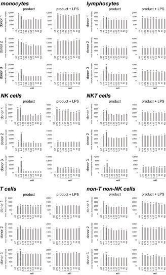
Immune cells harvested from human peripheral blood were used as a model for potential activities on immune activating and modulating. On the one hand, the direct activation of immune cells by the test substance, and on the other hand, the priming of immune cells to respond differently to a subsequent inflammatory insult was analyzed. Therefore, two sets of cell cultures were examined in parallel, on the one hand the highly inflammatory bacterial LPS from Escherichia coli and on the other hand the recombinant Interleukin-2 (IL-2) for activation was used as a positive control, and LPS was additionally used in one of the two cultures to induce inflammation after treating the immune cells with the test substance. As indicator for immune cell activation, the activation marker CD69 (cluster of differentiation 69) was chosen. In the case of lymphoid activation, CD69 is the earliest inducible surface glycoprotein and leads to lymphocyte proliferation and signal transmission at the cellular level [33,34]. Incubation of the cell culture with the test product (Figure 1) led to a slight increase in CD69 expression on monocytes (-6.3 to 28.8%) and lymphocytes (0.8 to 10.2%). Either no effect or else a slight increase was seen on Natural Killer (NK) cells (-4.1 to 24.7%), Natural Killer T (NKT) cells (-6 to 14.3%), non-T non-NK lymphocytes (-4.3 to 12.5%) and T cells (2.8 to 8.6%).

biomedres-openaccess-journal-bjstr

Figure 1: CD69 expression on immune cells in 24-hour cultures of peripheral blood mononuclear cells treated with products alone (column “product”) or pretreated with product prior to the addition of the inflammatory insult LPS (column “product + LPS”) plotted as CD69 mean fluorescence intensity. Statistical significance is indicated on the bar graph (*p<0.05, **p<0.01).

Under inflammatory conditions, treatment of cultures with the test product led to a reduced CD69 expression in monocytes of all three donors at lower concentrations of the test product (-0.4 to 14.4%). The highest concentrations of the test substance showed an induced expression of CD69 up to 5.9%. The lymphocytes possessed a reduction of CD69 expression from -2.7 to 19.19% for all three donors. A 38.6 to 42.0% reduction was seen in NK cell activation for both donor 1 and donor 3; donor 2 also showed a reduction in NK cell activation but the response was more variable (ranging from -1.1 to 12.0%). Incubation of NKT cells with the test product prior to LPS stimulation resulted in a -6.3 to 19.2% reduction of CD69 expression for all three donors. T cells possessed a slight reduction in CD69 expression for both donor 1 and donor 3 (-0.2 to 5.3%); donor 2 showed a slight increase in T cell activation (5.3%) at the highest dose tested with no change at lower concentrations of the test product. CD69 expression in non-T non-NK lymphocytes showed a 2.0 to 24.0% reduction in CD69 expression for all three donors. A dose-dependency was seen for donor 1 and donor 3 on NK cells, NKT cells and lymphocytes; on T cells only for donor 3 and for all donors in non-T non-NK cells.

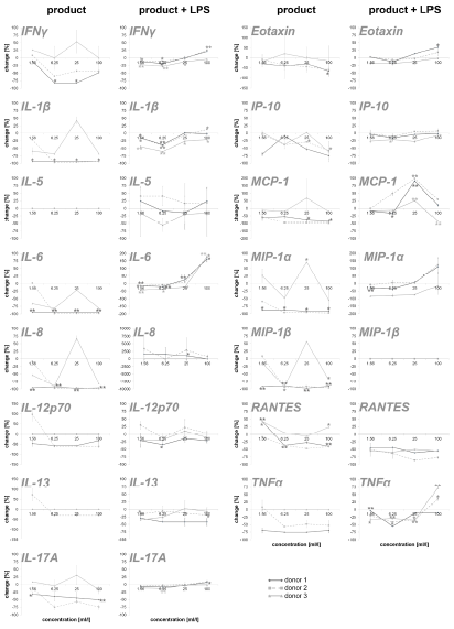
To obtain an overview of the effects of the test substance on the humoral components of the immune system, the test substanceinduced cytokine expression changes were analyzed on PBMC. In this part of the study, on the one hand, the direct effect of the test substance on PBMC cultures and, on the other hand, the effect of pre-incubation of PBMCs with the test substance and subsequent inflammatory stimulus (LPS) were tested. The exposure of immune cell cultures to the test substance without a subsequent inflammatory stimulus (LPS) resulted in a reduction in the cytokine IL-6 up to 98.9% (Figure 2). For two out of three donors (donor 1 and donor 2) the exposure of immune cell cultures to the test product resulted in consistent decreases in the following proinflammatory cytokines: INF-γ (8.5 – 82.2%), IL-1β (21.6 – 97.5%), IL-8 (8.6 –98.4%), IL-12p70 (46.5 – 61.4%; except lowest doses on cells of donor 2), IL-17A (12.0 – 75.7%), Eotaxin (28.4 – 65.4%; except lowest doses on cells of donor 2), IP-10 (3.9 – 74.7%), MCP-1 (55.0 – 97.7%), MIP-1α (58.8 – 97.3%), MIP-1β (13.8 – 97.2%; except lowest doses on cells of donor 2), RANTES (9.1 – 47.6%; in donor 1 and 2) and TNFα (52.4 – 75.6% in donor 1 and 2). The exposure of PBMC cultures to the test substance resulted in a reduction in the cytokine IL-13 in cultures from one donor (29.6%, without lowest dose) and no change in cultures from the other two donors. No change was seen for the cytokine IL-5 in PBMCs exposed to the test substance.

biomedres-openaccess-journal-bjstr

Figure 2: Percent change in proinflammatory cytokine levels in PBMC culture supernatants of three donors treated with serial dilutions of the test product in absence (column “product”) and presence (column “product + LPS”) of a subsequent inflammatory stimulus. Samples without inflammatory stimulus are compared to untreated control cultures; samples with inflammatory stimulus are compared to LPS-control (*p<0.05; **p<0.005).

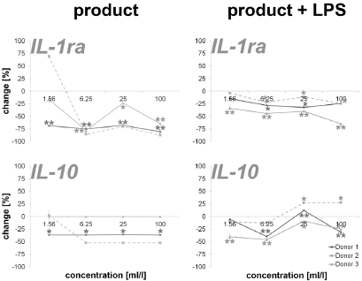
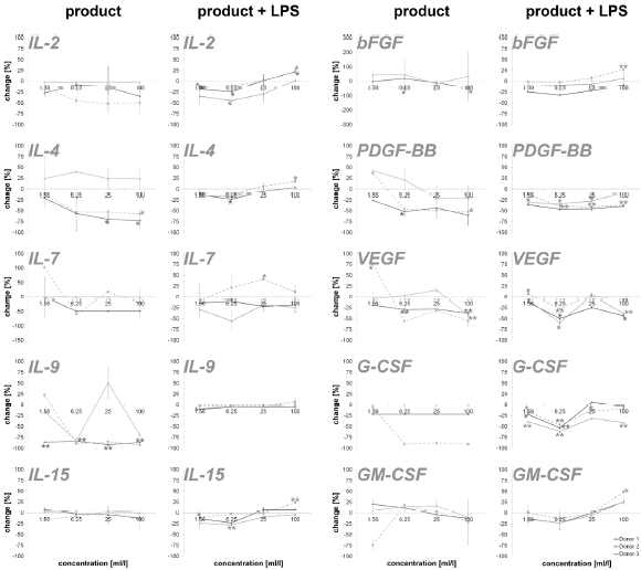
An incubation of PBMCs with the test substance resulted in a reduction of the anti-inflammatory cytokines IL-1ra (16.2 – 87.4%; except lowest doses on cells of donor 2) and IL-10 (35.8 – 52.2%; except lowest doses on cells of donor 2). The data is shown in Figure 3. Measuring cytokines with pro- and anti-inflammatory properties (Figure 4), the following reactions could be assessed: For two out of three donors (donor 1 and donor 2) the exposure of immune cell cultures to the test substance resulted in consistent decreases in IL-2 (8.6 – 51.9%), IL-4 (13.0 – 72.8%), IL-9 (85.1 – 92.5%; except lowest doses on cells of donor 2) and IL-15 (-8.0 – 16.2%) levels. The exposure of immune cell cultures to the test substance led to variable effects on IL-7 (-102.9 – 60.0%) levels. In the group of growth factors (Figure 4), for two out of three donors (donor 1 and donor 2) the exposure of PBMCs to the three highest doses of the test substance resulted in consistent decreases in PDGF-BB (26.2 – 60.3%; except lowest doses on cells of donor 2), VEGF (18.9 – 56.2%; except lowest doses on cells of donor 2) and G-CSF (18.9 – 56.2%; except lowest doses on cells of donor 2). Variable effects were detected in bFGF (-44.9 – 55.4%) and GM-CSF (-20.0 – 74.1%) levels. In total, a clear dose-dependency of cytokine expression could be seen in IL-17A, Eotaxin MCP-1, RANTES, IL-1ra, IL-4, IL- 15, bFGF, PDGF-BB, VEGF and GM-CSF in a minimum of two donors.

The exposure of PBMCs to the test substance with a subsequent inflammatory stimulus (LPS) led to consistent decreases in IL-13 (2.4 – 34.9%) and RANTES (43.8 – 85.7%). An incubation with the three lowest doses of the test substance led to consistent decreases in the following cytokines: INF-γ (0.0 – 26.3%), IL-1β (-1.4 – 41.4%), IL-17A (-9.1 – 7.8), IP-10 (-6.4 – 27.6%) and TNF-α (4.2 – 41.4; except highest doses on cells of donor 2 and 3). A consistent biphasic response, i.e. increases at higher doses and decreases at lower doses of the test substance, was seen for IL-6 (-177.4 – 19.3%) and Eotaxin (-35.2 – 12.8%). Variable effects were detected on IL-5 (-40.8 – 55.4%), IL-8 (-3356.6 – 79.8%), IL-12p70 (-97.6 - 58.8%), MCP-1 (-98.8 – 42.6%) and MIP-1α (-123.2 – 79.3 %) levels and no effects were seen on MIP-1β levels (Figure 2). Consistent decreases in IL-1ra (3.9 – 64.7%) and variable effects on IL-10 (-27-6 – 45.8%) levels were detected in PBMC cultures (Figure 3). The exposure of immune cell cultures to the three lower doses of the test substance led to consistent decreases in the following cytokines: IL-2 (12.0 – 44.5%), IL-4 (9.1 – 23.3%), IL-9 (1.8 – 10.2%) and IL-15 (2.8 – 27.9%); variable effects were found on IL-7 levels (-40.7 – 29.0%).

Under inflammatory conditions, the exposure of PBMC cultures to the test substance led to a decreased concentration of bFGF (9.3 – 24.4%; except donor 2), PDGF-BB (7.8 – 46.3%) and GM-CSF (-0.3 – 23.0%; except highest dose) in a concentration dependent manner. The growth factors VEGF (-7.5 – 58.8%) and G-CSF (-5.9 – 61.6%) showed variable results with a decrease in cytokine expression in two of three donors. According to the results from stimulation of PBMC without inflammatory stimulus, very consistent responses were seen for all three donors (Figure 4). Also in this group, a dosedependent cytokine expression was found in IFN-γ, IL-1β, IL-6, IL- 17A, Eotaxin, IP-10, MIP-1α, TNFα, IL-2, IL-4, IL-9, IL-15, PDGF-BB, GM-CSF and IL-10.

biomedres-openaccess-journal-bjstr

Figure 3: Percent change in anti-inflammatory cytokine levels in PBMC culture supernatants of three donors treated with serial dilutions of the test product in absence (column “product”) and presence (column “product + LPS”) of a subsequent inflammatory stimulus. Samples without inflammatory stimulus are compared to untreated control cultures; samples with inflammatory stimulus are compared to LPS-control (*p<0.05; **p<0.005).

biomedres-openaccess-journal-bjstr

Figure 4: Percent change in levels of cytokine with pro- and anti-inflammatory capacities and growth factors in PBMC culture supernatants of three donors treated with serial dilutions of the test product in absence (column “product”) and presence (column “product + LPS”) of a subsequent inflammatory stimulus. Samples without inflammatory stimulus are compared to untreated control cultures; samples with inflammatory stimulus are compared to LPS-control (*p<0.05; **p<0.005).

Discussion

In the present study, stimulation of immune cells with the test substance was done in the presence and absence of a subsequent inflammatory stimulus. Thereafter, CD69 expression, i.e., the activation of immune cells, was measured along with the cytokine expression. The data show no effects or only a slight induction of CD69 expression after stimulation of the immune cells with the test substance in the absence of an inflammatory stimulus. With the exception of the T cells, the immune cells showed only isolated inductions of CD69 expression compared to untreated controls. These inductions were distributed over the entire concentration spectrum of the test substance and no dose-dependency was seen. In addition, a reduced expression of proinflammatory cytokines was measured in groups without subsequent inflammatory insult. Noteworthy here is the particularly strong reduction of IL-1β, IL-6, IL-8, MIP-1α, MIP-1β and TNFα. The anti-inflammatory cytokines and growth factors also show a regulation in this context; in relation to the proinflammatory cytokines, however, this is more moderate. This suggests a shift of pro- and anti-inflammatory cytokines activity ratio towards an anti-inflammatory level. Together, the findings from CD69 and cytokine expression indicate an antiinflammatory effect of the test substance.

Upon incubation of immune cells in the presence of a subsequent inflammatory stimulus, a reduction in immune cell activation could be demonstrated for most of the samples in two out of three donors, indicating an anti-inflammatory effect of the test substance also under inflammatory conditions. Correlatively, a reduced expression of cytokines and growth factors the doses of 1.56 to 25 ml/l of test substance was measured. Interestingly, the results show an induction of cytokine expression in a concentration range of 25 to 100 ml/l of test substance, in particular of proinflammatory cytokines. This biphasic effect of the test substance (cytokine reduction at low levels and induction at high concentrations) is in contrast to the reduction of CD69 expression at this concentration. An association between reduced CD69 expression at high doses of the test substance and increased cytokine expression at the same dose, in addition to the shift in the balance between pro- and anti-inflammatory cytokines seen in the previous experiments, indicates an additional anti-inflammatory mechanism of action of the test substance. However, further investigations are needed to verify this.

Unfortunately, not all donors in this study showed an equal response to the test substance. In view of the individual variations in the immune status of the donors (e. g. genetic and epigenetic aspects, potential pre-existing conditions), high deviations in the data from primary cells of different donors are not unexpected [35- 37]. From this point of view, the results of this study are relatively consistent. The observed dose-dependent regulation of the CD69 and cytokine expression of individual groups clearly underlines the validity of the data. In addition, the data generated in this study is consistent with previously published data from other workgroups. For example, a significantly reduced in vitro LPS-induced expression of TNFα, IL-1 and IL-6 from the human whole blood culture and RAW-264.7 cells showed Mahajan et al. after incubation with different dilutions of A. montana and Bryonia species (6CH, 30CH, 200CH) [38]. Lussignoli et al. showed a significant reduction in systemic IL-6 expression in a traumatic animal model after the use of a homeopathic preparation containing A. montana and other plant extracts and minerals [39]. In another study, T. quercifolium in dilutions of 6CH, 12CH, 30CH and 200CH appeared to interfere with an histamine, prostaglandins and other inflammatory mediators driven inflammatory processes [40]. Anti-inflammatory actions due to inhibition of both lipoxygenase and cyclooxygenase metabolic pathways were also seen in homeopathic remedy containing A. montana and T. quercifolium [41]. Porozov et al. described reduced IL-1β, TNFα and IL-8 secretion without an effect on human T cell and monocyte proliferation by a homeopathic remedy containing A. montana and B. perennis [42]. In addition, various studies showed anti-inflammatory properties of various extracts out of A. montana [43-48], Bryonia species [48-50], T. quercifolium [40,51,52], B. perennis [53], L. palustre [54,55], R. graveolens [56-60] and V. album [61-65], which are also contained in the test substance of this study. In summary, the data of the present study possesses a clear antiinflammatory effect and thus a potential for the test substance for the treatment of acute or chronic inflammatory reactions.

For more Articles on: https://biomedres01.blogspot.com/

Antimalarial Aloe Compounds

  Antimalarial Aloe Compounds Introduction Among the most prevalent diseases caused by protozoan parasites, malaria is caused by parasites o...