Friday, July 21, 2023

Acute Appendicitis with Pyogenic Liver Abscesses

Acute Appendicitis with Pyogenic Liver Abscesses

Summary

Acute appendicitis is one of the most common surgical pathologies globally and its treatment has been well practiced for many years. However, clinically it can present in many different ways, from asymptomatic to septic shock and every variation in between. We present the case of a middle-aged man presenting with fever as his only symptom. Upon clinical examination, blood evaluation and imaging, an acute appendicitis with multiple pyogenic liver abscesses were found. The patient underwent surgery with removal of the appendix and drainage of the abscesses. With an antibiotic regiment over several weeks and drainage placement the abscesses could be drained successfully, and the patient remained asymptomatic during the follow-up over 6 weeks.

Background

Acute appendicitis is a very common surgical emergency. The laparoscopic removal is the standard of care all around the world. Despite the discussion of conservative treatment of acute appendicitis with antibiotics, the American College of Surgeons, the European Association of Endoscopic Surgery and the World Society of Emergency Surgery all recommend the treatment by appendectomy [1,2]. In an international collaboration, researchers found that 95.7% of patients with acute appendicitis were treated operatively [1]. The lifetime risk of acute appendicitis is 8.6 percent in males and 6.9 in females [3] and the mortality in developed countries lies between 0.09 and 0.24 percent [4]. Typical complications include the perforation of the appendix (observed in 13-20% of acute appendicitis cases), which can result in peritonitis [5]. More rarely, a perityphlitic abscess can form in the proximity of the appendix. The hematogenic spread of bacteria due to an acute appendicitis is possible and has been described. This can lead to the formation of abscesses in the liver through pyemia of the portal vein [6].

Liver abscesses are relatively rare and present 2.3 cases per 100,000 people [7-9], but they account for 48 percent of visceral abscesses and 13% of intraabdominal abscesses [10]. In half of the cases, an underlying disease of the biliary tract can be identified, and the abscesses are often formed following a pyemia of the portal vein due to bowel leakage and peritonitis [6,7,11]. The most common pathogens are Streptococci, especially the Streptococcus milleri group (S. anginosus, S. constellatus, S. intermedius) [12]. Other pathogens include E. coli, K. pneumoniae and S. aureus [13,14]. The symptoms include fever (90%) and unspecific abdominal complaints (50-75%) (6–8,15). Blood work findings usually include elevated alkaline phosphatase (70-90%), Bilirubin and liver enzymes (50%) [6,7,15].

Case Presentation

A 51-year-old male presented in our emergency department with a 4-day history of fever up to 39.0°C. He did not complain of pain, denied any nausea or diarrhea, and had no urinary symptoms. He had no trouble breathing, no coughing and no throat pain. A Covid-19- PCR-Test was negative. He presented with normal blood pressure, tachycardia with a pulse rate of 126/min, a body temperature of 37.7°C, an oxygen saturation of 97% and a breathing rate of 20 bpm. The patient had no personal history of illnesses, no previous operations and did not take regular medication. A blood sample revealed a leucocyte count of 21.48 G/l with predominantly neutrophil granulocytes (18.85 G/l) and a thrombocytosis of 731 G/l. The Acute-phase proteins were significantly high with a C-reactive protein of 237 mg/l, Procalcitonin of 10.48 ng/ml and Ferritin of 4593 μg/l. He also showed elevated liver enzymes (GOT 97 U/l, GPT 87 U/l) and elevated cholestasis parameters (y-GT 472 U/l, ALP 242 U/l and Bilirubin 47.7 μmol/l) upon which we performed a sonography of the abdomen.

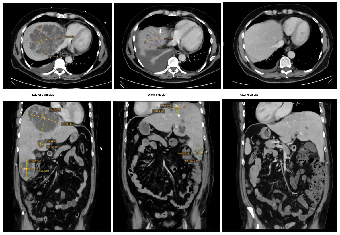
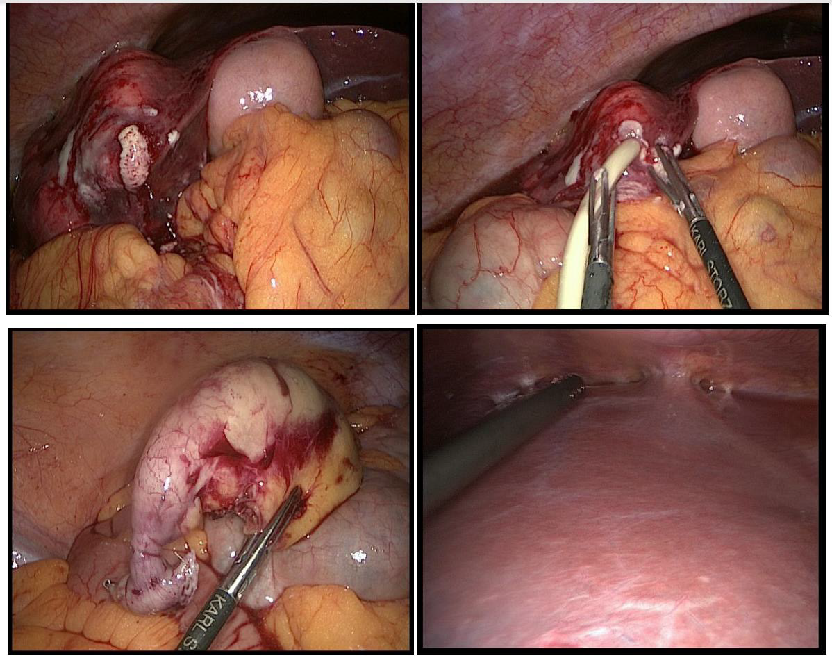
The ultrasound scan showed no evidence of cholecystitis or cholezystolithiasis, but multiple liver lesions, which warranted further investigation by computer tomography. The findings of the CT scan corresponded with the ultrasound images and showed multiple liver lesions and a possible appendicitis. The lesions were located in segment VI (6.4 x 7.2 x 6.1 cm) and subcapsular in the segments VII/VIII (11.0 x 12,1 x 7.3 cm) and were suggestive of liver abscesses. The working diagnosis of acute appendicitis with pyogenic liver abscesses was established. The patient was hospitalized. Blood samples with bacterial cultures, serology for parasites (amoeba, echinococcosis) and viruses (HIV, HAV, HBV, HCV) were obtained and the patient underwent surgery. The diagnostic laparoscopy showed an acute appendicitis without perifocal abscess formation. The appendix was removed at the cecal pole using a linear stapling device (Endo-GIA, 45 mm).

Upon inspection and palpation of the liver perforation of the caudal abscess with purulent discharge occured. A microbiotic sample was obtained and a drainage was placed in the abscess cavity, which quickly collected 300ml of pus. The cranial (subdiaphragmal) liver abscess was left untouched for later interventional drainageplacement. The blood culture showed growth of Streptococcus milleri, which coincided with the findings of the intraoperative sample from the abscess. An intravenous antibiotic regiment with Metronidazol and Ceftriaxon was initiated. 7 days after drain removal the antibiotic treatment with Ceftriaxon was changed to Amoxicillin and Probenecid according to the antibiogramm results.

The histology of the appendix showed pyogenic inflammation with no signs for malignancy, further supporting the working diagnosis of acute appendicitis with hematogenous liver abscesses. The serology (viruses and parasites) results all turned out to be negative. Upon interdisciplinary discussion and according to well established treatment practice [16], the cranial abscess was drained percutaneously (25cm, 10 French), which immediately released 135ml of pus. Further bacteriological samples were obtained, which again confirmed Streptococcus milleri. After a few days the drain was removed after complete drainage.During the hospitalization period, the patient remained afebrile and without any symptoms apart from general fatigue. The patient was discharged from hospital after 15 days with an oral antibiotic regiment containing Metronidazol, Amoxicillin and Probenecid.

Investigation Images (Figures 1 & 2)
biomedres-openaccess-journal-bjstr

Figure 1: 6 weeks course with evident size reduction of the liver abscesses.
biomedres-openaccess-journal-bjstr

Figure 2: Intraoperative situs: Top left and right: Spontaneus perforation of the caudal liver abscess with consecutive drainage insertion. Bottom left: Appendix (stapled and resected, apex on the right, basis on the left). Bottom right: View of the subdiaphragmal abscess with adhesions

Treatment

Antibiotic Treatment

Ceftriaxon i.v. 2g, 17.05-01.06.2020
(total duration 14d).
Metronidazol i.v. 1500mg, 17.05-20.05.2020
(total duration 3d).
Metronidazol p.o. 500mg, 20.05-08.07.2020
(total duration 49d).
Amoxicillin p.o. 3000mg, 20.05-08.07.2020
(total duration 49d).
Probenecid p.o. 1000mg, 20.05-08.07.2020
(total duration 49d).

Surgical Treatment

1. 17.05.2020: Laparoscopic appendectomy and drainage of liver abscess (total duration of drainage: 8 days).
2. 20.05.2020: Ultrasound-guided drainage of liver abscess (total duration of drainage: 6 days).

Supportive Treatment

1. Respiratory physiotherapy

Outcome and Follow-Up

The patient was seen weekly for follow-up examinations over the course of 6 weeks after discharge. During this period, the patient presented himself once with tachycardia and episodes of sweating in the emergency department. Computer tomography ruled out a pulmonary embolism but showed a thrombus of the medial hepatic vein. Oral anticoagulation with Rivaroxaban 20mg daily was established for a planned total duration of 3 months. The antibiotic treatment was continued until the abscesses could no longer be identified in the CAT scan. The patient remained afebrile and did not show any symptoms, apart from fatigue, during followup. The antibiotic treatment could be discontinued as planned, 7 weeks after initiation.

Discussion

The treatment of pyogenic liver abscesses consists of abscess drainage and antibiotic therapy [17-20]. The therapeutic approach depends on the number and size of liver abscesses. For single, unilocular abscesses smaller than 5cm in diameter, a percutaneous drainage is indicated. This can either be performed through needle aspiration or catheter placement, both bearing similar success rates [17,21-23]. However, needle aspiration has to be repeated in approximately half of the cases [22-24]. In the case of a single, unilocular abscess bigger than 5cm in diameter, a percutaneous drainage with a catheter placement is recommended. With needle aspiration of these larger abscesses the results show slower abscess drainage, longer time to clinical improvement and more often the need for surgical intervention compared to patients with catheter placement [17]. This management should also be applied to very large abscesses (larger than 10cm in diameter), so called “giant abscesses”. With a size of over 10cm in diameter, the risk of complications, including drainage failure and even sepsis leading to death, become significantly higher [8,24,25]. There have been attempts to manage these abscesses surgically, which have shown a lower rate of treatment failure.

However, no change in mortality, duration of clinical manifestation or rate of complications could be identified in the comparison between percutaneous and surgical drainage [26,27]. The therapeutic approach to multiple abscesses should be made by a multidisciplinary team according to the various capabilities and experiences. The successful percutaneous drainage of multiloculated and multiple abscesses has been described [28], which led to a shift in treatment strategy from surgical to an interventional approach. The empiric antibiotic regiment with ceftriaxone and metronidazole is recommended, such that Streptococci, enteric gram-negative bacilli and anaerobes are covered. Alternative regiments should be applied according to local resistances or probable infection pattern. Antibiotic regiments should generally be continued for a total duration of 4-6 weeks, in patients with incomplete drainage the antibiotic application should be intravenously for the whole duration, whereas patients that showed a positive response to drainage can continue the antibiotic therapy orally after 2-4 weeks of parenteral application [21,29,30].

In the case of our patient, the more caudal abscess perforated spontaneously during a laparoscopy. A drainage was inserted, and microbiological samples were taken, which helped identify the responsible pathogen. According to the stated guidelines, we initiated the antibiotic treatment and inserted a percutaneous drainage of the cranial abscess, which was left untouched during surgery. Our case shows the importance of a multidisciplinary approach and shows the efficacy of surgical as well as interventional percutaneous drainage of liver abscesses.

For more Articles on: https://biomedres01.blogspot.com/

Thursday, July 20, 2023

How Can We Track COVID-19 Hotspots and Prevent its Spread?


How Can We Track COVID-19 Hotspots and Prevent its Spread?

Short Communication

The outbreak of novel coronavirus disease 2019 (COVID-19) was declared a public health emergency by the World Health Organization (WHO) on 30th January 2020, due to its spread across the globe [1,2]. Because of continued waves of severe acute respiratory syndrome coronavirus 2 (SARS-CoV-2), it is essential to introduce efficient monitoring and screening techniques Virus containment remained challenging in spite of the various advancements in the research field. The most common modes of virus transmission include droplet, contact or fomite, and fecal transmission [3,4]. Fecal transmission can be a serious risk for both humans and animals in case of aerosolization of fecal waste contaminated with the virus [5]. In diarrhea patients, severe acute respiratory syndrome coronavirus (SARS-CoV) was found stable in feces at room temperature for a minimum of 1-2 days and could survive for up to 4 days in the stool [6]. This detection increases the chance of fecal-oral transmission [7] because flushing may aerosolize fecal matter and cause airborne transmission [8]. This type of transmission could be high where toilets are shared for example in quarantine centers and hospitals. From these toilets, flushed water enters into sewerage systems. Consequently, the sewerage system becomes a carrier of this virus. Countries are trying hard to maximize the testing of the virus to avoid community spread of the COVID-19 and after around two years it looks still difficult. Therefore, it is crucial to locate the COVID-19 hotspots to plan mitigation strategies And sewerage system monitoring could be an efficient strategy for finding the virus hotspots. This probable route of virus transmission may worsen the problem of community transmission [9]. Though, this environmental surveillance, the magnitude, and duration of the virus spread may be measured in specific populations. Further, bacterial and viral community interactions can be easily studied in wastewater systems. And this model has already been successfully working for monitoring of poliovirus and Aichi virus towards elimination [10,11].

Coronaviruses can survive up to 2–3 days in sewage water and up to10 days in tap water at 23 °C [10]. Further, it was also observed that temperature, organic matter levels, and the presence of antagonistic bacteria and oxidants such as chlorine may affect the virus survival [10]. Some studies reported the presence of ribonucleic acid (RNA) of SARS-CoV-2 in sewage water [12], however, the persistence of the virus in the sewage system is not yet exactly determined [13]. Fecal matter’s chemical components are mostly organic in nature and may stimulate the extended survival of the virus in the system [14]. Previous studies on SARS-CoV and Middle East respiratory syndrome coronavirus (MERS-CoV), determined that coronaviruses (single-stranded RNAs) are less resistant and more fragile to water treatment procedures. It was found that the virus can be grown with the help of bacteriophage in cell culture media for propagation. After the RNA isolation, it was tested for SARS-CoV-2 activity by using a real-time reverse transcriptionpolymerase chain reaction (RT-PCR) assay and untreated samples tested positive. However, the treated samples showed the presence of viral RNA, it was unclear whether the virus retained infectious properties after the routine treatment [11].

Standard methods are not so far established for COVID-19 detection in wastewater, however, environmental surveillance of sewersheds helps to track COVID-19 hotspots in different areas [15]. Factors that affect the efficiency of monitoring tools include geographical location, general sanitary, climatic conditions, sampling methods like trap sampling, precipitation methods, charge-based filters, and detection methods [11]. The main challenge is that the virus’ genetic markers may easily get lost during the flow of sewage [15]. Water treatment plants perhaps impact the virus signals. Researchers are investigating the procedures to understand the data collected from sewage samples. These results help to create an accurate map of how the virus is spreading and show the emergence of the next wave of the pandemic. Sampling and sample storage is a very important part; it can be dangerous for the wastewater workers due to exposure to the virus. This would help in to make policy decisions [15] find out the virus hotspots, and identify the communities with virus carriers to prevent further spread of COVID-19, and also for surveillance purposes.

Arthropods such as cockroaches and houseflies which are major vectors of some pathogens may play a role in transmitting coronaviruses mechanically by contact with contaminated surfaces and/or with the feces of infected individuals [4]. Though, SARSCoV- 2 spread to healthy individuals through inhalation of droplets of infected individuals’ coughs and sneezes. To investigate and measure the potential role of houseflies and cockroaches in the transmission of COVID-19 is crucial in the countries with open sewerage systems because these arthropods feed on human feces, wastes, and carcasses [16,17] and can mechanically carry microbes including viruses in their moth parts and with their legs to transmit to a healthy individual. Previously, through nested RT-PCR, coronavirus was detected in surface wipe to study the spread of coronaviruses by cockroaches [18]. Hence, in an open wastewater or sewerage system, these arthropods could carry and transmit SARS-CoV-2.

Therefore, it is crucial to study the survival rate of SARSCoV- 2 in both surface wastewater and sewage water because of asymptomatic infections. For virus surveillance, existing wastewater treatment systems and evaluation methods should be improved. Consequently, the sensitivity of tools is very important, so that these can capture even the smallest amount of viral infection during the initial stages to prevent the spread of viral particles. In wastewater pre-treatment, the use of nanofiber filters especially electrospun nanofiber membranes can screen diseasecausing pathogens. Virus detection methods such as enzyme-linked immunoassay (ELISA), RT–PCR, multiplex PCR, complementary deoxyribonucleic acid (cDNA) microarrays, isothermal nucleic acid amplification-based methods, or the newly discovered paperbased device for coronaviruses could be used in these wastewater treatment facilities. Metagenomic approaches could also be helpful to study the entire microbiota of wastewater towards management strategies. Owing to the physical stability of coronaviruses in the environment, the absence of protective immunity in humans, infection control against SARS-CoV-2 remains the primary means to prevent person-to-person transmission. Further, there is a possibility of the re-emergence of SARS-CoV-2 and other novel viruses; therefore, there is a need for preparedness for the next pandemic.


For more Articles on: https://biomedres01.blogspot.com/

Wednesday, July 19, 2023

Methods for Determining the Energy Function of Mitochondria

Methods for Determining the Energy Function of Mitochondria

Introduction

Energy exchange in the cell is associated with mitochondria, which play an important role in vital processes, participating not only in the formation of ATP but also in the storage and transmission of hereditary information, apoptosis and plastic processes [1,2]. Mitochondria are very mobile and plastic organelles that constantly change their shape, merge, and then separate again. The movement of mitochondria in the cytoplasm is associated with microtubules, which determines their orientation and distribution in the cell. In some cells, mitochondria form long mobile filaments or chains, while in others, they are fixed near the places of consumption of ATP [3,4]. Each mitochondrion contains highly specialized membranes that play a key rolein its activity. The membranes form two isolated mitochondrial compartments: the inner matrix and the narrow intermembrane space. Each section contains a unique set of proteins [5,2]. The outer membrane contains the protein porin, which forms wide hydrophilic channels in the lipid bilayer, resulting in a membrane-like sieve, permeable to all molecules weighing less than 10,000 daltons.

These molecules can penetrate the intermembrane space, but most of them are unable to pass through the impermeable inner membrane [1,6]. The main functional part of mitochondria is the matrix and the surrounding inner membrane. The matrix of mitochondria has a more viscous consistency than the cytoplasm of the cell. It contains enzymes, mitochondrial DNA, ribosomes, organic compounds, ions, including calcium and magnesium. Matrix enzymes are involved in the Krebs cycle, oxidative phosphorylation, pyruvate oxidation, and beta-oxidation of fatty acids [2]. The inner membrane forms a complex system of folds in the mitochondrial matrix – cristae, which significantly increase its area. For mitochondrial cristae in cells of various organs, morphological features and different enzymatic compositions are characteristic [7]. The most characteristic feature of organelles that the presence of enzyme complexes involved in oxidative phosphorylation and energy supply to the cell.

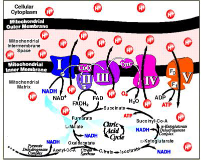
Most of the enzyme proteins are components of the electron transport chain that maintains a proton gradient across the membrane. Another large protein complex is the enzyme ATP synthase, which catalyzes the synthesis of ATP [8]. In mitochondria, oxidative metabolism takes place, the substrate for which is mainly fatty acids and pyruvate, formed as a result of glycolysis in the cytosol. These substances are transported from the cytosol to the mitochondrial matrix, where they break down into twocarbon groups, combined with acetyl coenzyme A (acetyl-CoA). In the composition of the acetyl-CoA molecule, each acetyl group is included in the Krebs cycle as a source of high-energy electrons. Electrons are transferred to the respiratory chain of the inner mitochondrial membrane, where energy is generated as a result of their transfer (Figure 1).

biomedres-openaccess-journal-bjstr

Figure 1: Complexes of the mitochondrial respiratory chain.
Note: complex I (NADH-ubiquinone oxidoreductase; NADH-dehydrogenase), complex II (succinate dehydrogenase; succinate-ubiquinone reductase), complex III (cytochrome bc1 complex; ubiquinone-cytochrome c oxidoreductase, complex IV (cytochrome c oxidase, complex V (mitochondrial ATP synthase) [9]

Disorders of energy metabolism in the cell are one of the key links in many diseases. This makes it necessary to study the work of the electron transport chain of mitochondria, both of its complexes and the entire chain as a whole [9]. The purpose of this review is to analyze and systematize literature data on methodological approaches to the study of the energy function of mitochondria.The Oxidative Phosphorylation System (OxРhoS), localized in the inner mitochondrial membrane, consists of five membrane enzymes. Four of the five protein complexes make up the “respiratory chain” and are involved in the transfer of electrons, which at three points is coupled with the translocation of protons across the inner mitochondrial membrane. The resulting proton gradient is used by the ATP synthase complex (the fifth enzyme complex) to phosphorylate ADP [10,11,1]. For a long time, a fluid-state model has been used to describe the organization of the OxPhoS system. According to this model, the complexes of the respiratory chain freely diffuse in the membrane, and the transfer of electrons occurs as a result of random chaotic collisions.

This model is based on the fact that all protein complexes of the OxPhoS system can be isolated while maintaining enzymatic activity [8,9]. In the last decade, there is more and more evidence indicating stable interactions of OxPhoS complexes in the form of supercomplexes. It is assumed that the OxPhoS supercomplexes and their single complexes coexist in the inner mitochondrial membrane. The association of complexes into super-complexes and the dissociation of super-complexes into OxPhoS complexes is a dynamic process that dependson the physiological state of the cell. Recent studies of mitochondria show that ATP synthase in mitochondrial membranes is organized into long strips of dimers and mitochondrial cristae act as proton traps, and ATP synthase can optimize its activity when there is a lack of protons [12,13]. Complex I, or NADH dehydrogenase, is the main entry point for electrons into the respiratory chain. Complex I oxidizes NAD-H, taking two electrons and reducing one ubiquinone Q molecule, which is released in the membrane. Ubiquinone Q is lipid-soluble; inside the membrane, it diffuses to complex III.

Complex I plays a central role in cellular respiration and oxidative phosphorylation, providing up to 40% of the proton gradient for ATP synthesis. During the oxidation of one NADH molecule, the NADH-dehydrogenase complex transfers four H+ protons from the matrix to the intermembrane space of the mitochondria through the membrane. The formation of reduced NADH is associated with the conversion of malate to oxaloacetic acid and glutamate to α-ketoglutarate. The transfer of electrons through the I complex is associated with the release of 3 ATP molecules [14,1]. Complex II, or succinate dehydrogenase, is another entry point for electrons into the respiratory chain, it is not associated with the translocation of protons across the membrane, transfers electrons from succinate to ubiquinone, and directly binds the Krebs cycle with the respiratory chain. In this case, succinate is oxidized to fumarate with subsequent reduction of ubiquinone Q.

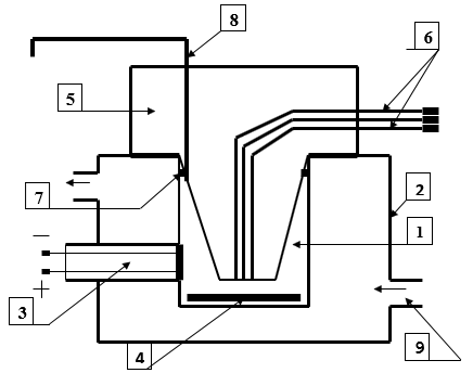
Electrons from succinate are first transferred to flavinadenine dinucleotide, and then through Fe-S clusters to ubiquinone Q. Electronic transport of the II complex is not accompanied by the creation of a proton gradient. The H + protons formed during the oxidation of succinate remain in the matrix and then are reabsorbed during the reduction of the quinone. Complex II works as a carrier of electrons with the formation of 2 ATP molecules [15,1].Respiration of mitochondria and the work of complexes I and II are assessed by registering the rate of oxygen consumption in a polarographic cell using a built-in Clarke electrode (volume 3 ml, at a temperature of 25 ° C (Figure 2).To study the operation of complexes I and II, the rat brain is removed in the cold (0-4 ° C), dried with filter paper, and homogenized in an isolation medium (0.32 M sucrose, 10 mM Tris-HCl, 1 mM ethylenediaminetetraacetic acid, pH 7.4 in the ratio 1:10) using a Potter-Evelheim homogenizer with a Teflon pestle according to a modified method [16,17].

biomedres-openaccess-journal-bjstr

Figure 2: Polarographic cell for studying the respiratory activity of mitochondria
1. Cell
2. Thermostatically controlled chamber
3. Clarke electrode
4. Magnetic stirrer
5. Sealing plug
6. Channels for dosed anaerobic administration of substrates and ADP
7. A sealing ring
8. Channel for removing air and excess liquid
9. Fitting for connection to an ultrathermostat.

Mitochondria are isolated by differential centrifugation. The nuclear fraction is separated by centrifugation of the brain homogenate at 600 g for 10 min (4 ° C). The resulting supernatant is centrifuged at 8500 g for 10 min (4 ° C), the mitochondrial pellet is washed twice in the isolation medium and resuspended to a protein concentration of 35-40 mg/ml in the isolation medium and stored in a short tube on ice. Protein concentration is determined by the Lowry method [16]. A concentrated suspension of mitochondria is introduced into a thermostatted sealed polarographic cell with an incubation medium (0.17 M sucrose, 40 mM KCl, 10 mM Tris-HCl, 5 mM KH2PO4, 8 mM KHCO3, 0.1 mM ethylenediaminetetraacetic acid, pH 7.4) in an amount providing a final protein concentration in the cell of 1 mg/ml. Registration of changes in oxygen tension (pO2) in the mitochondrial suspension is carried out using an electronic recorder. The Clarke electrode is calibrated by sequentially blowing ai) and gaseous nitrogen through the cell.
Basal respiration is assessed, as well as respiration stimulated by the introduction of substrates: malate/glutamate to assess the work of complex I and succinate to assess the work of complex II. The following indicators of mitochondrial respiration are recorded: V1 - basal respiration rate, V2 - substrate-dependent respiration rate, V3 - respiration rate associated with phosphorylation (after ADP introduction), V4 - respiration rate after completion of added ADP phosphorylation. Indicators characterizing the conjugation of oxidation and phosphorylation processes in mitochondria are determined: the acceptor control coefficient (V3 / V2), the respiratory control coefficient (V3 / V4), and the phosphorylation coefficient - ADP / O. After recording the rate of basal (endogenous) respiration in the absence of a substrate (V1), respiration substrates (malate - 2 mM/glutamate - 5 mM or succinate - 5 mM) are alternately introduced into the mitochondrial suspension, and then ADP in an amount of 200 nmol/ml.
The obtained polarograms are used to calculate the respiration rate of mitochondria in different metabolic states and the coefficients characterizing the conjugation of oxidation and phosphorylation processes. The use of solutions of substrates of succinate and malate/glutamate complex makes it possible to assess the degree of functional activity of complexes I and II of the electron transport chain [16]. The central component of the OxPhoS system is cytochrome c-reductase, or complex III, which functions as a dimer. It transports electrons from reduced ubiquinone (ubiquinol) to cytochrome c, a small mobile electron carrier bound to the outer surface of the inner membrane. This multiprotein transmembrane complex is encoded by the mitochondrial (cytochrome b) and nuclear genomes [18]. Electronic transport in complex III is associated with the transfer of protons from the matrix to the intermembrane space and the generation of a proton gradient on the mitochondrial membrane. Cytochrome c is a component of the electron transport chain, the function of which is to transfer electrons between complex III (ubiquinonecytochrome c-reductase or cytochrome bc complex) and complex IV (cytochrome c-oxidase).
For every two electrons passing along the chain of transfer from ubiquinone to cytochrome c, two photons are absorbed from the matrix, and four more protons are released into the intermembrane space. The reduced cytochrome c moves along the membrane in an aqueous medium and transfers one electron to the next respiratory complex, cytochrome oxidase [19,9]. It is the only peripheral protein that interacts with the outside of the inner mitochondrial membrane.Cytochrome c is a water-soluble protein of low molecular weight (about 12,000 Da), the primary structure of which contains about 100 amino acids. Cytochrome c can catalyze hydroxylation and aromatic oxidation reactions and has peroxidase activity by oxidizing various electron donors [20]. Cytochrome c is a metalloprotein that functions in electron transfer reactions and contains heme c (or several hemes) as a prosthetic group, covalently bound to a protein molecule through one or two thioether bonds between the cofactor and the sulfhydryl group of cysteine squirrel. The ligand in the 5th coordination position of the iron ion is always histidine.
Cytochrome c is localized in the intermembrane space. All cytochromes c can be divided into four classes [10].The first class is the low-spin form of soluble cytochrome c of mitochondria and bacteria, in which histidine bound to the heme is located in the N-terminal part of the protein molecule, and the ligand in the 6th coordination position - methionine is located 40 residues towards The C-terminus of the molecule [18].The second class is the high-spin form of cytochrome c and several low-spin forms (for example, cytochrome c 556), which have heme-binding sites in the C-terminal region of the protein molecule. The protein contains four helical regions of the polypeptide chain [19]. The third class includes cytochromes containing several hemes and having a low redox potential (cytochrome c7 (three hemes), c3 (four hemes), and high molecular weight cytochrome c (hexadecahaem)), in which there are 30-40 residues per heme molecule. Hemes, coordinated by two histidines, are structurally and functionally nonequivalent and are characterized by different redox potentials from 0 to 400 mV [19,18].
The fourth class is necessary to maintain complex proteins with heme and other prosthetic groups, for example, flavocytochrome c, cytochromes cd. Cytochromes of this class are proteins containing four hemes, which in the 5th and 6th coordination positions have either two histidines or histidine and methionine. Mitochondrial cytochrome c is one of the three redox subunits of the third complex of the respiratory chain (cytochrome bc complex) [11,19]. Mitochondrial cytochrome c is anchored in the membrane by one membrane segment near the C-terminus. A water-soluble cytochrome C1 preparation can be obtained by removing the hydrophobic C-terminal region. Water-soluble cytochrome c1 cannot participate in the assembly of the bc complex [11,18]. Cytochrome c, in addition to the function of a carrier of electrons in the respiratory chain, can separate from the inner mitochondrial membrane, be transported into the cytoplasm of the cell, and trigger a chain of events in the cytosol, which accelerates apoptosis [11].
The ability of cytochrome c to exhibit various functions inside mitochondria and in the cytosol is associated with the cellular localization of the hemoprotein. Cytochromes are an important class of metal proteins involved in electron transfer and redox catalysis. Redox enzymes and metal proteins play an important role in signaling processes, are responsible for the regulation of genes and their expression, provide the conversion of energy in the processes of respiration and photosynthesis. Thus, cytochrome c in the electron transport chain of mitochondria acts as an electron acceptor for complex III (cytochrome c reductase) and an electron donor for complex IV (cytochrome oxidase) [20,21]. Determination of the activity of complex III The activity of complex III is determined spectrophotometrically at a wavelength of 550 nm by antimycin A-sensitive reduction of ferri- and ferrocytochrome with decylubiquinone in the presence of Tween 20, albumin, and sodium azide.To measure the activity of complex III, the mitochondrial suspension is diluted to a concentration of 0.05 mg/ml of protein in a medium containing 35 mM KH2PO4, 2 mM NaCN, 0.5 mM ethylenediaminetetraacetic acid, pH 7.25. The samples are then sonicated in a water bath for 30 s.
The activity of the complex is determined by the rate of antimycin-sensitive reduction of cytochrome c (550 nm, molar extinction coefficient 18,500 M4 cm4) with the addition of 60 μM decilubiquinone, 50 μM cytochrome c, and 5 mM MgCl2 to a suspension of mitochondria exposed to ultrasound (the content of mitochondrial protein in the sample is 0.01 mg/ ml). Spectrophotometric determination of the concentration of cytochromes c and b in aqueous solutions. The molar extinction coefficients for cytochrome c A vost-oxide = 18,500 M4 cm-1 at 550 nm. Recovery is carried out with ascorbate or dithionite. In the presence of cytochrome b, the content of cytochrome c was determined from the differential spectra of the forms reduced by ascorbate, minus the forms oxidized by ferricyanide. Cytochrome b is determined from the differential spectra of the forms reduced by dithionite minus the spectra of the forms reduced by ascorbate. The activity of enzyme systems seems to be the most important parameter characterizing the work of the electron transport chain of mitochondria and the bioenergetic status of the cell.
Extraction of cytochrome c from the mitochondrial membrane and the reconstruction of the respiratory chain makes it possible to assess the transfer of electrons from complex III to IV. The principle of the method for extracting cytochrome c from the mitochondrial membrane consists of the destruction of the outer membrane using detergents or hypotonic processing and extraction of proteins with saline solutions. Reagents for isolation of mitochondria include: 0.25 M sucrose solution, 0.15 and 0.015 M KS solutions, 5 mM potassium succinate solution, pH 7.4, 5 mM potassium glutamate solution, pH 7.4, 0.005 M solution 2.4 dinitrophenol, pH 7.4, incubation medium (0.15 M sucrose, 0.075 M KC1, 0.01 M potassium phosphate, pH 7.4), cytochrome c solution - 1 mg / ml and reagents for protein determination. All solutions are prepared with bidistilled water. Isolated mitochondria are suspended in 3 ml of 0.25 M sucrose solution. Three conical flasks are filled with 40 ml of solutions:
1) 0.25 M sucrose;
2) 0.15 M KC1
3) 0.015 M KC1. 1 ml of a thick suspension of mitochondria is added to each flask. The contents of all flasks are gently mixed for 10 minutes at 0 ° C and then transferred to three 50 ml centrifuge beakers.
The mitochondria are separated by centrifugation at 10,000 g, suspended in 0.5 ml of 0.15 M KC1, and transferred into three flasks containing 40 ml of 0.15 M KC1. The contents of the flasks are again stirred in the cold for 10 minutes and centrifuged again under the same conditions to separate the mitochondria. The resulting sediments of mitochondria are resuspended in 0.3-0.5 ml of 0.3 M sucrose solution. The rates of oxygen consumption are determined polarographically, using succinate and glutamate (5 mM) as substrates, and after 1-2 minutes dinitrophenol (50-100 μM) is added. For a preparation washed with a hypotonic solution of KCl, the dependence of the rate of succinate oxidation on the amount of cytochrome c added to the incubation medium (concentration from 0 to 100 μg / 2 ml, 5-6 points) is determined [15,4 ,11]. Complex IV, or cytochrome c oxidase, is the final complex of the respiratory chain. Cytochrome c oxidase catalyzes the transfer of electrons from cytochrome c to molecular oxygen, reducing the latter to water.
Complex IV is the terminal oxidase of the aerobic respiratory electron transport chain, which catalyzes the transfer of electrons from cytochrome c to oxygen to form water. Complex IV sequentially oxidizes four cytochrome c molecules and, accepting four electrons, reduces O2 to H2O. During O2 reduction, four H+ are captured from the mitochondrial matrix to form two H2O molecules, and four more H+ are actively pumped across the membrane. Thus, cytochrome oxidase contributes to the creation of a proton gradient for ATP synthesis and is part of the oxidative phosphorylation pathway [11,4]. Complex IV activity Cytochrome oxidase activity is assessed by the polarographic method according to the rate of oxygen consumption by mitochondria. It should be noted that all procedures for the isolation, operation, and storage of mitochondria must be carried out observing the temperature regime: the samples must be stored in ice, the media and the instrument must be pre-cooled to a temperature of 0-4 ° C. A suspension of intact mitochondria or mitochondria destroyed by detergent is introduced into a polarograph cell containing a medium of the following composition: 0.125 M sucrose, 60 mM KCl, 10 mM Tris-HCl, 0.1 mM 2,4-dinitrophenol, 40 μM cytochrome c.
The content of mitochondrial protein in a polarographic cell is 0.5-1.0 mg/ml. The cytochrome oxidase reaction is started by introducing a solution of ascorbic acid neutralized to pH 7.4, creating a concentration of 10 mM in the sample. Measure the rate of oxygen uptake and calculate the activity of cytochrome oxidase in nanomoles of absorbed 02 for 1 min per 1 mg of protein [17]. ATP synthase, complex V, uses the resulting proton gradient to synthesize ATP. ATP synthase is an integral protein of the inner mitochondrial membrane that carries out the reaction of ATP formation from ADP [20]. Mitochondrial ATP synthase plays an important role in stem cell differentiation, promotes the maturation of mitochondrial cristae by dimerization and specific regulation. The enzyme belongs to the alpha/beta ATP synthase family. It consists of two structural domains (F1 - extramembrane catalyst and F0 - membrane proton channel), connected by a central rod, consisting of γ, δ, and ε subunits, and together with the membrane subunit oligomer representing the rotary domain of the enzyme [15].
Determination of the content of ATP synthase (V complex) is carried out by the immunohistochemical method using monoclonal antibodies. For this purpose, after decapitation and extraction of the brain, the material is fixed in zinc-ethanol-formaldehyde at + 4 ° C (overnight), then embedded in paraffin [22]. Paraffin sections with a thickness of 5 μm are prepared using a microtome, mounted on glass slides. The preparations are processed according to the protocol of immunocytochemical reaction for light microscopy, excluding the procedure of thermal unmasking of antigens. To determine the immunoreactivity of the molecular marker of mitochondria ATP synthase (complex V, which forms ATP from ADP), primary monoclonal antibodies (Anti-ATP5A antibody) are used at a dilution of 1: 2400 at + 4 ° C, with an exposure of 20 h in a humid chamber [22]. Thus, the above methods for studying the activity of the electron transport chain of mitochondria, especially their use in combination, can significantly detail the understanding of the pathogenesis of disorders of cell energy exchange that occurs in various diseases, which will improve the prevention and correction of mitochondrial dysfunction.


For more Articles on: https://biomedres01.blogspot.com/

Tuesday, July 18, 2023

Post-ACL Rupture Integrated Rehabilitative Pathway: Factors Influencing Economic Resources Absorption


Post-ACL Rupture Integrated Rehabilitative Pathway: Factors Influencing Economic Resources Absorption

Introduction

In the healthcare setting, randomized controlled trials (RCTs) usually represent the main approach to create new knowledge [1]. Nowadays the use of real-world data (RWD) is becoming more and more relevant, even from a strategic point of view, to optimize and determine the best patients’ pathway. Using RWD gives the opportunity to generate new knowledge assets, mixing points of view from different actors (i.e. hospitals or patients) together with outcome and efficiency data, [2,3] thus supporting the delivery of healthcare services improvement and the outcomes’ optimization [4]. In this growing scenario, where RWD assume more and more importance all around the world, there is the opportunity to gather different dimensions and information, to create larger and integrated databases. Subsequently, they can be elaborated and merged at different levels, and could become available for both scholars, and policy makers research, thus achieving improvements in the quality, safety, and value of healthcare services delivery.

RWD could thus represent a useful support tool to assess the consequences of disease management interventions, to design an integrated clinical pathway based on knowledge derived from real world data and facts, implementing, and improving, the healthcare services that are satisfactory, in terms of effectiveness and efficiency [5]. The use of RWD could also provide a robust support for making customized decisions, concerning the treatment of patients, and for developing strategies for the delivery of care that respect the principles of fairness and coverage, without overshadowing the dimensions of quality and services efficiency/productivity. One of the most important knowledge assets that should leverage on RWD, are the costs related to specific rehabilitative programs or integrated clinical and rehabilitation pathways, [6] that could rely on real-life and specific country-oriented evidence, always following international and national guidelines suggestions and indications, and for which poor evidence is still available.

According to the above, integrated clinical and rehabilitative pathway could be the perfect example to define the significance to observe real-world healthcare data, beyond clinical trial evidence, thus providing stakeholders with valuable information about the safety and effectiveness of rehabilitative programs in a large and heterogeneous populations. Rehabilitation is an essential part of universal health coverage along with promotion of good health, prevention of disease, treatment, and palliative care, thus being defined as “disability in individuals with health conditions in interaction with their environment” [7]. The above consideration is strengthened because literature evidence focused their attention, on the inpatient’s integrated clinical pathway, without considering the outpatients’ activities and programs, after discharge. Moving on from these premises, the present paper aims at investigating the knowledge assets that might influence the creation and the design of a proper patient integrated rehabilitative pathway, after the Anterior Cruciate Ligament (ACL) rupture, assuming the hospital’s perspective. Coherently to the above, the study addressed the following two research questions.

1. Which are the main factors influencing the efficiency of the integrated rehabilitative pathway, in terms of costs’ optimization?
2. What configurations of such factors are sufficient to generate the emergence of a certain level of efficiency, in the absorption of resources?

Theoretical Backgrounds

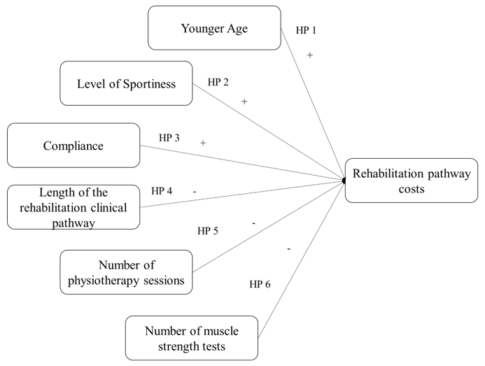
The deep investigation about the above research questions requires the analysis of different independent variables, and their relations with the optimization of the entire integrated clinical and rehabilitative pathway. Based on an extensive literature review, a set of original hypotheses was developed, to produce a specific framework, declaring the main characteristics of a rehabilitative pathway that affect the absorption of economic resources: patient’s age, patient’s compliance, duration of the rehabilitation pathway, level of sportiness, number of physiotherapy sessions performed, number of muscle strength and threshold tests performed were the factors investigated [8-12].

Age

De Valk stated that age is a key factor for the success of the integrated rehabilitative pathway: younger patients are more likely to achieve a better clinical outcome, with a positive impact on satisfaction [8]. Since, in other contexts, younger patients are usually associated with a lower absorption of economic resources, [13] it is possible to assume that also in this case age could be a factor able to reduce the overall costs of the clinical rehabilitation pathway. Based on the above considerations, the following hypothesis was displayed.
• HP 1: Younger age has a positive impact on rehabilitative pathway management costs.

Level of Sportiness

As reported in the study of De Valk et al., a high level of sportiness enables a fast and successful integrated rehabilitative pathway [8]. Patients who are used to practice sports at high levels (e.g. professional athletes), are more likely to fully recover knee functions, achieving levels of mobility and resistance to pre-injury efforts The level of sportiness, therefore, has a strong impact on the outcome of rehabilitation, and consequently also on the resources’ absorption as well as on the rehabilitation pathway efficiency.
The following hypothesis was formulated.
• HP 2: High level of sportiness, positively impacts on rehabilitative pathway management costs.

Compliance

Patient compliance is a fundamental component of rehabilitation activity of which the main goal is improved recovery outcome. Without compliance to the treatment regimen, the expected outcomes cannot be achieved. According to this consideration, one of the most frequent obstacles to the healthcare professionals work, is low level of compliance and adherence in all integrated clinical pathways, but particularly in physical rehabilitation.

In fact, many patients do not follow the instructions of the clinicians and physiotherapists, who plan the timing and exercises of rehabilitation sessions, and often do not respect them or do them partially. Literature estimated that sport injury rehabilitation adherence rates could be as low as 40%. [14-16]. On the contrary, being adherent to the integrated rehabilitative pathway plays a key-role in the achievement of the expected results, with a positive impact in the overall economic resources absorption [17].
Thus, the following hypothesis was proposed.
• HP 3: Compliance has a positive impact on rehabilitative pathway management costs.

Length of the Rehabilitative Clinical Pathway

The duration of the rehabilitative pathway presents a direct impact on the overall clinical results, and it is closely related to adherence [12]. In general terms, the presence of adequate clinical pathway would enhance the overall patients’ journey optimization. [18] Moving on from the above consideration, the more the patients are satisfied and adhere to the proper appointments, fewer follow-up activities would be required, with a positive impact on the possibility to free up economic resources [19]. The following hypothesis was defined.
• HP 4: The length of rehabilitation negatively impacts on rehabilitative pathway management costs.

Number of Physiotherapy Sessions Performed and Number of Muscle Strength Tests

Two other variables related to the overall duration of the integrated rehabilitative pathway, impacting on the rehabilitation management costs, are the number of sessions and the number of muscle strength tests performed. Risberg and colleagues (2004) revealed a correlation between prolonged rehabilitation and improvement in patient’s physical conditions, leading to increased costs to support intensive rehabilitation programs [20].
According to this, the following hypotheses were displayed.
• HP 5: The number of physiotherapy sessions negatively impact on rehabilitative pathway management costs.
• HP 6: The number of muscle strength tests negatively impact on rehabilitative pathway management costs.
A synthesis of the research frameworks developed, is proposed in Figure 1.

biomedres-openaccess-journal-bjstr

Figure 1: Conceptual framework for rehabilitative pathway costs.

Methods

An observational study, designed in accordance with the STROBE guidelines, [21] was conducted in two private rehabilitation hospitals in Italy, involving a total of 118 patients, to retrieve RWD, in relation to the integrated rehabilitative pathway. The informed consent forms were signed during the taken in charge of the patients, and they were informed concerning the use of data with an anonymous and aggregated methodological approach. In particular, the study involved economic data referring to all the adult patients (age ≥ 18 years old) requiring an integrated rehabilitative pathway after ACL rupture, in the year 2018, after having signed the informed consent form for dissemination activities.

Definitions of the Measures

As previously mentioned, the study was structured considering just one dependent variable, the rehabilitative pathway costs, and six independent variables, described below.

Dependent Variable

Costs: For the estimation of the integrated rehabilitative pathway average costs, the following variables were considered: the number of physiotherapy sessions and the number of procedures / tests performed during the entire programme. Economic data were evaluated, in accordance with outpatient visits and tests Regional Reimbursement Tariffs, valid for the years 2021/2022.

Independent Variables

Age: The patient’s age when rehabilitation program begins.

Level of Sportiness: It is the declared level of sportiness when the integrated rehabilitative pathway begins, defined by clinician and physiotherapist taken in charge the patient, in terms of number of training sessions performed within a week. In particular, the level of sportiness was classified as:
1. None (no training sessions performed).
2. Practitioner (at least 1 or 2 training sessions a week).
3. Amateur (at least 3 or 4 training sessions a week).
4. Athlete (at least 4 or 5 training sessions a week, with participation at sports competitions).

Compliance: The adherence/compliance to the integrated rehabilitative pathway was esteemed by attendance and participation in scheduled sessions. Patients were classified as compliant patients and not-compliant patients, depending on these two cases:
1. Patients completed the pathway prescribed at the beginning, even considering any modifications in the integrated rehabilitative pathway, and
2. Patients abandoned the rehabilitative pathway earlier.

Length of the Rehabilitation Clinical Pathway: This variable indicates the duration of the pathway, expressed as number of days occurred between the beginning and the end of the integrated program.

Number of Physiotherapy Sessions Performed and Number of Muscle Strength: This variable indicates the number of tests and sessions performed by each patient, during the whole rehabilitation pathway.

Statistical Analyses

The above variables were first analysed considering descriptive statistics, frequencies, and distributions. To ensure the relevance of the assumptions of normality, linearity and homoscedasticity, preliminary analyses were carried out. Three methodological approaches were used to answer the research questions underlying the study.
1. An analysis was carried out on the relationships among variables, verifying the existence of correlations among them. In particular, the correlation coefficient “person-product-moment” was evaluated, to verify the existence of small (from 0.10 to 0.29), medium (from 0.3 to 0.49), or large (from 0.5 to 1) correlations [22]. The exact value of -1 or +1 indicated a perfect correlation among variables.
2. In order to test the proposed hypotheses, a hierarchical sequential linear regression model was implemented (with enter methodology), that defines the predictors of the dependent variable (level of economic resources’ absorption). This approach was used to identify the impacts of the independent variables. One parameter of attention was the Adjusted R2, useful to control the explanatory power of each model. Thanks to this approach it is possible to test the hypotheses, through incremental models, to establish the specific impact of each input variable on the dependent variable. The “exclude case pairwise” option has been implemented, as it represents the preferred methodology to be applied for a small sample size, avoiding data exclusion. All the statistical analyses, referring to the coefficients test, and the development of the hierarchical sequential linear regression model were performed using the Statistical Package for Social Science (IBM SPSS Statistics Viewer - Version 22).
3. A qualitative-configurative analysis - QCA - was carried out to integrate the statistical approaches previously described [23,24]. The QCA is a comparative case-oriented methodology, useful to find, through an in-depth comparison of real-world cases, consistent configurations of causal conditions (the independent variables in our study), sufficient to determine the emergence of a specific outcome [25]. In the QCA, a symmetric relationship is disarticulated into two asymmetric analyses formalized by set and sub-set relationships [21]: one, related to the necessity of the conditions, with respect to the dependent variable, and the other on the sufficiency. This approach allows researchers to deal with the complexity of real phenomena. The QCA assumes the nonlinearity of phenomena under investigation and is based on the principle of causal complexity. This means that, in most cases, it does not make sense to isolate the effect (positive or negative), of a single independent variable on the outcome, but configurations of variables are identified, being related to the dependent variable. Moreover, several different configurations can be recognized as “causal recipes” of the same dependent variable [23].
In accordance with Vis, [26] which discussed the advantages of using the QCA technique to complement regression analyses for moderately large samples (between 50 and 100), the 118 empirical cases collected in this research were studied through the QCA to better understand the relationships between combinations of independent variables (the effects of complex interactions between causal conditions) and dependent variables, i.e. the results presented above. In QCA approach, the variables can be considered crisp or fuzzy. The crisp set variant (csQCA) is the version in which the variables of the study are dichotomous, and the empirical analysed cases are classified as alternatively “fully in” or “fully out” in the sets representing causal conditions and the outcomes. The fuzzy-set variant (fsQCA) is characterized by the fact that the empirical cases are classified in terms of membership degrees in the fuzzy sets of causal conditions, and of the outcome. In this analysis, we adopted the crisp set QCA.

Results

The Sample Under Assessment

The sample was composed of 118 patients, requiring a specific integrated rehabilitative pathway after ACL rupture. It should be noted here that all patients required an outpatient integrated rehabilitative pathway, starting the activities program immediately after surgery. The sample consisted mainly of males (66%) and is 34 years old on average. As far as the level of sportiness is concerned, only 3% of the patients in the sample had previously practiced sport at competitive level, while 64% of the patients belonged to the “practitioner” category. In general, the average absorption of economic resources associated with the rehabilitation of the patients in the sample was equal to € 3,334.07 ± 269.28. Compliant patients are associated with a higher absorption of financial resources than non-compliant patients (€ 3,634.47 vs € 2,157.50 p-value=0.026).

Hypotheses Testing

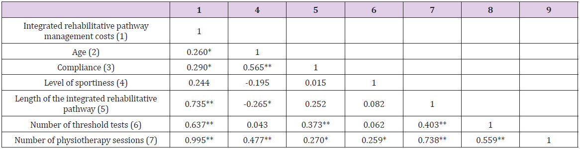
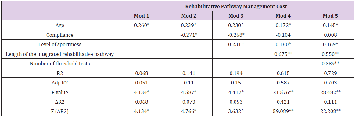
Table 1 shows that age (p-value = 0.047), compliance (p-value = 0.026), length of the rehabilitative pathway (p-value = 0.000) and number of threshold tests (p-value = 0.000) significantly influenced the integrated rehabilitative pathway costs. The great relationship between total costs and the number of physiotherapy sessions (β = 0.995, p-value = 0.000) depicts that between the two variables there is the collinearity phenomenon, thus being two aspects explaining the same concept. This is the reason why the variable “number of physiotherapy sessions” was not included in the regression model. The independent variables (age, compliance, and the length of the rehabilitative pathway) have a significant p-value < 0.05. Moreover, the same relationships emerged among the length of the integrated rehabilitative pathway, the number of threshold tests and the number of physiotherapy sessions (p-value = 0.000). After testing the correlation among all variables, a regression analysis was conducted to test hypotheses (Table 2). Older age (β=0.145, p=0.049), higher sportiness level (β=0.169, p=0.022), as well as the length of the rehabilitative pathway (β=0.550, p-value=0.000), and threshold tests (β=0.389, p-value =0.000) are antecedents of higher rehabilitative pathway management costs (Adjusted R²=0.703 and F=24.482).

biomedres-openaccess-journal-bjstr

Table 1: Relationships between variables.

biomedres-openaccess-journal-bjstr

Table 2: Regression model for integrated rehabilitative pathway management costs.

Configuration of Factors Through QCA

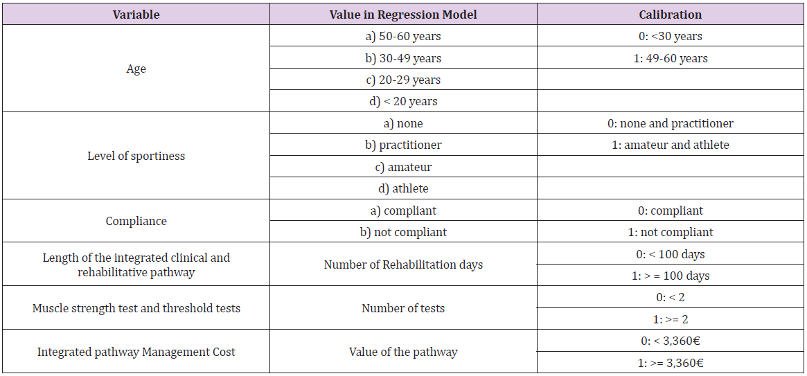
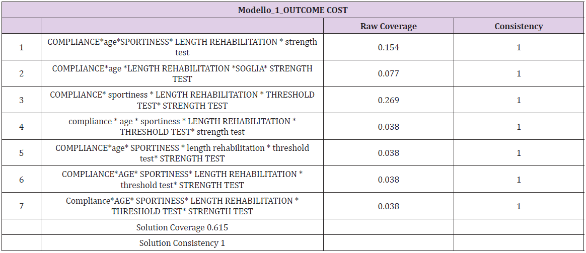
The QCA analysis requires a preliminary step for calibration of variables transforming them in crisp ones (Table 3). After performing the calibration process, consistency, and coverage of a set of configuration variables were evaluated. Consistency is the extent to which the results are in line with statements of need or sufficiency. Coverage provides information about the empirical importance of necessary and sufficient conditions. Each configuration has a raw coverage value that “measures the relative importance of several combinations of causally relevant conditions” [23]: the proportion that a configuration covers the outcome. It is assessed by the sum of consistent scores of the configuration divided by the sum of outcome scores [27]. In the QCA analysis, the crisp value 1 is reported in uppercase whereas the crisp value 0 is reported in lowercase. According to the regression analysis, the variables that most influence the management costs are the compliance and the length of the integrated rehabilitative pathway (Table 4). In particular, the most representative recipes show that level of sportiness associated with young age affects the management, as well as the length of rehabilitation, also for the patients with a lower level of sportiness.

biomedres-openaccess-journal-bjstr

Table 3: Calibration of the variables.

biomedres-openaccess-journal-bjstr

Table 4: QCA analysis for rehabilitative pathway management cost.

Discussion and Conclusion

In the current era, where the availability of large amounts of clinical data gathered during care delivery is rapidly increasing, as well as the ability to access, process, link, and analyze these data in efficient ways, alternative sources to supplement evidence from RCTs look promising. Among all the information that could derive from RWD, one of the most important topics to analyze consist of the management costs related to a specific integrated clinical or rehabilitative pathway. The knowledge of the economic resources’ absorption could contribute to fully understand the pathways as well as the choices and behaviors involved in those pathways. Economic information provides the tools for developing effective and efficient policy strategies and addressing potential tradeoffs between the goals of increasing social welfare and improving the distribution of healthcare delivery across individuals and population groups.

Within the setting of ACL integrated rehabilitative pathway, results reported that strong compliance and a high level of sportiness could be considered among the main factors that cause an increase in the total cost of managing the rehabilitation pathway for a patient with ACL rupture, confirming the information found in literature [14-16]. In fact, orthopedic surgery often requires many months of rehabilitation to achieve a successful outcome, regardless of subspecialty, with an important impact on the overall resources’ absorption [28]. For patients practicing sports by profession, the rehabilitation pathway could be more intense and costly, as it is their priority, to recover their normal motor and sports conditions as soon as possible [8]. Thanks to the QCA it is possible to integrate these results, identifying all combinations of factors that are determining the emergence of a certain results. For example, more consistent QCA analysis solutions show that compliance has an important influence on the absorption of economic resources by patients, but also higher levels of sportiness related to young age of patients, determining higher pathway costs. The results obtained and the findings of this study could give a contribute useful to enlarge the existing research, regarding the use of RWD in the healthcare sector, with particular focus on the rehabilitation programs, thus considering a larger sample and producing results that could be replicable. Although the relevance of this topic, healthcare stakeholders often do not have enough information on outcomes to take decisions, being well-informed. In this context, knowing which are the factors that have a positive or negative influence on the integrated patients’ rehabilitative pathway, could be useful to optimize economic resources, as well as for the achievement of an adequate effectiveness, thus becoming more efficient and effective [6]. This, in the Italian setting, is becoming more and more important, due to the recent re-consideration of the integrated clinical and rehabilitative pathways, after the COVID-19 pandemic, for the re-organization of the healthcare delivery of services.


For more Articles on: https://biomedres01.blogspot.com/

Antimalarial Aloe Compounds

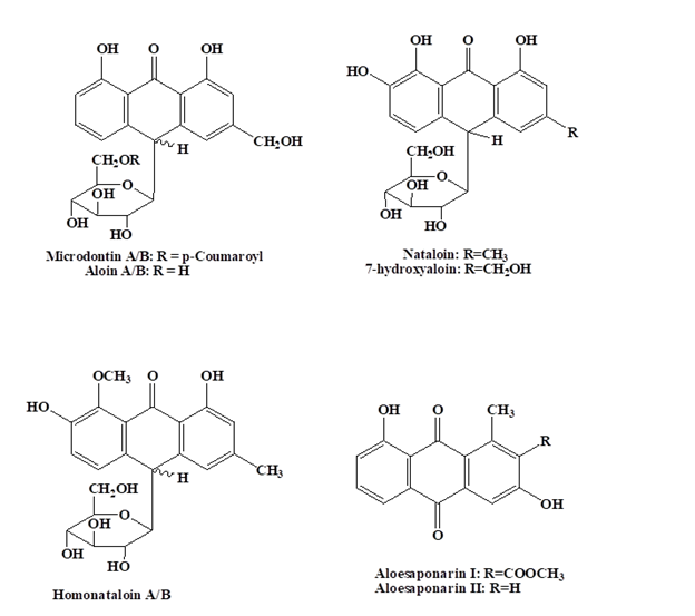
  Antimalarial Aloe Compounds Introduction Among the most prevalent diseases caused by protozoan parasites, malaria is caused by parasites o...