Monday, November 25, 2024

Does Access to Remittance Improve Welfare Status of Households in Nigeria?


Does Access to Remittance Improve Welfare Status of Households in Nigeria?

Introduction

Remittance is defined as the proportion of migrants’ earnings sent from their destination of employment to their origin or communities (Samal [1]). They also are considered as compensation (brain gain) for the loss of human capital (brain drain) by a net labour exporting country (Ratha, et al. [2]). Remittances can be categorised as monetary versus non-monetary remittances; domestic versus international remittances, and inward versus outward remittances. Remittances have attracted attention in empirical studies with some concentration on their determinants and developmental impacts (Adenutsi [3]). Developing countries as a whole have consistently been the largest recipient of international remittances in the world. Between 1995 and 2005 the total amount of official migrant remittances received by developing countries increased by more than 300% (Adenutsi [3]). Remittances to developing economies reach US$338 billion in 2008, higher than its estimated value of US$328 billion (World Bank [4]). They are person-toperson flows, well targeted to the needs of the recipients, who are often poor, thereby assisting poor families deal with negative economic shocks (World Bank [5]). This makes remittances very important source of finance for the rural households traditionally known for high level of poverty (low welfare level) and low access to foreign aid, government grants or bank loans.

Remittances enable the hitherto risk averse farming households insured by remittances, to shift their portfolios towards riskier enterprises, an indicator of welfare (Chukwuone, et al. [6]). Adams [7] noted that increased workers’ remittance inflow reduced income inequality among households in rural Egypt. Adams [8] further established that remittances reduce the severity of poverty in Guatemala, with households spending greater percentage of income on durable goods than on non-durable goods. Taylor, 2005, submitted that international migrants’ remittances increased per capita income of households and reduced the incidence and depth of poverty in Guatamala. In addition, it improves human capital indicators (education and health) of the recipient countries Fajnzylber, et al. [9]. Increased per capita income, reduced poverty incidence, improved health, asset acquisition and human capital developments are all indicators of Welfare, which is synonymous with good quality of life (Narayan, et al. [10]). However, there are empirical facts that households domiciled in the rural sector received significantly less remittances than their urban counterpart did. It was also established that only between 30 and 40 per cent of all remittances are destined to rural areas (Netri, 2010), where the majority of the population lives. Such disparity in remittances distribution among household categories makes the effect of remittance on welfare unequal.

Thus there is need to examine the effect of migrants’ remittance on the welfare status of households in Nigeria. This encompasses examining the sectoral disparity in migrants’ remittances and welfare status of the households; and the contribution of remittances and indicators of welfare to the overall welfare status of the households. Lastly, the study will establish whether migrants’ remittances have significant effect on the welfare status of households in Nigeria.

Methodology

The study area is Nigeria, West Africa. The source of data is from the Living Standard Measurement Survey (LSMS, 2015/2016) collected by National Bureau of Statistics. Total respondents of 4,068 households were sampled and used for the analysis. The methods of analysis of data adopted are descriptive statistics, Multidimensional Welfare Index and Tobit regression model. Descriptive statistics was used to analyse the socioeconomic characteristics of the respondents and the amount of remittance received by households. Fuzzy set was adopted to estimate the multidimensional welfare status of each household in Nigeria. The Multidimensional Welfare Index (MWI) is a measure of acute global welfare developed by the Oxford Welfare and Human Development Initiatives (OWHI) with the United Nations Development Programme’s Human Development Report (UNDP, et al. [11-14]. The index method belongs to the family of measures developed by Alkire [15]. This method requires determining the unit of analysis (i.e. household), identifying the set of indicators in which they are deprived at the same time and summarizing their welfare profile in a weighted deprivation score. The households are identified as multidimensional worse off if their deprivation score is below a cross-dimensional welfare cut-off. The number of worse off people and their deprivation score (i.e. the percentage of simultaneous deprivations they experience) become part of the final welfare measure.

The constructed Multidimensional Welfare Index (MWI) is based on five dimensions: education, sanitation, standard of living, housing and asset, which were carefully selected as guided by literatures (following Pampalon, et al. [16-18]):

1. Education

2. Sanitation

3. Standard of living

4. Housing

5. Assets

Fuzzy set involves constructing an index from array of items that could be linked to welfare (attributes of welfare). Costa [19], citing Dagum, (2002), gives an exposition of the methodological framework of fuzzy set theory. Given a population A of a households, A = {a1, a2, …, an}, the subset of worse off households B includes any household a1∈B which presents some degree of welfare in at least one of the m attributes of X, The degree of membership to the fuzzy set B of the i-th household (i=1,…, n) with respect to the j-th attribute (j=1,…, m) is defined as;

X = 1, if the i-th household possesss the j-th attribute and ij x = 0, if the i-th household does not possess the j-th attribute. The multidimensional welfare ratio of the i-th household ( ) B i μ a , i.e the degree of welfare of the i-th household as a weighing function of the m attributes, is defined as the weighted average of ij x ,

Where Wj is the weight attached to the j-th attribute. The weight wj attached to the j-th attribute stands for the intensity of deprivation of Xj . It was proposed by Cerioli, et al. [20] and can be represented with the following expression:

The multidimensional welfare ratio of the population μ B is obtained as a weighted average of the welfare ratio of the i-th household μB(a1).

Fuzzy set framework also follows obtaining one-dimensional welfare ratio for each of the j attributes. This is the weighted average of xij with weight ni.

It is also possible to obtain the multidimensional welfare ratio of the population μ B as the weighted average of μ B( Xj) (unidimensional welfare ratio of the attributes), with weight wj .

In order to determine the effect of migrants’ remittances on the welfare status of households, Tobit regression model was used since the values of the endogenous variable lies between zero and one with none having zero value. The household welfare threshold was put at 0.5 according to (UNDP [11]) the model for the regression analysis is specified thus,

1. Y1 = ∝1+β1 X¬1+β2 X2+……….βnXn+ ei

2. Yi* = Yi if 0.5 ≤Yi≤1 (better off)

3. Yi* = 0 if Yi < 0.5(worse off)

4. Y = dependent variable

5. ∝= Intercept term

6. β i = parameters to be estimated

7. Xi = Vector of explanatory variables

8. ei= Disturbance term assumed to be independently and normally distributed with zero mean and constant variance

The dependent variable (Y) is the welfare index while the explanatory variables are as follows:

1. X1 = Age of household heads (years)

2. X2 = Gender (dummy) – male 1, female 0

3. X 3 = Household size (number)

4. X4 = Household remittance (Naira)

5. X5 = Sectors (rural 1, urban 0)

6. X6= Household income (Naira)

Geopolitical Zones

1. X7 = south-west 1, 0 otherwise

2. X8 = south-south 1, 0 otherwise

3. X9 = south-east 1, 0 otherwise

4. X10 = north-east 1, 0 otherwise

5. X11= north-central 1, 0 otherwise

6. X12= north-west 1, 0 otherwise
biomedres-openaccess-journal-bjstr

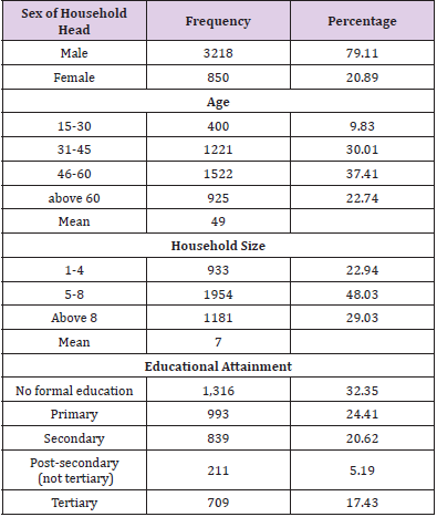
Table 1: Socioeconomic Characteristics of Households in Nigeria.

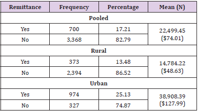
Table 1 profiles the socioeconomic characteristics of the households in Nigeria. Male respondents head almost eight out of every 10 households in Nigeria. The mean age of the respondents (49 years) revealed that majority of the respondents are in their energetic and resourceful age. This is in consonance with the submission of Ashagidigbi, et al. [21] who reported that average age of household heads in Nigeria is below 50 years. Households in Nigeria have an average of seven members indicating a relatively high household size, corroborating the finding of Ashagidigbi, et al. [22] who stated similar value. About one-third of the entire population does not possess formal education. In other words, significant proportion of households possesses one form of formal education or another. The access of households to remittance is depicted in Table 2. In the pooled data, 17% of the entire households received remittance from their kith and/or kin in Nigeria. However, 13 out of every 100 respondents have access to remittance in the rural sector of the country, while a quarter receives remittance in urban sector. This reveals that access to remittance by households in Nigeria is low. A household on the average earns $74 as remittance in Nigeria; value of urban remittance is 73% higher than that of the pooled data, while that of rural sector is 34.3% lower. The figures are considerably low due to the inaccessibility of majority of Nigerian residents to remittance, specifically those residing in rural areas of the country.
biomedres-openaccess-journal-bjstr

Table 2: Distribution of Households based on their Remittance Status.

Note: N304 = $1

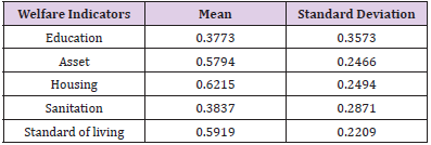
This supports the finding of (Kshetri, et al. [23]), who established that rural households received between 30-40 percent of total remittance in Africa. As relayed in Table 3, five indicators contributing to the overall welfare status of households in Nigeria were considered. Housing is the leading contributor, responsible for 62.15% of the households’ welfare status. However, education and sanitation are the least contributing factors contributing 37.73% and 38.37% respectively to the welfare status of households in Nigeria. Thus, emphasising that education and health/sanitation issues still pose considerable challenge to households resident in Nigeria.
biomedres-openaccess-journal-bjstr

Table 3: Contributions of Welfare Indicators to the welfare Status of Households.

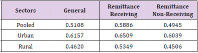
In the pooled data, the welfare index of the households is 0.51, similar to that reported by Ashagidigbi et al, 2019 (0.58). The value is higher for households resident in urban sector of the country, while the rural resident is far below the country’s mean value. Across the sectors, the welfare status of remittance-receiving households in urban sector is better off than those in rural sector and Nigeria as a whole (Table 4). Similar trend is observed for the non-remittance receiving households. However, households that receive remittance have better welfare status in the pooled data and across the sectors than non-remittance receiving respondents. The finding aligns with that of Taylor, et al. [24], who posited that remittances reduce the incidence and depth of poverty among households in rural Mexico.
biomedres-openaccess-journal-bjstr

Table 4: Welfare Status of Households based on their Remittance Status in Nigeria.
biomedres-openaccess-journal-bjstr

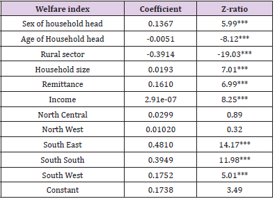
Table 5: Effect of Remittance on Households’ Welfare Status in Nigeria.

Note: Log likelihood = -2832.7946

Prob > chi2 = 0.0000

This clearly shows that remittance is a significant factor positively influencing welfare status of households in Nigeria. Tobit regression model was adopted to analyse the effect of remittance on welfare status of households in Nigeria Table 5. Age, sex, household size, income, remittance and sector where the respondents reside are the factors that significantly influence households’ welfare status at one percent level of probability. Others are northcentral, southeast, southwest and south-south zones. A naira increase in income and amount of remittance received increases the welfare status of households by 2.91e-07 and 0.16 respectively. This is an indication that households that receive remittance have higher likelihood to be better off in relation to their welfare status compared to the households that do not receive. This submission is in tandem with (World Bank [4,5] where it was reported that access to remittance by households avail them the opportunity to cope with negative economic shock. Likewise, (Pfua, et al. [25], Adams [8]) submitted that remittance assists in improving the living condition and reduce severity of poverty of the recipients, most especially the poor households. The welfare status of higher income earning households are better off relative to the lowincome earners. A unit increase in age of the respondents reduces households’ welfare status by 0.51%, while an additional member to a household increases households’ welfare status by 1.93%. This submission establishes age as a welfare-reducing factor, while household size is regarded as welfare enhancing variable.

Furthermore, the welfare status of residents in the northcentral, southeast, southwest and south-south zones of the country increases by 2.99%, 48.10%,39.49% and 17.52% respectively, relative to the northeast residents. This aligns with Ashagidigbi, et al. [26] who concluded that inadequate access to better welfare is prominent among the northeast and northwest residents in Nigeria [27] Likewise, rural dwellers’ welfare status reduces by 39.14% in comparison to urban residents. This implies that the welfare status of households residing in urban sector and other zones with the exception of northeast are far better off. Hence, rural and northeast residents should be of primary focus when implementing poverty reduction measures in Nigeria.

Conclusion

The facts deduced from the study are that majority of households in Nigeria do not receive remittance (83%). The scenario is worse in rural areas compared to the urban sector. Among the indicators of welfare, housing contributes the most to the overall welfare of the households, while education and health/sanitation contribute the least. The welfare status of households receiving remittance is considerably higher than non-remittance receiving households in the pooled data and across the sectors. However, the welfare status of households that receive remittance in urban sector is better off than rural sector residents. Remittance positively influences the welfare status of the households. On the contrary, households residing in rural sector and northeast zone of the country are worse off in relation to their welfare status. Sector and zone specific policy measures that could ease and enhance the access of residents in the rural sector and northeast zone to remittance is paramount in ensuring improved welfare status among households in Nigeria.


For more Articles on: https://biomedres01.blogspot.com/

Friday, November 22, 2024

Types and Treatments of Leishmaniasis


Types and Treatments of Leishmaniasis

Introduction

The Leishmaniasis are a cluster of parasitic diseases produced by morphologically alike parasites in the genus Leishmania with Order Kinetoplastida and Family Trypanosomatida. The disease is transferred through the bite of infected Phlebotomine Sandfly, which becomes infected by taking blood meal from infected mammalian host. A total of about 30 species in Phlebotomus genus and Lutzomyia genus have been recognized as vectors (Desjeux P [1]). Sandflies are comparatively weak, silent flyers; they rest in dark, moist places, and are usually most active in evening and at night-time hours. Adventure travelers, bird spectators, priests, army employees, building workers, and researchers on night time tasks are at higher risk of being exposed to sandflies. The clinical spectrum of leishmaniasis ranges from a self-resolving cutaneous ulcer to a mutilating mucocutaneous disease and even to a lethal systemic illness. Therapy has long been a challenge in the more severe forms of the disease, and it is made more difficult by the emergence of drug resistance. With the exception of Australia, the Pacific Islands, and Antarctica, the parasites have been identified throughout large portions of the world. The WHO (world health organization) has rated leishmaniasis as the sixth largest infective disease (WHO [2]).

Epidemiology of Leishmaniasis in the World

Recurrent epidemics of visceral leishmaniasis in East Africa (Ethiopia, Kenya, South Sudan and Sudan) have caused high morbidity and mortality in affected communities. Likewise, major epidemics of cutaneous leishmaniasis have affected different parts Afghanistan and the Syrian Arab Republic. In 2017, 20 792 out of 22 145 (94%) new cases reported to WHO occurred in seven countries: Brazil, Ethiopia, India, Kenya, Somalia, South Sudan and Sudan. In the WHO South-East Asia Region, the kala-azar elimination programme is progressing satisfactorily, and countries such as Bangladesh that reported more than 9000 cases in 2006 reported 255 and 192 new cases in 2016 and 2017, respectively. The majority of cutaneous Leishmaniasis cases occur in Afghanistan, Algeria, Brazil, Colombia, the Islamic Republic of Iran, Pakistan, Peru, Saudi Arabia and the Syrian Arab Republic. Anthroponotic cutaneous leishmaniasis (where humans are the major reservoir of the parasite) is predominantly urban and periurban, and shows patterns of spatial clustering similar to those of anthroponotic visceral leishmaniasis in South-East Asia. The disease is usually characterized by large outbreaks in densely populated cities, especially in war and conflicts zones, refugee camps and in settings where there are large-scale migration of populations. The epidemiology of cutaneous leishmaniasis in the Region of the Americas is complex, with intra- and inter-specific variation in transmission cycles, reservoir hosts, sandfly vectors, clinical manifestations and response to therapy, and multiple circulating Leishmania species in the same geographical area. Almost 90% of mucocutaneous leishmaniasis cases occurs in the Plurinational State of Bolivia, Brazil and Peru (WHO [2]).

Clinical Forms of Leishmaniasis

Leishmaniasis currently threatens 350 million men, women and children in 88 countries around the world. The leishmaniasis are parasitic diseases with a wide range of clinical symptoms:

1) Cutaneous Leishmaniasis is the most common form. The parasite species are divided into old world (Southern Europe, the Middle East, Asia and Africa): Leishmania tropica, leishmania major, and leishmania aethiopica and new world leishmaniasis (Latin America): leishmania mexicana and leishmania braziliensis as reported by Umi Azizah and Yadhu Nurdian. Cutaneous forms of the disease normally produce skin ulcers on the exposed parts of the body such as the face, arms and legs. The disease can produce a large number of lesions sometimes up to 200 causing serious disability and invariably leaving the patient permanently scarred, a stigma which can cause serious social prejudice. The incubation period of this ranges from 2 to 6 weeks approximately.

2) Diffused cutaneous leishmaniasis is very rare even in countries where leishmania is endemic. It is caused by L.mexicana and L.aethiopica as reported by Consuelo V. David and Noah craft. Disease begins with a primary, painless small pimple at the spot of inoculation and proceeding to diffuse, non-ulcerating, erythematous to violet color macules, nodules, and plaques excessively infiltrated with amastigotes. The face, upper and lower extremities, and buttocks are most affected (David, et al. [3]).

3) Mucocutaneous leishmaniasis is a life threatening in contrast to cutaneous leishmaniasis and requires treatment. This disease is caused by leishmania species of viannia subgenus leishmania viannia braziliensis, leishmania viannia amazonesis, leishmania viannia panamensis, and leishmania viannia guyanesis. Clinical progress of this disease depends on the collaboration of host cell-mediated immunity and parasite virulence. The patient has scars from the prior occurrence of CL. Early ML starts with the erythema and ulceration of the nostril. After that, there is continuous destruction of the cartilaginous facial and upper airway structure, and oronasopharyngeal mucosa, following in secondary infection, disfiguration, and airway obstruction. Indication of intracellular amastigotes and biopsy are needed for diagnosis (Ahluwalia, et al. [4]).

4) Visceral leishmaniasis is caused by the species Leishmania Donovani. Post-kala-azar dermal leishmaniasis is the dermal action of VL. PKDL ranges from hypopigmented macules to infiltrated papules. Depending on the species and geographic area, the infection may or may not be developed. PKDL lesions may serves as a reservoir of parasites. Diagnosis is focused on trends of epidemiology and clinics. The gold standard diagnoses are the culture of the tissue and slit smear.

Morphology of Leishmania

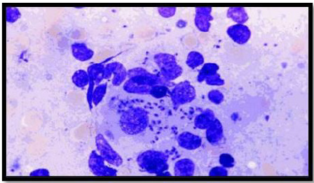
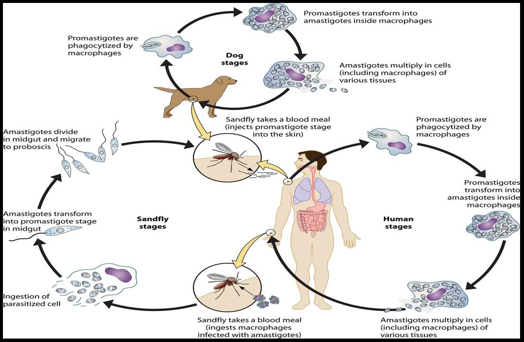
Leishmania has two forms, the amastigote and the promastigote. Amastigote forms are found in the vertebrate host whereas promastigote forms are found in the vector (Unat, et al. [5-7]). During feeding, sandflies inject infectious promastigotes into a susceptible mammal. Promastigotes are phagocytes, converted into amastigotes in the tissue level, and multiplied by simple division within these cells. The parasite continues to infect phagocytic cells either at the skin infection site or in secondary lymphoid organs. Sandflies are infected with an active skin lesion in CL or parasitemia in VL by feeding on the host. In the sandfly midgut, parasites transform to promastigotes. Promastigotes migrate from the midgut and become heavily infected (Esch and Petersen 2013). The life cycle of Leishmania is shown in Figure 1. Amastigotes, shown in Figure 2, are 2-4m in length, are ovoid or round in shape. In addition, they are usually found in monocytes, polymorphonuclear leukocytes and endothelial cells. When stained with Giemsa, the cytoplasm appears in blue and nucleus in pink or dark red, respectively (Unat, et al. [5,7]). Kinetoplast is rod shaped and stained in dark red, shiny red or purple. Amastigotes are nonmotile, feed on via osmosis and get nutrient from tissues. They are aerobes and proliferate longitudinal by binary fusion in macrophages. Firstly, Kinetoplast and blepharoplast and then nucleus and cytoplasm are divided. There is a large nucleus close to the cytoplasm and the Kinetoplast adjacent to the nucleus (Unat, et al. [5,7]). Additionally, there are vacuoles, blepharoplast and axonem in the cytoplasm. Flagellum does not come out of the cell freely. In all Leishmania species, there is only one mitochondrion, the Golgi apparatus and lysosome that helps to feed on parasites by various enzyme activities (Unat, et al. [5,7]).

biomedres-openaccess-journal-bjstr

Figure 1: Life cycle of Leishmania.

biomedres-openaccess-journal-bjstr

Figure 2: Amastigotes.

biomedres-openaccess-journal-bjstr

Figure 3: Promastigotes.

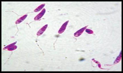
Promastigotes, shown in Figure 3, are 15-28m in length and 1.5- 3.5m in width. One end is sharper and the other end is ovoid. Flagella come up from the front edge. As a result of dying with Giemsa, cytoplasm is stained in blue, inside the cytoplasm nucleus is stained in pink or red (Ozbel, et al. [7]). Kinetoplast is stained in lilac or shiny red in front of the nucleus. Blepharoplast is present before Kinetoplast. Promastigotes are found in the midgut of the vector and the culture medium when amastigotes develop into promastigotes. There are free flagella and axonem which is located near to blepharoplast (Ozbel, et al. [7]). Additionally, kinetoplast, nucleus, nucleolus and pores located in nucleus membrane are present. Moreover, the Golgi apparatus and the endoplasmic reticulum are found in the cytoplasm (Unat, et al. [5,7]).

Leishmanolysin (gp63)

Leishmania species express a membrane glycoprotein on their surface which is called leishmanolysin. The Leishmania ‘s major surface glycoprotein, referred to as gp63, is a 63k Dalton zinc metalloproteinase containing a glycosyl phosphatidyl inositole (GPI) membrane, the major component of promastigotes (5x10⁵ molecules / cell), but at a lower surface density (Casgrain, et al.). It has been stated by Elfaki, et al. [8] that gp63 mainly act in receptor mediated uptake of the promastigotes in the mammalian host by macrophages (Elfaki, et al. [8]). Because of its abundance, surface location and proteolytic activity, the importance of the gp63 in binding promastigotes to macrophages has been inferred. It is one of the parasite receptors that is reported to enhance phagocytosis and increases the survival of extracellular promastigote stage in the presence of host complement and promotes amastigote survival within macrophage phagolysosomes (Chang, et al.). Gp63 prevents parasite from lysosomal cytolysis, exert control over complement activation and degradative activities of macrophages by its protease activity (Alexander, et al. [9]). With the aid of macrophages it will protect the liposome-encapsulated proteins from phagolysosomal degradation. Therefore, gp63 is primarily active in binding macrophage to the Leishmania, in replication and in intramacrophage survival (Razzazan, et al. [10]). It has been suggested that gp63 may interact directly with macrophage receptors, including receptor type 3 macrophage complement, and receptor fibronectin (Rizvi, et al.). This cleaves a number of substrates found on human T-cells, including the CD4 differentiation molecules cluster.

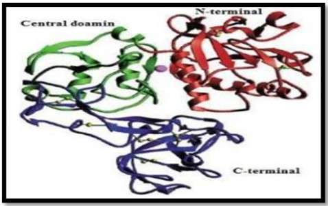
In addition, Lieke et al, noted that gp63 influences proliferation and interferon-gamma unconfined by natural killer cells (NKs) in humans, thus limiting immune response (Lieke, et al.). It has many features that mimic matrix members such as fibrinogen, Zn2 + requirement, cell surface presence, and the sequence resemblance of the proposed active site (Button, et al.). In 1998, the crystal structure of L.major gp63, reported by Schlagenhauf et al, revealed that gp63 with the β-sheet secondary structures belongs to the metzincin class zinc proteinase. The gp63 455070 dimensions have 478 amino acid residues. The protein structure consisted of three domains; N-terminal, Central and C-terminal as shown in Figure 4.

biomedres-openaccess-journal-bjstr

Figure 4: Ribbon representation of the gp63 structure with N-terminal domain, Central and C terminal domain shown in red, green and blue color respectively. Disulfide bonds shown in yellow and active site zinc atom is represented as sphere.

The N-terminal domain of protein contains Zn and the active site have catalytic residues such as His264 and His268. In addition, in metal ligation His264 and His268 are involved with the atoms of Zn+2. The central domain encompasses His334 (third catalytic residue), a Met-turn (Asp342-Asp348), α-helix C (H12), and 62 amino acid insertions (Phe272-Ser333) between Gly271 and His334. The extensive C-terminal domain of gp63 protein exist mainly the anti- p ll l β-strands and the random coil structure with only the slightly helical assistance. In the catalytic active site of gp63, zinc atom is correlated with the nitrogen atoms of His264, H 268, nd H 334 d nc of 2 18Ȧ, 2 18Ȧ nd 2 12Ȧ p c v ly (Schlagenhauf, et al. [11]). If the 3-dimensional structure of gp63 of Leishmania specie does not exist, there would be no correlations between the 3D structure and function of its defence as a vaccine. Razzazan, et al. [10], reported that in view of the development of molecular modeling bioinformatics, a model of gp63 structure with the help of homology modelling with the high precision can be predicted. Afterwards, analysis of the 3D structure of gp63, which can depict the exact information about its structure, function and its interactions (Razzazan, et al. [10]). Gp63 plays a central role in number of host cell molecular events that likely contribute to the infectivity of Leishmania. Furthermore, due of its abundance and ability to mediate resistance against infectious promastigotes, it has been suggested as a promising candidate for vaccination against leishmania infection.

Treatment for Leishmaniasis

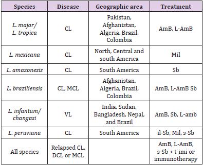
Treatment options used for leishmaniasis disease syndromes are intra-lesional stibogluconate, systemic stibogluconate, ketoconazole, fluconazole, Miltefosine, topical paromomycin, intramuscular paromomycin, liposomal amphotericin B, amphotericin deoxycholate, cryotherapy, heat therapy, Pentamidine, pentoxyfylline, allopurinol, topical imiquimod reported by McGwire and Satoskar. Table 1 Shows the Leishmanial diseases, parasites and treatment regimens by regions (McGwire, et al. [12]).

biomedres-openaccess-journal-bjstr

Table 1: Leishmanial diseases, parasites and treatment regimens (McGwire & Satoskar) [12].

Plants as Remedy

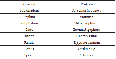
Nature has provided many things for humankind over the years, including the tools for the first attempts at therapeutic intervention. Folk society use plant extracts for the treatment of various conditions. Nowadays, plant constituents stay a vital source for contesting illnesses, comprising contagious ailments. Variety of plants have been investigated for novel drugs or used as templates for the isolation of new therapeutic agents, food flavorings, agrochemicals and industrial chemicals (Lamchouri, et al. [13]). Almost all parts (leaves, stem, roots, seeds, flower and fruits) of the plant are helpful and used for the isolation of natural ingredient. The phytochemical is a natural bioactive compound found in plants, such as vegetables, fruits, medicinal plants, flowers, leaves and roots that work with nutrients and fibers to act as an resistance system against disease or more accurately, to protect against disease. Phytochemicals are categorize as primary like, common sugars, proteins, amino acids and chlorophyll while the second group include, alkaloids, carotenoids, terpenoids and phenolic compounds and many more such as flavonoids and tannins. The beneficial medicinal effects of plant materials typically result from the combinations of secondary products present in the plant. So the systematic screening of plant species with the purpose of discovering new bioactive compounds can help us to cure many fungal and bacterial diseases of economically important crops. The plant chemicals have been found to possess biocidal activity against several pests and pathogens. These are superior to synthetic pesticides in a number of ways like low mammalian toxicity, target specificity and biodegradability (Chugh, et al. [14]). Development in phytochemical studies of medicinal plants for pharmacological as well as nutritional purpose has ruling long time ago. Phytochemicals isolated from plants include essential oils, fixed oils, proteins, flavonoids, phenolic compounds and antioxidants serving as biocontrol agents. Extensive research has been conducted to identify the activity of medicinal plants against various diseases (Samiullah, et al. [15]) (Table 2).

biomedres-openaccess-journal-bjstr

Table 2: Taxonomic Classification of L. tropica.

According to world health organization (WHO) more than 80% of the world’s population relies on traditional medicine for primary health care needs. 250,000-500,000 plant species only a small percentage has been investigated phytochemicals and even few number submitted for biological and 2 pharmacological screening (Zain, et al. [16]). Access to hospitals and other medical abilities is inadequate and therefore, population depends mainly on local customary healers, when they get sick. These healers use various methods in their practice but use preparations of plants which is very successful and nominated practice since ages (Boulanour, et al. [17]). These preparation serves as real drugs, which have direct action on the body and can help to heal (cure) a certain disease (Sameulsson, et al. [18]). Almost every rural communities depending absolutely on medicinal plants for treating diseases and now most often urban population starts recurring to medicinal plants remedies, due to more expensive modern medicines (Boulanour, et al. [17]). Actually, more than 50% of drugs help for years as direct source or may as chemically altered natural products. China (consider as inventor of herbal medicine), India (the Middle East), especially Arab-Muslim World, Egypt, Greece and Rome those civilization used medicinal plants as a huge impact. Herbal-derived components with certain alteration (synthetic / chemical) serve as commercial medications used now-a-days world-wide against various treatments (Miguel, et al.). Due to climatic and phytogeographic conditions in Pakistan help to increase floral diversity containing many medicinal plant species. According to reports 6000 verity of floral species exist in Pakistan. Herbal remedies used in folk medicine provide an interesting and still largely unexplored source for the creation and development of potentially new drug for chemotherapy.

Conclusion

In the new world, leishmaniasis is a serious public health problem. Considering the drug development resistance worldwide, it is vital to achieve new, effective, safe and affordable drugs for parasite disease treatment. Natural source such as plants could be used for production of new antileishmanial agents against leishmania.


For more Articles on: https://biomedres01.blogspot.com/
The Effect of Interface Design and Perceived Risk of Customers’ Satisfaction and Loyalty on Online Booking Application

  The Effect of Interface Design and Perceived Risk of Customers’ Satisfaction and Loyalty on Online Booking Application Introduction The ra...