MRI Imaging of Osmotic Demyelination Syndrome
Case Report
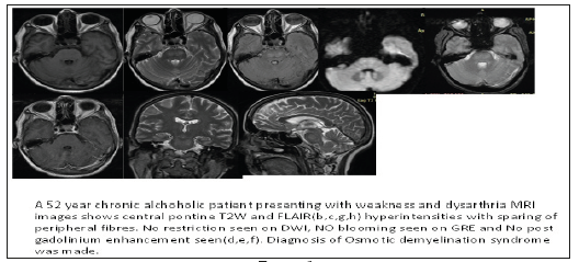
We present a case of a 52 year old chronic alcoholic patient presented with history of weakness in limbs and dysarthria for 10 days. He was admitted in our institute with hypotension, so IV fluids were also rushed to him. MR Scan was done which shows central pontine T2W and FLAIR hyperintensities with sparing of peripheral fibres. No restriction seen on DWI, NO blooming seen on GRE and No post gadolinium enhancement seen. Diagnosis of Osmotic demyelination syndrome was made.
Osmotic demyelination syndrome encompasses central pontine demyelination as well extrapontine demyelination and is seen in alcoholics and malnourished individuals associated with rapid correction of hyponatremia [1-4].
Patient presents with spastic paralysis, pseudo bulbar palsy and disorientation [5,6].
1) Symmetric central pontine T2W hyper intensity with sparing of periphery with trident shaped appearance [6,7].
Pontine ischemia-Asymmetric involving central and peripheral pontine fibers [7,8].
 Demyelinating disease-look for lesion elsewhere, horse-shoe type in particular [1,8] (Figure 1).
Osmotic demyelination is common entity in alcoholics and malnourished individual. It is a treatable entity, so strong clinical suspicion and Typical diagnostic imaging feature on MR scan can lead to the diagnosis.
More BJSTR Articles : https://biomedres01.blogspot.com

No comments:
Post a Comment
Note: Only a member of this blog may post a comment.