Preventing Spread of Vancomycin-Resistant Enterococci in a Hospital by Using a Nanotechnology-Based Disinfectant
Introduction
VRE (vancomycin-resistant enterococci) colonization is a serious problem in hospitals. Because of their resistance to environmental conditions and their natural resistance to antibiotics, VRE is easily spread in the environment. The contamination of pathogenic microorganisms on certain surfaces and their direct or indirect contact with these surfaces contribute to the formation and spread of nosocomial infections. Therefore, the cleaning and disinfection of these surfaces and devices are admitted as a major issue [1,2]. The proper selection and application of disinfectants and antiseptics make it possible to obtain much more effective results than the use of antibiotics in combating hospital infections [3]. Bacoban® (Adexano, Germany) produced with nanotechnology has a Nano sponge layer which prevents microorganisms from building surfaces and also has antimicrobial effect with biocidal deposits. The Fresenius Institute in Germany has approved that Bacoban® showed the disinfectant effect in five minutes and continued for ten days. Bacoban® contains ethanol, benzalkonium chloride, isopropanol, pyridine-2-thiol-1-oxide sodium salt (sodium pyrite), inorganic/organic polymer and distilled water [4,5]. In the study, firstly, the bactericidal effect of Bacoban® on VRE was investigated in laboratory conditions, then the disinfectant effect of Bacoban→ on VRE contamination and colonization were studied with environmental and rectal swab samples, respectively. As far as we know, it is the first study to demonstrate the efficacy of Bacoban® in the clinical setting.
A prospective study was conducted at Istanbul University Cerrahpasa Medical School, a 1,300-bed tertiary care teaching hospital. First, the long-acting disinfectant ( Bacoban®, Cheshire, UK) was examined on various surfaces/materials in a laboratory setting. Next, its efficiency on contamination and colonization with VRE was investigated in several intensive care units (ICUs), as well as other units in which vancomycin-resistant enterococcal outbreaks were reported by the Hospital Infection Control Committee.
Two series of materials/surfaces were used for the main and the control groups, including tile, wood, fabric, plastic (flat, slight and rough surfaces), metal, and Formica in sizes of 10 x 10 cm. After these surfaces were cleaned with alcohol and dried, 200μl of 1Mc Farland VRE suspension was spread over the surfaces/materials with sterile swabs. After drying the surfaces, Bacoban® solution was sprayed on them (for the main units) at a distance of 30 cm. The same amount of sterile saline solutions was sprayed on the surfaces for the control groups. Five minutes later, swab samples from the surface series were cultured onto the VRE agar plates (Oxoid, Ottawa, Canada). Along with the continuation of the study, these surfaces received equal amounts of VRE suspension, and in 15 minutes, swab samples were taken with sterile saline solution and cultured onto the VRE agar plates. This practice was continued until all material surfaces were reproduced with VRE. The first days of VRE reproduction on the surfaces/materials were recorded.
Diluted household bleach contains ~ 5-6% sodium hypochlorite (1:100 or 1⁄4 cup:1 gallon, 525-615ppm chlorine) was used as a surface disinfectant in the pre-Bacoban® period of the study [6]. During this period, a total of three times a week, environmental samples were collected from the main units. In General Surgery, Infectious Diseases and Bone Marrow Transplantation Hematology Units, the peripheral swab samples were taken only from the rooms of VRE-positive patients, while the samples were taken all environmental surroundings in Anesthesiology and Reanimation ICU. In the Bacoban® period, practical information of the Bacoban® application was given to the staff, and the other disinfectants were not used. Bacoban® was applied once a week at Anesthesiology and Reanimation ICU, twice a week at Bone Marrow Transplantation Hematology Unit, and once a week for the other two units. Disinfection was repeated in case of contamination with patient wastes. During this period, the environmental swab samples were taken on average six times per week from the previously sampled areas and sent to the laboratory as soon as possible.
During the environmental disinfection applications, patients admitted to the units were included in the study for VRE colonization. Rectal swab samples from the patients were taken during the initial hospital admission and then once a week. The samples were sent to the laboratory as soon as possible. Patients with no colonization in the first admission, but were VRE positive in their later samples were identified as “new VRE colonization”.
In the experimental study, swab samples from the surfaces were cultured on VRE agar plates (Oxoid, Ottawa, Canada) and incubated at 37 °C for 24-48 hours. The environmental and rectal swabs samples were firstly cultured with VRE Broth (Oxoid, Ottawa, Canada) and incubated at 35-37 °C for 24 hours. After the incubation, passages were taken on VRE agar and incubated at 35-37 °C for 24- 48 hours. VRE strains isolated from the agars were identified by conventional microbiological methods [7,8]. Susceptibility of the isolates to glycopeptide was determined by disc diffusion method, and the confirmation was performed by E-test strips (Liofilchem, Italy). All breakpoints were applied according to the CLSI (Clinical and Laboratory Standards Institute) guidelines [9,10]. Quality control was performed by using Enterococcus faecalis ATC 29212 reference strain.
Biostatistical evaluation of the study results was conducted. Fischer’s exact test was used to compare frequency and percentages for the pre- and post- Bacoban® periods with control groups, and the Pearson Chi-Square test was used for group comparisons of continuous data and appropriate criteria of the normal distribution. All analyses were performed using SPSS 16.0 package program (SPSS, Chicago, IL, USO). The significance value was considered as p < 0.05.
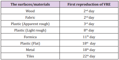
In the experimental study with Bacoban®, VRE strains were isolated from the first-day samples of the control surface / materials. After the Bacoban® application, VRE growth was not detected during the 22 days on tile, 18 days on metal and flat plastic, 11 days on Formica, 8 days on slightly roughened plastic, 3 days on roughened plastic, and 2 days on wood and fabric surfaces. The experimental study showed that Bacoban® had a bactericidal effect on VRE, and was long-acting on preventing VRE growth, especially on tile, metal and flat plastic surfaces. The first day of the VRE growth on different surface/materials after the application of Bacoban® are shown in Table 1. A total of 969 environmental samples, 362 and 607 from the pre-and post-Bacoban® periods were studied, respectively. All samples were taken from the main units (Anesthesiology and Reanimation ICU, General Surgery, Infectious Diseases, and Bone Marrow Transplantation Hematology Units). 38 (10.5%) of the 362 and 13 (2.1%) of the 607 environmental samples were VRE positive in the pre- and post-Bacoban® periods, respectively.
 *: Vancomycin- resistant enterococci.
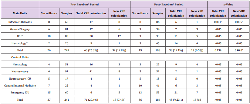
In the pre-Bacoban® period, the highest VRE contamination rates were seen on the edges of the bedside (23.2%) and tables near the patients (13,6%). Compared with pre- and post- Bacoban® periods, the decrease in VRE isolation rates was found statistically significant (p ≤ 0.001). VRE positivity and negativity in peripheral swab samples in the pre- and post- Bacoban® periods are shown in Table 2. In the rectal swab samples from the patients, VRE contamination rates were 25.3% and 19.1% during the preand post- Bacoban® periods, respectively. Although the difference was not statistically significant (p = 0.139), there was a significant difference between the newly acquired VRE colonization rates in pre-Bacoban® (12.8%) and post-Bacoban® (6.5%) periods (p = 0.028). The results of the total and newly acquired VRE colonization in the pre- and post-Bacoban® periods are shown in (Table 3). There wasn’t a significant difference between the rates of total VRE colonization (29.4% and 23.1%) and newly acquired VRE colonization (7.4% and 8%) at control units without Bacoban® application. In this study, it was reported that the application of Bacoban® was easy, it dried quickly and did not leave any residues after the application. On the other hand, some side effects such as burning sensation, shortness of breath, and slight itching were reported by three staff members within direct contact with Bacoban®.
 *: Vancomycin - resistant enterococci, 1: p<0.05.
Table 3: VRE* colonization’s in pre- and post- Bacoban® periods.
 *: Vancomycin - resistant enterococci
**: Anesthesiology and Reanimation Intensive Care Units
***: Bone Marrow Transplantation Hematology Unit
1: p < 0.05
Hospital-acquired infections (HAIs) are one of the major patient safety problems in hospitals, especially in ICUs [11]. Approximately, 20-40% of hospital-acquired pathogens are spread with the hands of healthcare professionals or by colonized patients or contaminated environmental surfaces [12,13]. The risk of infectious disease of environmental microorganisms depends on the pathogenicity as well as the ability to survive on the surface [14]. Among environmental pathogens, VRE pathogenicity isn’t a high factor, but it is confused as a causative agent that can lead to outbreaks that are difficult to control [15]. VRE can stay alive for days, weeks, even on dry surfaces, and this can cause them to be particularly persistent in a hospital environment [16]. Hands of healthcare professional play a major role in the spread of VRE among patients [17,18]. Even if the handwashing is effective, the hand can be recontaminated with the contact of the surface. Therefore, cleaning is very important for environmental surfaces with VRE [19,20]. However, cleaning with water and detergent is not sufficient for reducing of VRE colonization or contamination. Frequently touched surfaces and patient surroundings must be disinfected in addition to cleanliness. An effective method of disinfection leads to a significant reduction in environmental contamination [21,22].
More BJSTR Articles : https://biomedres01.blogspot.com


No comments:
Post a Comment
Note: Only a member of this blog may post a comment.