Breast Cancer: The Accuracy of The Paus in Detecting pN2 and Factors that Lead to the True- and False-Negative Results
Introduction
The condition of Axillary Lymph Nodes (ALN) is the most important prognostic factor in breast cancer and is relevant to the choice of breast cancer treatment tactics [1,2]. There is a strong correlation between the number of damaged axillary lymph nodes and the risk of breast cancer regeneration [3]. Since 1994, Sentinel Lymph Node Biopsy (SLNB) has become a gold standard for assessing the status of the axillary lymph nodes. The sentinel lymph node is hypothetically the lymph node or group of lymph nodes that the primary cancer cells reach first. If the sentinel lymph node is intact, the probability that the tumor will not spread is high. According to several studies, when metastases are detected and the secondary Axillary Lymph Nodes Dissection (ALND) is performed, in 40-70% of cases, additionally removed lymph nodes were healthy [4]. When isolated tumor cells or micrometastases are found after SLNB, the removal of other lymph nodes in the axilla is not recommended [5]. A randomized ACOSOG Z0011 study was presented at the annual conference of the American Society of Oncology (ASCO) in 2010, which found that survival and the risk of recurrence was very similar in patients with T1 or T2 tumors and during SLNB 1 or 2 damaged lymph nodes who had ALND and those who did not [5].
About 70% of the early breast cancer cases have intact axillary lymph nodes and surgical axillary intervention is unnecesAbstractsary [6]. Although SLNB complications are rare and less damaged than ALND, this is a risky procedure. Several prospective studies (ACOSOG Z0010, NSABP B-32, ALMANAC) identified the incidence and frequency of SLNB complications: allergic reactions (0.1-1.0%), wound infection (1.0-10%), seroma (7.1 %), paresthesia (8.6-11%) and hematoma (1.4%) [7]. Accordingly, there is an ongoing search for non-invasive methods to evaluate the status of axillary lymph nodes in order to prevent ALND or SLNB [6]. In the guidelines of the European Union, Preoperative Axillary Ultrasound (PAUS) is a routine examination for all breast cancer patients with palpable or impalpable axillary lymph nodes [4]. It is possible to assess whether the lymph node is damaged by ultrasound (PAUS-positive) according to the morphological features of the axillary lymph node [7]. In different studies, the sensitivity and accuracy of the ultrasound examination varied between 23% to 87% and 67.9% to 90.2% [2,5,6-18]. One of the weaknesses in the ultrasound examination is the false-negative results (~21%) [15], but in the determination of metastases in three or more axillary lymph nodes, false negative tests are found to account for about 4% [2].
Symptoms of the disease are less favorable and the prognosis is poorer in patients who are PAUS- positive [4] versus patients who are PAUS negative and metastases were found rarely in more than three axillary lymph nodes [2]. There are studies suggesting that an ultrasound examination can replace SLNB when the axillary surgery is no longer curative and only biomarkers are important for adjuvant chemotherapy [7]. The purpose of this study was to determine the accuracy and reliability of the ultrasound examination in detecting more than three metastatic axillary lymph nodes (pN2) and to assess the clinicopathological factors that lead to the true-negative and false-negative results of the ultrasound study.
Materials and Methods
The present study is a retrospective analysis of the medical documentation of all newly diagnosed breast cancer patients between 1 January 2011 and 31 December 2012 in the Hospital of Lithuanian University of Health Sciences Kauno klinikos. The study included women (n=667) who underwent a preoperative axillary ultrasound examination. When normal-appearing or suspicious axillary lymph nodes were found during the ultrasound study, SLNB was performed. If there were definitely pathological axillary lymph nodes by ultrasound examination, an immediate ALND was performed or treatment started with neoadjuvant chemotherapy (these women were not included in the study). The criteria for assessing the lymph nodes are shown in Table 1. The cases of breast cancer found on both breasts were analyzed as two separate cases. Rejection criteria: Neoadjuvant chemotherapy, recurrent breast cancer, non-invasive breast cancer, unexplored or poorly documented PAUS study. PAUS study data were compared to the results of postoperative histological examination. The study design is shown in Figure 1). PAUS was performed using an Acuson S2000 ultrasound unit with a 14MHz linear Siemens transducer.
Figure 1: Study design - axillary assessment protocol.
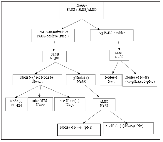 Table 1: PAUS lymph node evaluation criteria.
 The sentinel lymph nodes were marked by injection periareolarly of 0.2ml - 50MBq of 99mTC- labeled nanocole one day before surgery. The location of the sentinel lymph node was detected using the 2010 Philips BrightView Gamma Camera and detected during the operation with the Crystal Probe-automatic-handheld gamma detector. Surgically removed radioactive lymph nodes were sent for pathological examination (hematoxylin and eosin staining and immunohistochemistry). PAUS data were compared with histological examination results. The following histopathological data were analyzed: Tumor Size (T), Tumor Grade (G), Lymphovascular Invasion (LV), the presence of Estrogen (ER) or Progesterone (PR) receptors, Human Epidermal Growth Factor (HER2) status and Lymph Nodes (pN) status.
Statistical analysis was performed using IBM SPSS Statistics 24 package. Descriptive statistics methods were used to systemize the research data. The accuracy, sensitivity, specificity, Positive Predictive Value (PPV) and Negative Predictive Value (NPV) of the ultrasound examination were evaluated and the estimates of these characteristics were given along with their 95% confidence intervals. The Chi-square test was used to compare effects of clinicopathological factors and the correlation of outcomes from PAUS and SLNB pathology. The results were considered statistically significant if p< 0.05.
The study analyzed 667 females aged from 27 to 88 years old, with an average age of 60.07 (SD=12.451). By age, patients were divided into three groups: young (< 40 years, n=30 (4.5%)), middle age (40-69 years, n=476 (71.4%)) and elderly (≥70 years, n=161 (24.1%)). Patient demographic and tumor clinopathological data are presented in Table 2. Small tumors were found more frequently (T1 - 52.2%). The majority of tumors were moderately- differentiated (G2), in 63.7% of cases. Tumor spreading in blood vessels and lymphatic vessels was found in 36.2% and 49.6% of cases. More frequently, ER (+) (43.3%), PR (-) (44.7%) and HER2 (-) (57.0%) tumors were detected (Table 2). By PAUS, pathological lymph nodes were found in 86 cases and lymphodectomy was performed; for all remaining cases (N=581), SLNB was performed. Metastatic lymph nodes (node-positive) were diagnosed in 230 cases (34.5%) (pN1 - 28.4%, pN2 - 6.1%), and the axillary lymph nodes were undamaged (node-negative) (pN0) in 437 cases (65.5%). After SLB, metastases were detected in three sentinel lymph nodes in 68 cases (11.7%) and secondary ALND was performed. Of all of the additional removed lymph nodes, metastases were found in one or two lymph nodes (pN2) in 24 (35.3%) cases, while the additional removed nodes were undamaged in the remaining 44 cases (64.7%). Therefore, of the 581 SLB, heavy nodal disease burden (pN2) was only found in 24 cases (4.1%) (Figure 1).
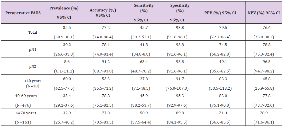
 PAUS-negative axillary lymph nodes were found in 535 cases, of which heavy nodal disease burden (pN2) was detected in only 15 cases (2.8%). PAUS-positive axillary lymph nodes were evaluated in 132 cases: 86 as pathological (3 cases were node-negative, 83 cases were node-positive), and 46 as suspicious (24 cases were node-negative, 22 cases were node-positive) (Figure 2). In general, PAUS was not very sensitive (45.7%) but was very specific (93.8%), sufficiently accurate (77.2%) and had rather high positive and negative predictive values (79.5% and 76.6%). According to age groups, metastases were most commonly found in young women (60%), but the accuracy of PAUS was the lowest (53.3%); in middle and older women, metastases in the axillary lymph nodes were found less frequently (33.4% and 32.9%) and the PAUS was quite accurate (78.8% and 77.0%). Determining more than three metastatic lymph nodes (pN2), the accuracy of PAUS was 91.2% (95% confidence interval [CI]=88.7-93.8), sensitivity was 63.4% (95% CI=48.7- 78.2) and NPV was 96.5% (95% CI=94.7-98.2) (Table 3). Of the 535 patients who were PAUS-negative, metastases were detected in 125 cases (18.7%) in the histological examination (false-negative results).
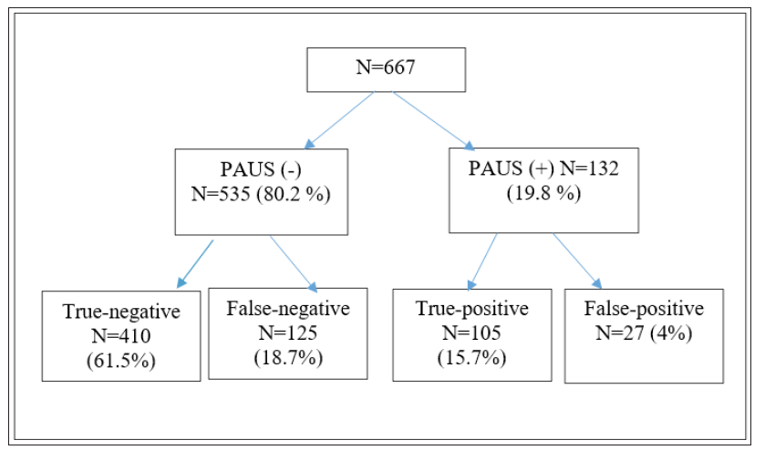 Table 3: PAUS statistical results according to lymph node status and patient’s age.
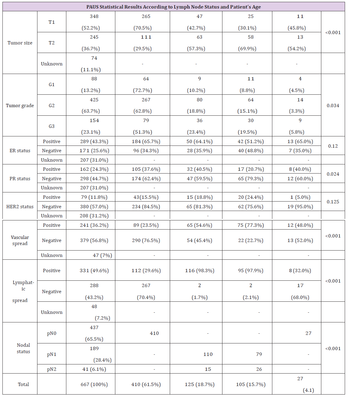 In younger patients, false-negative PAUS results (10.4%) were more frequently found than true-negative (2.7%) (p=0.006). In cases when the tumor size was larger (T2), false-negative (57.3%) and true-positive (69.9%) results of the study were more often detected (p< 0.0001), in contrast to cases when the Tumor Size was smaller (T1), when true-negative (70.5%) results were more often detected. Statistically significantly more frequent false-negative results of ultrasound examination were obtained in cases with poorly-differentiated tumors (G3), in 23.4% of cases (p=0.034). In cases with tumor spreading in blood vessels and lymphatic vessels, false-negative and true-positive PAUS results were higher (V1 54.6% and 77.3%, L1 98.3% and 97.9%), otherwise true-negative PAUS results were lower (V1 - 23.5%, L1 - 29.6%) (p< 0.0001). Expression of the estrogen (ER), progestin (PR) and human epidermal growth factor (HER2)) was not statistically significant for PAUS results (Table 2). After surgery in PAUS-positive cases, heavy nodal disease burden (pN2), larger tumor (T2) (p< 0.0001), and poorly-differentiated tumors (G3) (p=0.034) were more frequently found and tumor spreading in the blood and Lymphatic Vessels (LV1) was more often detected (p< 0.0001) (Table 2).
According to the literature, metastatic lymph nodes are detected in about 30-40% cases of newly diagnosed breast cancer [18]. In order to evaluate the status of axillary lymph nodes, all patients undergo surgical intervention (SLNB or ALND) to this day. Although SLNB is less risky than ALND, about 60-70% cases of newly diagnosed breast cancer are node-negative and axillary surgical intervention is unnecessary. In our study, metastatic lymph nodes were detected in 230 patients (34.5%), but in all 667 cases, either SLNB or primary ALND, or SLNB and secondary ALND were performed; however, for 437 patients (65.5%), axillary surgical interventions were unnecessary. In the guidelines of the European Union, PAUS is a routine examination for all breast cancer patients with palpable or impalpable axillary lymph nodes [4]. Recently, if there is definitely pathological axillary lymph nodes by ultrasound examination, neoadjuvant chemotherapy has been initiated. During the study period, our clinic had two possible solutions: either primary ALND or neoadjuvant chemotherapy; therefore, in 86 cases, primary ALND was performed. Patients with normal-appearing or suspicious lymph nodes on PAUS (n=581) underwent SLNB.
 (PAUS: preoperative axillary lymph node; PPV: positive prognostic value; NPV: negative prognostic value)
Most often, metastases were found in young women (60%), but the accuracy of PAUS was the lowest (53.3%), while metastases were found less often in the axillary lymph nodes in middle-aged and older women (33.4% and 32.9%), and the PAUS study was quite accurate (78.8% and 77.0%). These results suggest that the PAUS study can be more reliable for middle-aged and elderly women, but should be considered more cautiously when it is performed in young women (Table 3). Few studies have been conducted to analyze the causes of false-negative results of an ultrasound examination. Jalin’s study suggests that the negative results of PAUS study should be evaluated with caution when the tumor is larger (≥ T2), poorly differentiated (G3), or when tumor spreading in blood vessels and lymphatic vessels is detected, as that increases the probability of false-negative results [3]. Nwaugu’s study also suggests that the probability of false-negative outcomes increases when the tumor is larger and the tumor spreading in blood vessels and lymph nodes is diagnosed, but the tumor differentiation degree is not statistically significant [15]. In our study, false-negative PAUS results were found in 18.7% of cases (Table 5).
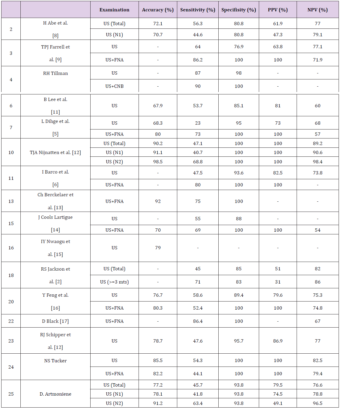 (US: Ultrasound; CNB: Core Needle Biopsy; FNA: Fine Needle Aspiration; PPV: Positive Predictive Value; NPV: Negative Predictive Value)
In the young age group, the false-negative results of the PAUS study were statistically significantly more frequent (10.4%) than the true-negative (2.7%) (p=0.006), and there were no statistically significant differences in other age groups. In our study, in the case of a larger tumor (T2), false-negative (57.3%) and true-positive (69.9%) PAUS results were statistically significantly more frequent (p< 0.0001); in the case of smaller tumors (T1), true- negative study results were more often received (70.5%). Also, false-negative results of the PAUS study were found statistically significantly more frequently (p=0.034) in poorly-differentiated tumors (G3) (23.4%) than in well-differentiated tumors (G1) (10.2%). In the presence of tumor spreading in blood vessels and lymphatic vessels, false-negative and true-positive PAUS study results were statistically significantly (p< 0.0001) more frequent (V1 - 54.6% and 77.3%, L1 - 98.3% and 97.9%) than true-negative results (V1 - 23.5%, L1 - 29.6%). ER, PR and HER2 expression were not statistically significant for PAUS study results.
An ultrasound examination is sufficiently accurate to detect a heavy nodal disease burden (pN2) and an additional axillary surgery can be avoided in most cases. Ultrasound examination is more accurate and more sensitive to middle-aged and older women and the sensitivity of the young women’s ultrasound examination is rather low. In the middle-aged and elderly patients, when the tumor is small (T1), well-differentiated (G1) and the tumor spreading in blood vessels (V0) and lymphatic vessels (L0) is not detected, we can rely on PAUS study results and avoid SLNB. On the contrary, in the younger age group with larger (T2), poorly-differentiated (G3) tumors, and with the detection of tumor spreading in blood vessels (V1) and lymphatic vessels (L1), the probability of false-negative results is increased. Therefore, in these cases, the results of the PAUS study should be evaluated more cautiously. In PAUS-positive patients there is a greater possibility that the tumor will be more aggressive and more widespread, so these patients require more aggressive treatment of the axillary lymph nodes (operational or radiotherapy).
Molecular Diversity in Bulgarian and Cypriot Barley Germplasm Collections – A Reference Point for Better Understanding, Exploitation and Improvement of Adaptiveness to Agro-Climatic Conditions-https://biomedres01.blogspot.com/2020/11/molecular-diversity-in-bulgarian-and.html
More BJSTR Articles : https://biomedres01.blogspot.com


No comments:
Post a Comment
Note: Only a member of this blog may post a comment.