Low-Molecular Weight Heparin/Protamine Micro-Nanoparticles Augmented Viability of Human Adipose-Derived Regenerative Cells
Introduction
Recently stem cell therapies have been widely applied for various diseases refractory against conventional medical as well as surgical treatments. Critical limb ischemia due to peripheral artery disease or Bürger disease is a rational indication for the stem cell implantation to induce neovascularization. Bone marrow-derived stem cells (BMSCs) can improve limb ischemia after injection in experimental limb ischemia models [1] and patients with critical limb ischemia [2]. Mesenchymal stem cells (MSCs) are also known to induce neovascularization in a hindlimb ischemia model [3]. Adipose tissue is abundant in the human body and is constantly reorganized through angiogenesis. Therefore, this tissue is an ideal source of angiogenic MSCs. It has been shown that adipose-derived regenerative cells (ADRCs), which contain not only MSCs but also various regenerative cells such as endothelial progenitor cells, have characteristics similar to those of BMSCs [4]. The implantation of in vitro cultured ADRCs in the mouse hindlimb ischemia model was effective for its proangiogenic action [5]. In addition, the safety and effectiveness of autologous ADRCs implantation in patients with critical limb ischemia has been reported [6].
ADRCs were isolated from periumbilical subcutaneous adipose tissues collected by a liposuction procedure in 3 patients with critical limb ischemia (Case 1: 51yo male; progressive systemic sclerosis, Case 2: 70 yo female; progressive systemic sclerosis and Case 3: 45yo male; Bürger disease), who were planned to undergo the stem cell therapy for ischemic limbs. For the isolation of ADRCs, we used an automated cell-processing system, Cytori Celution device (Cytori Therapeutics Inc., San Diego, California). The stem cell therapy was performed under the approval of local committee for regenerative medicine and an informed consent regarding the present study was obtained from each patient. The LH/P-MPs were synthesized as described previously [9]. Briefly, 0.3mL of protamine sulfate solution (10mg/mL; Mochida Pharmaceutical Co., Tokyo, Japan) was added dropwise to 0.7mL of a low-molecular-weight heparin solution, Dalteparin sodium (6.4 mg/mL; Kissei Pharmaceutical Co., Tokyo, Japan) and vortexed for approximately 2min. The mixed LH/P-MPs were then washed twice with phosphate-buffered saline to remove nonreactants using centrifugation at 4,900g for 5min, and the precipitates were finally resuspended in 1mL of Dulbecco’s modified Eagle’s medium (DMEM; Life Technologies Oriental, Tokyo, Japan).
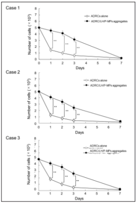
Patients’ derived ADRCs promptly decreased cell number in suspension culture, eventually they were eliminated through cell death after seven days. Addition of LH/P-MPs produced aggregation with ADRCs, remarkably slowed down the cell death. Such a protection effect against cell death was reproduced in three independent patients. The number of survived ADRC was significantly higher in ADRC/LH/P-MPs aggregates compared to ADRCs alone at the next day (Case 1: 4.53±0.34 vs 1.49±0.29 ×10³, P< 0.01; Case 2: 4.10±0.44 vs 1.28±0.39 ×10³, P< 0.01; and Case 3: 4.33±0.35 vs 1.83±0.56 ×10³, P< 0.01), at the 2 days (Case 1: 4.11±0.38 vs 1.00±0.28 ×10³, P< 0.01; Case 2: 3.38±0.37 vs 0.75±0.26 ×10³, P< 0.01; and Case 3: 3.75±0.41 vs 1.04±0.48 ×10³, P< 0.01) and at the 3 days (Case 1: 3.13±0.37 vs 0.62±0.26 ×10³, P< 0.01; Case 2: 2.45±0.30 vs 0.40±0.19 ×10³, P< 0.01; and Case 3: 2.72±0.50 vs 0.51±0.26 ×10³, P< 0.01) (Figure 1).
 Discussion
In the present study, we observed that cell viability of ADRCs isolated from human subcutaneous adipose tissues was augmented, when the LH/P-MPs were added to the ADRCs and ADRC/LH/PMPs aggregates were formed, compared with the ADRCs alone in all of 3 patients with critical limb ischemia. Survived cell number was reduced to approximately 30% at the next day, 20% at the 2 days and only 10% at the 3 days after incubation in case of ADRCs alone, indicating that ADRCs hardly sustain cell number due to lack of extracellular scaffold. On the other hand, addition of LH-P-MPs rescued such a rapid cell death and the ratio increased to 90% (next day), 80% (2 days) and 70% (3 days), respectively. The ADRCs have the potential to differentiate into various cell lineages, including vascular cells [10]. In addition, cultured ADRCs secrete various angiogenic growth factors, such as basic fibroblast growth factor, hepatocyte growth factor, and vascular endothelial growth factor as well as cytokines [11-13]. Angiogenetic mechanism of stem cell therapy such as ADRCs therapy is mainly based on paracrine effects by these angiogenic factors [14].
More BJSTR Articles : https://biomedres01.blogspot.com


No comments:
Post a Comment
Note: Only a member of this blog may post a comment.