Saturday, January 30, 2021

An Introduction to Bioactivity of Fucoidan

An Introduction to Bioactivity of Fucoidan


Fucoidan
Fucoidan is a polysaccharide, which is derived from brown algae and some marine invertebrates, consisting mainly of L-fucose and sulfate ester groups [1]. Fucoidan is particularly found in the cell wall of marine brown algae. This polysaccharide is named as fucoidin when it is derived from the first marine brown algae. Kylin gave Fucoidin in 1913. However, this name has been changed to fucoidan according to IUPAC rules [2]. Fucoidan related different studies have performed in the literature [1,2]. It has different bioactivities such as anticoagulant, anti-thrombotic, antiinflammatory, antitumoral, immunomodulatory, anti-inflammatory, antioxidant, anti-hepatopathy, anti-uropathy, anti-inflammatory. These activities depend on the source of fucoidan samples taken from different species [1,2]. In addition, fucoidan is non-toxic or any adverse effects on the healthy tissues so that it can be used safety. There are many forms of fucoidan but the simplest molecular structure of fucoidan is obtained from Fucus vesiculosus consists mainly of 44.1% fucose, 26.3% sulphate and 31.1% ash and a small proportion of aminoglucose [3,4].

Bioactivity of Fucoidan
Anticancer effect of fucoidan on various cancer cells has been showed thus far. The researchers have showed that its’ anticancer effect induced by inhibiting angiogenesis, metastasis and invasion and by cell accumulation in sub G0 / G1 phase [5-8]. In a study, examining about the effect of fucoidan on human metastatic PC-3 prostate cancer cells showed that fucoidan increased the levels of p21 / WAF1 / CIP1 in cells and suppressed the E2F transcription factor. In another research related to effect of Fucoidan on Burkitt’s lymphoma cells have also been showed to suppress metalloproteinase-9 secretion and migration [9]. Fucoidan induces apoptosis by extrinsic and intrinsic pathways in cancer cells. It is reported that the activation of caspase-3 due to the permeability of the mitochondrial membrane is reduced [6]. Besides, Fucoidan stimulates ER stress mechanisms by induces Toll-like receptor-4 regulated reactive oxygen species and promotes endoplasmic reticulum stress-mediated apoptosis in lung cancer cell lines and tumors [10]. Consequently, it would be suggested that fucoidan induces apoptosis in cancer cells, but its effect depends upon dose and time. One study has been reported that fucoidan has significantly increased drug efficacy because of synergistic effects with anticancer drugs. Furthermore, the immune response has been shown to increase the activity of T cells and reduce the effect of free oxygen radicals as a protective effect of fucoidan against side effects of chemotherapeutic drugs [9]. Fucoidan coated doxorubicin nanoparticles for the transmission of doxorubicin, shows a significant increase in the cell is also determined in the literature. According to this effect, it is thought to be an important agent in increasing the effectiveness of cancer drugs [11]. Masahide et al. indicated that fucoidan regulated the incidence of fatigue for the period of chemotherapy.

The study outcomes showed that chemotherapy with fucoidan group was endured for a longer period than chemotherapy without fucoidan group. Besides, Fucoidan inhibited toxicity of chemotherapeutic agents. As a result, the survival of patients with fucoidan treatment group was longer than that the patients without fucoidan [12]. Fucoidan is a safely agent both increasing the antitumoral effects of chemotherapeutics beside it has no adverse effects on healthy tissues. Fucoidan has the potential to be a high influence drug without damaging normal tissue and by reducing the side effects of anticancer drugs. However, it is still needed to study understanding the mechanism of fucoidan.

FMTVDM©℗ Provides the First Patented Quantification of Myocardial Perfusion Imaging (MPI) Yielding Theranostic Benefit for Individuals with Suspected Coronary Artery Disease (CAD)-https://biomedres01.blogspot.com/2021/01/fmtvdm-provides-first-patented_30.html

More BJSTR Articles : https://biomedres01.blogspot.com

FMTVDM©℗ Provides the First Patented Quantification of Myocardial Perfusion Imaging (MPI) Yielding Theranostic Benefit for Individuals with Suspected Coronary Artery Disease (CAD)

FMTVDM©℗ Provides the First Patented Quantification of Myocardial Perfusion Imaging (MPI) Yielding Theranostic Benefit for Individuals with Suspected Coronary Artery Disease (CAD)


Introduction
Myocardial perfusion imaging (MPI) using either single photon emission computed tomography (SPECT)/Planar or positron emission tomography (PET) has been limited by the utilization of qualitative image interpretation. This qualitative approach has restricted the use of SPECT/Planar and PET to assessment of coronary artery disease (CAD) as either present (sensitivity) or absent (specificity). Consequently, determination of treatment success has been constrained. The introduction of the first truly quantitative method [1-8] for measuring CAD and regional blood flow differences (RBFDs), providing artificial intelligence (AI)/machine learning (ML) application has been compared with conventional qualitative imaging [9-10] and was recently demonstrated at the 2018 American Society of Nuclear Cardiology [11] Conference. The clinical significance and application of this method for BOTH diagnostic assessment and treatment monitoring and adjustment (viz. theranostics) is shown here in two cases. The first where conventional MPI missed critical CAD (lack of sensitivity), which would have cost the veteran his life had the critical disease not been caught by FMTVDM and the second where conventional MPI resulted in invasive coronary angiography, which produced adverse vascular consequences resulting in the patient being placed on a balloon pump (lack of specificity).

Case One
FMTVDM finds critical CAD missed using qualitative MPI. A late middle-aged Caucasian male presented to his community hospital for evaluation of intermittent retrosternal chest discomfort. He was scheduled for diagnostic evaluation comparing [9-12] qualitative MPI imaging with quantitative FMTVDM (Figure 1). Figure 2a shows conventional MPI qualitative image display with and without attenuation correction. The clinician/physician interpretation of the qualitative study was “no evidence of ischemic CAD”. Figure 2b shows the results displayed quantitatively, in vertical and longaxis views. FMTVDM quantification showed wash-in of the isotope (redistribution) demonstrating critical anterior, anteroseptal and anterolateral CAD. The patient was referred for coronary angiography and PCI (Figure 2c) treatment based upon MPI quantitative analysis.

Note: Sequential FMTVDM quantification of isotope redistribution, measuring metabolic and regional blood flow differences (RBFDs).
Figure 1: True FMTVDM Quantification of Myocardial Perfusion Imaging and RBFDs.
biomedres-openaccess-journal-bjstr
a) Standard display of qualitative imaging including attenuation correction (AC images), reveal no evidence of CAD.
(b) Quantification of the patients MPI demonstrates isotope redistribution, with the left panel demonstrating isotope measurement at 5-minutes post stress and the right panel demonstrating 60-minutes post stress imaging.
(c) The quantified images resulted in percutaneous coronary intervention (PCI) of the left anterior descending (LAD), 1st diagonal and 1st obtuse marginal arteries as demonstrated in the before (left) and after (right) panels.

Figure 2: Critical CAD missed using conventional qualitative MPI.
biomedres-openaccess-journal-bjstr
Case Two
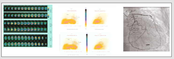
FMTVDM quantification demonstrates patent coronary artery bypass graft (CABG) without RBFDs/CAD. A late middle-aged Caucasian female presented with dyspnea on exertion and a history of CAD previously treated by CABG. Her physician ordered a “stress-rest” qualitative MPI study. She also underwent FMTVDM quantitative imaging [9-12] using the same protocol [1-6, 8-11] employed in case one (Figure 1). Physician interpretation of her qualitative MPI (Figure 3a) reported multivessel CAD, involving the LAD and circumflex marginal system. Quantitative FMTVDM measurement (Figure 3b) revealed no evidence of coronary ischemia, which was confirmed angiographically (Figure 3c) with a patent left internal mammary artery (LIMA) graft to her 1st diagonal (D1), 1st obtuse marginal (OM1) and posterior descending (PDA) arteries. During the course of the patient’s coronary angiogram, vascular complications resulted from the procedure and she ended upon on an intra-aortic balloon pump (IABP) for several days in the coronary care unit. She made a complete recovery without further coronary artery intervention or treatment.
(a) Standard display of qualitative imaging was interpreted as showing multi vessel LAD and circumflex CAD.
(b) Quantification of patients MPI revealed no evidence of CAD with a total 5-minute measurement (upper left) of 280,026 and a 60-minute measurement (lower left) of 236,089, revealing a washout of 5.7%, excluding ischemic CAD.
(c) Coronary angiography reveals no evidence of ischemia, with patent LIMA bypass graft to her 1st diagonal, 1st obtuse marginal and posterior descending arteries.

Figure 3: Qualitative MPI interpreted as demonstrating ischemia, while quantitative measurement revealed no ischemia.
biomedres-openaccess-journal-bjstr
Discussion
Limitations in qualitative assessment of disease [12] including assessment of coronary artery disease from MPI and coronary arteriography [13] has resulted in the call for the development of quantitative methods to accurately measure [1-8] outcomes to provide for better clinical care and management of patients. Documentation of the diagnostic benefit of this FMTVDM has been well established [9-11]. These two case studies demonstrate the needed benefit of using FMTVDM quantitative imaging to better manage patient diagnostics, treatment and clinical outcomes. The first example revealed the importance of using FMTVDM to find CAD in a patient whose critically narrowed coronary arteries were missed using two-injection “stress-rest” qualitative imaging; the wrong imaging sequence promulgated by misrepresentations made by the pharmaceutical company to the FDA regarding Sestamibi redistribution [3,5,6,8]. In the second case a woman who had previously undergone successful left internal mammary artery (LIMA) bypass surgery, demonstrated complete patency and regional blood flow through the LIMA graft to her native coronary arteries, quantitatively measured by FMTVDM imaging, while the conventional “stress-rest” two injection method promulgated by the same pharmaceutical company and the resultant physician qualitative interpretations, indicated a need for further invasive testing and intervention, which in the end was not needed and consequently resulted in the rare but statistically recognized interventional vascular complications.

Conclusion
The introduction of FMTVDM quantitative MPI provides enhanced patient management and treatment superior to qualitative image interpretation of MPI, using the first patented truly AI quantitative measurement of CAD and regional blood flow differences (RBFDs) to initially define the extent of CAD and then to measure the truly quantitative affect of patient-specific, patientoriented treatment; saving time, money and lives. COI: FMTVDM; B.E.S.T. Imaging©℗ patent is issued to the Primary Author.

Identification and Isolation of the Yeasts in Traditional Yogurts Collected From Villages in Gaziantep,Turkey-https://biomedres01.blogspot.com/2021/01/identification-and-isolation-of-yeasts_30.html

More BJSTR Articles : https://biomedres01.blogspot.com

Identification and Isolation of the Yeasts in Traditional Yogurts Collected From Villages in Gaziantep,Turkey

Identification and Isolation of the Yeasts in Traditional Yogurts Collected From Villages in Gaziantep,Turkey


Introduction
During the fermentation of foodstuffs, the main physical and chemical changes occur due to the growth and fermentative activity of the LAB that are used as starter cultures. LAB are used as starter cultures for the fermentation of milk [1]. Yogurt is a milk product that is obtained by the bacterial fermentation of milk. Lactic acid is produced via the fermentation of milk sugar, also known as lactose. Lactic acid is the substance that confers the typical textural and sensory characteristics of yogurt and acts on milk protein [2]. Besides low asid is a suitable environment for the life of yeasts. Fermented products also contain yeasts. They have created positive effects on the bacteria via PH changing and biological substances releasing [3]. If a matter of caution is to be taken, they may be causing of the spoilage in fermented product.
Yogurt contains high concentrations of LAB and the smaller yeast have several potential health benefits and probiotic potential; it can increase lactose tolerance, balance the intestinal microflora, act as an antimicrobial, stimulate the immune system, induce antitumor effects, and induce anti-cholesterolemic effects. Yeast may be some aroma components and interact with LAB that are primer starter cultures. Yeast growth stabilizes LAB which are starter culture [4]. Knowledge of the biochemical properties of the starter cultures is necessary to determine the final characteristics of the fermented food product, such as its acidity, aroma and flavor. To gain such knowledge, it is crucial to identify and characterize the yeast in a starter culture. The aim of this study was to identify of the yeast that could be isolated from traditional yogurts collected from villages in Gaziantep Province. Additionally, preliminary information was prepared to create reliable yogurt in respect to pathogen microorganisms.

Material and Methods
Sample Collections and PH Measurement
Yeasts were isolated from traditional yogurts collected in villages in Gaziantep Province (from Sahinbey, Sehitkamil, Oguzeli, Yavuzeli, Nizip, Karkamis, Araban, Islahiye and Nurdagi townships) between 2015-2016 years. The PH values of 208 the yogurt samples were measured with a PH meter (Martini Instruments, Romania) and recorded.
Isolation and Total Yeast Counts
The samples were diluted before culturing on MRS (de Man, Rogosa and Sharpe) agar (Oxoid, England). The MRS plates were incubated for 3 days in aerobic environments at 30°C [5-7]. All of the suspect colonies in petri dishes were recorded as CFU/g [6].
Stock Culture Preparation
A sterile loop was used to sample colonies and to inoculate them into Eppendorf tubes containing 1ml of MRS Broth (Oxoid, England). Yeast samples inoculated into MRS were incubated for 48 hours. Stocks were prepared from 500μl of yeast cultured in MRS broth 500μl of sterile liquid glycerol that were mixed in a 1ml Eppendorf tube and stored at -20°C [8].
Identification of Yeasts Using MALDI-TOF MS
Identification of yeast colonies isolated from traditional yogurts was conducted according to their protein and peptides database analysis using MALDI-TOF MS. Matrix-assisted laser desorption ionization-time of flight mass spectrometry (MALDI-TOF MS) is a new technology application for identify yeast cultures. Avantages of this application have got faster and routine properties [9]. For these tests, stock cultures were grown on MRS agar. The isolates were tested with respect to their various surface proteins. Twenty-five isolates were selected for yeasts identification by the MALDI-TOF MS (BioMérieux, France) test kit. Data were subjected to evaluation as a percentage (%).

Results
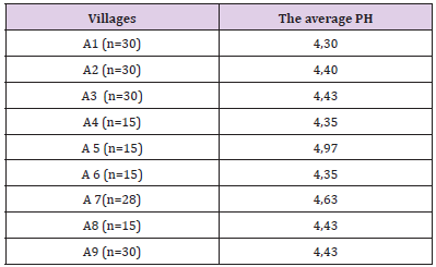
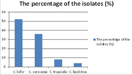
According to the results of this study, the pH values of 208 yogurt samples ranged from 3.9 to 6.0. The average PH of the yogurts ranged between 4.30 and 5.14 as seen Table 1. The total yeast counts on MRS agar medium were 1-1.8 x 10⁸ CFU/g following the isolation of yeasts from the yogurt samples. Seventy four (74) colonies out of 25 analyzed yogurt samples were identified on MRS medium as the yeast. The yeast identification results revealed that the mycoflora in yogurt from Gaziantep Province, Turkey contained 52% Candida kefyr, 36% Saccharomyces Cerevisiae, 8% Candida tropicalis, 4% Candida lypolitica. The distribution of different yeasts (%) among yogurts collected from townships in Gaziantep Province is shown in Figure 1.

Table 1: The PH values of yoghurts collected from villages in Turkey.
biomedres-openaccess-journal-bjstr
Figure 1: The distribution of different yeasts (%) among yogurts collected from townships in Gaziantep Province.
biomedres-openaccess-journal-bjstr
Discussion
The present study is the first to specifically analyze the yogurt mycoflora in Gaziantep Province. In this study, the results showed that the PH levels of traditional yogurts from Gaziantep were acidic (below 7) as a result of lactic acid fermentation. The PH of the yogurts ranged from 3.9-6.0. The animal sources of the yogurts varied; they included cow milk, sheep milk and goat milk. It has been determined that the PH levels of yogurts produced from different milk types ranged between 4.02 and 4.26 and reported that these PH values were related to the metabolic activity of the starter cultures and the fermentation time of the yogurt [10]. Meanwhile, it has been showed that the PH values of yogurts obtained from villages in Nigeria were between 3.80 and 4.48 [11]. Additionally, they reported that these levels were lower than the PH levels of commercial yogurts. Yeast isolation studies were carried out from fermented milk products in several countries. C. tropicalis has been isolated from Tanzania fermented milk. But, the same yeast species has not isolated from yogurt samples.

Researches said that yeast contamination has been prevented and controlled is importance on fermented dairy producing [12]. Kavas et al. [4] have isolated these yeast species from isolated from Ä°zmir, Turkey [4]. They have identified the yeast species which inhibit yogurt starter cultures growth. For this reason yeast contamination should been blocked. In present study, [12] percentage of two hundred and eighty (208) were the yeast species. But, these yeasts are not also indigenous in yogurt normally. For this reason, the yeasts isolated from the yogurts are worrying for consumer health and their control is necessary. Contamine yeasts needs to be controlled. On the other hand these yeasts may be play a severity role the ending of fermentation. Besides it is gratifying that these yogurt samples are not contain toxigenic yeasts such as Penicillum, Aspergillus. While Omokaro et al. [13] 20% 20% Saccharomyces Cerevisiae have isolated from Nigeria yogurt samples, this rate was 36% in present study [13]. Saccharomyces may be related to suitable flavor development and Candida spp. yeasts that have got protease activity would long shelf life of milk products, also enhancing activation in LAB fermentation [12]. The Candida species were the predominant among 25 yeasts species including 16 Candida (64%) and 9 Saccharomyces (36%).
In this study, Candida kefyr, Candida lypolitica, Saccharomyces cerevisiae isolated from traditional yogurts were also isolated from traditional fermented camel milk in a previous study [3]. The present study has showed that yogurt fermented dairy food comprise yeast microflora. Yeasts interact with other microorganisms [12] and yeasts are consist secondary flora in fermented milk products [14] isolated sixteen species belonging to Candida and Saccharomyces genus from Pakistan yogurt samples. But these yeast species are out of our isolated yeast species [15]. Biberoglu and Ceylan (2013) found as 50% Candida kefyr,23.95% Saccharomyces Cerevisiae, 4% Candida lypolitica same results with our present study from yogurt samples of Erzurum and Kars regions, Turkey [14]. Soulides (1956) isolated lactose fermented Torulacremoris from yogurt. New name of this species in binominal named is Candida pseudotropicalis [16].
We isolated several Candida species in too, in present study. Moreira et al. [17] found 19% Saccharomyces Cerevisiae rate from Brazil yogurts [17]. This rate was 36% in traditional yogurt samples, Gaziantep, Turkey in our study. Saccharomyces Cerevisiae and Candida spp. which are contained yeasts causing spoilage in traditional yogurt production, were isolated in this study. However, the yeast species that we isolated have previously been demonstrated to have the potential harmful ability to produce yogurt. These species may represent factors that is to be need underlie hygiene properties of traditional yogurts. These contaminants must be struggled. In a sense important results have been obtained for the provision of traditional yogurts to consumers in our city. This study may represent a fundamental investigation for eliminated contained yeasts during the yogurt production process, as isolated bacterial species that have previously been isolated in other studies. The four different kinds yeast of in these yogurts samples have been isolated in this study. In the data obtained is a preliminary study and firstly study in this region which will be useful information to another region for this non-commercial yogurt.

Conclusion
Our study researches the basic development of suitable hygiene conditions for traditional yogurt production. So far, there has been little specific research on yogurt contained microflora in Turkey. Our study is the first to specifically analyze the yogurt contaminants in Gaziantep Province, Turkey.

Pigmented Lesions of Ocular Fundus: Clinical Aspects-https://biomedres01.blogspot.com/2021/01/pigmented-lesions-of-ocular-fundus.html

More BJSTR Articles : https://biomedres01.blogspot.com
Friday, January 29, 2021

Pigmented Lesions of Ocular Fundus: Clinical Aspects

Pigmented Lesions of Ocular Fundus: Clinical Aspects


Introduction
Circumscribed pigmented lesions of the ocular fundus are not uncommon. They can be congenital, developmental, neoplastic or the result of infectious or inflammatory processes. Pigmented neoplastic lesions of ocular fundus, both benign and malignant, can arise from the retinal pigment epithelium (RPE) or from choroidal melanocytes. It is important for the ophthalmologist to properly diagnose these lesions to avoid erroneous diagnosis and misdirected treatment.
Discussion
Lesions Arising from the Retinal Pigment Epithelium
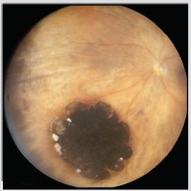
Congenital hypertrophy of the Retinal Pigment Epithelium (CHRPE) is an uncommon benign fundus lesion. The typical unifocal, unilateral lesion occurs in about 0.5% of individuals [1]. CHRPE lesions are usually asymptomatic and detected on routine ophthalmic examination. The typical unifocal CHRPE is a gray to black, well-defined, minimally elevated fundus lesion having a diameter in the range of 2–5 mm (Figure 1). The lesion frequently undergoes at least partial depigmentation with aging, developing discrete intralesional atrophic foci (lacunae that tend to enlarge and coalesce over the years). No diagnostic studies are generally indicated for characterization of these retinal lesions. Fluorescein angiography (FA) and indocyanine green angiography (ICGA) show a well-defined blocking defect at the retinal pigment epithelial level. Transmission of choroidal fluorescence takes place through lacunae in the lesion [2].
Figure 1: Fundus photography of CHRPE.
biomedres-openaccess-journal-bjstr
The unilateral multifocal CHRPE, also known as congenital grouped pigmentation of the retina or “bear tracks”, is characterized by multiple, flat, gray to black RPE clustered in one region of the fundus. These lesions are hypofluorescent on FA and hypoautofluorescent on fundus autofluorescence (FAF) [3]. Atypical multifocal bilateral CHRPE presents oval or irregularly shaped lesions, variable in size (usually between 0.1 and 2.0 mm in maximal diameter), widely separated rather than clustered; it is associated with familial colonic adenomatous polyposis syndrome.1 Reactive Hyperplasia of the Retinal Pigment Epithelium is a result of an ocular insult, such as inflammation or trauma. It can appear as a diffuse or sessile lesion or as a nodular mass. Its dark black color and associated signs of prior inflammation or trauma can help to differentiate the lesion from choroidal melanoma. Major complications or progression are quite unusual [4].
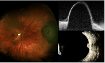
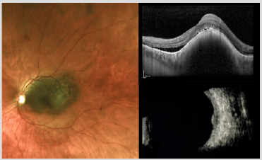
Adenoma and adenocarcinoma of the RPE have respectively benign and malignant histopathologic features and they are generally diagnosed in adulthood, with a mean age of 53 years [5] Unlike uveal melanoma, these tumors seem to have no predilection for race. They may have a slight predilection for females [6] Clinically, these lesions are usually solitary and unilateral and begin as a small, deep retinal tumor that is dark brown to black in color. They usually grow very slowly and invade the overlying sensory retina. In some instances, they have arisen from solitary congenital hypertrophy of the RPE [7] FA can document the retinal feeder vessels and show early hypofluorescence and late minimal hyperfluorescence of the tumor, without visibility of choroidal vessels. Ultrasonography (US) typically demonstrates the tumor to be elevated and to have medium to high internal reflectivity. Enhanced depth imaging optical coherence tomography (EDI-OCT) shows the tumor surface as irregular and dense posterior optical shadowing (Figure 2) [6].
Figure 2: Fundus photography (Left), EDI-OCT (Top Right) and US imaging (Bottom Right) of adenoma of the RPE.
biomedres-openaccess-journal-bjstr
Combined hamartoma of the retina and RPE is a congenital, nonhereditary lesion with an uncertain etiology. This lesion is composed of an admixture of pigment epithelial cells, proliferating blood vessels and glial tissue. It appears as an ill-defined gray retinal mass with tortuous or straightened retinal blood vessels and it is characteristically located on or adjacent to the optic disc, but it can be seen in extrapapillary areas of the fundus. FA shows abnormal retinal blood vessels in the mass and gradual late staining of the lesion. Optical coherence tomography (OCT) shows an irregular lesion with vitreoretinal traction [8].
Lesions Arising from Choroidal Melanocytes
Choroidal nevus is the most common primary intraocular tumor. In Caucasians, it has been estimated that approximately 2 to 10% of persons older than 50 years have at least one choroidal nevus [9].It is much more common in persons of lightly pigmented races and it appears to be equally common in men and women. The typical choroidal nevus appears as a small gray to brown tumor. Many choroidal nevi exhibit characteristic surface alterations such as drusen and RPE pigment clumping. Most choroidal nevi are 5 mm or less in basal diameter and 1 mm or less in thickness. Blurred or distorted vision attributable to a choroidal nevus may be the result of an accumulation of serous subretinal fluid [10], cystic degeneration of the retina overlying a macular choroidal nevus [11] or choroidal neovascularization [12] Diagnosis of a typical choroidal nevus is based almost exclusively on clinical features; fundus photograph can be useful to compare subsequent follow-up evaluations.
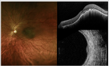
US is commonly used for the measurement of tumor thickness; most choroidal nevi are nearly flat and can be difficult to detect. Choroidal nevus is characterized by high internal reflectivity on US. On FA nevus remains typically hypofluorescent. EDI-OCT can image choroidal nevi, even those undetectable by US: choriocapillaris thinning over the nevus, partial or complete choroidal shadowing deep to the nevus, RPE atrophy, photoreceptor loss and inner segment–outer segment junction irregularity are some of the EDI-OCT features of choroidal nevus (Figure 3). On FAF imaging pigmented nevus generally appears more hypoautofluorescent from the intrinsic melanin that absorbs light compared with less pigmented nevus that appears more hyper-autofluorescent. The estimated annual rate of malignant transformation of a pre-existing nevus into melanoma reported in several studies varied from 0,2 to 1% [13].
Figure 3: Fundus photography (Left), EDI-OCT (Top Right) and US imaging (Bottom Right) of choroidal nevus.
biomedres-openaccess-journal-bjstr
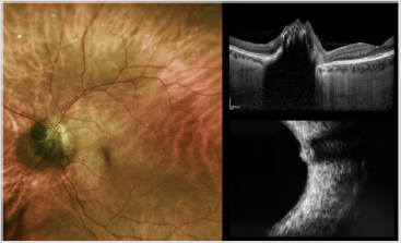
A special type of uveal nevus is the melanocytoma (magnocellular nevus) of the optic disc. This lesion is usually composed entirely of maximally pigmented, polyhedral nevus cells (magnocellular nevus vcells). Clinically, the melanocytoma is a dark brown tumor that involves the substance of the optic disc. The surface of the tumor commonly appears striated because of insinuation of the darkly pigmented cells of the tumor between axons in the nerve fiber layer. If the melanocytoma compresses the optic disc, it can produce prominent visual field defects. Lesion enlargement can occur but is usually slow. The diagnosis can be made by ophthalmoscopic recognition of its characteristic clinical features. In most cases, FA of a melanocytoma of the optic disk demonstrates hypofluorescence throughout the angiogram, because the cells are deeply pigmented and closely compact with relatively little vascularity. In cases with optic disk edema, there is hyperfluorescence of the edematous portion of the optic disk. ICGA also shows the lesion to be generally hypofluorescent. US shows high reflectivity of the lesion (Figure 4). Malignant change is estimated to occur in about 1-2% of cases [14].
Figure 4: Fundus photography (Left), EDI-OCT and US imaging (Bottom Right) of melanocytoma of the optic nerve.
biomedres-openaccess-journal-bjstr
Choroidal melanoma is the most common primary intraocular malignant tumor, with an incidence of 5.1 cases per million per year [15] Choroidal melanoma can originate from a pre-existing choroidal nevus. In 1995, Shields et al. identified several risk factors predictive of nevus growth into melanoma: thickness more than 2 mm, subretinal fluid, symptoms of flashes/floaters and blurred vision , orange pigment, margin less than or equal to 3 mm from optic disk, ultrasonographic hollowness and absence of halo . Tumors with three or more risk factors likely represent small melanoma, demonstrate malignant transformation in 50% of cases at 5 years and treatment should be considered early [16]. The classic appearance of choroidal melanoma is pigmented domeshaped or mushroom-shaped tumor with an associated exudative retinal detachment.
The mushroom-shaped configuration is due to the breaking of the overlying Bruch’s membrane and RPE, forming a nodular eruption beneath the retina that progressively enlarge. Although some choroidal melanomas are detected on ophthalmic evaluations prompted by visual symptoms (e.g., blurred vision, visual field defect, flashes, floaters), many patients who have such a tumor are asymptomatic at the time of detection. Small choroidal melanomas can be detected by EDI-OCT and it is also a useful tool for differentiating choroidal nevus from small choroidal melanoma. It has limited use with heavily pigmented tumors, which cast a significant posterior shadow. US is an important diagnostic tool used to define tumor extent and shape, and to measure tumor dimensions of choroidal melanoma. Typical features of a posterior uveal melanoma on US include acoustic hollowing, choroidal excavation and orbital shadowing (Figure 5). Spontaneous vascular movement may be noted in a highly vascularized tumor.
Figure 5: Fundus photography (Left), EDI-OCT and US imaging (Bottom Right) of choroidal melanoma.
biomedres-openaccess-journal-bjstr
On A-scan the four cardinal acoustic hallmarks of malignant melanoma are a regular internal structure with similar height of the inner tumor spikes or regular decrease in height, low to medium reflectivity, solid consistency with no after movement of tumor spikes, and echographic sign of vascularization with a fast, spontaneous, continuous, flickering vertical motion of single tumor spikes. The characteristic features on FA include gradually increasing fluorescence that starts at or before the arterial phase, increases in intensity well into the recirculation phase, and persists for at least 45 min. The tumors that have broken through Bruch’s membrane characteristically reveal “double circulation” pattern because of superimposition of the intravascular fluorescence of the intact retinal vasculature over the fluorescence of large caliber vessels within choroidal tumor. ICGA allows better visualization of intrinsic vasculature in choroidal melanoma with maximal fluorescence at an average of 18 min from injection. Choroidal melanoma generally shows on FAF slight intrinsic hyperautofluorescence and the brightness increases with pigmented tumours, larger tumours, and those associated with disrupted RPE [17, 18].
Pseudomelanoma
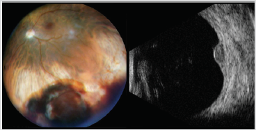
There are conditions that can clinically simulate melanoma, leading to diagnostic ambiguity. The most frequent pseudomelanomas included choroidal nevus, peripheral exudative hemorrhagic chorioretinopathy (PEHCR), congenital hypertrophy of the retinal pigment epithelium and idiopathic hemorrhagic detachment of the retina or RPE [18] This section covers selected nonneoplastic-simulating conditions that are not discussed elsewhere in this article. Peripheral exudative hemorrhagic chorioretinopathy (PEHCR) is a peripheral retinal degeneration of the elderly manifesting with exudative and hemorrhagic changes. This condition can closely simulate melanoma with a homogenous deep brown mass in the periphery (Figure 6). However, PEHCR shows blockage on FA, dark hypoautofluorescence on FAF, echodensity with retraction cleft on US and spontaneous resolution [19].
Figure 6:Fundus photography (Left) and US imaging (Right) of PEHCR.
biomedres-openaccess-journal-bjstr
Idiopathic hemorrhagic detachment of the retina occurs after bleeding into the subretinal space and in some cases large amount of blood may accumulate, creating a hemorrhagic detachment of the retina, whereas idiopathic hemorrhagic detachment of the retinal pigment epithelium occurs in a variety of conditions such as senile disciform macular degeneration and angioid streaks, and it may complicate other conditions characterized by breaks in Bruch’s membrane. In many of these cases there is a clinically detectable source of the blood, such as an intraretinal microaneurysm [20].
Conclusion
A variety of pigmented lesions can simulate choroidal melanoma. Suspicious choroidal nevus is still the lesion most difficult to differentiate from choroidal melanoma. Most other pseudomelanomas can lead to diagnostic ambiguity. Some elements can still generate confusion, but careful assessment of the clinical and instrumental characteristics can help to avoid diagnostic errors.

Assessment of Motor Proficiency in Atypically Developing Children: Measurement and Clinical Applications-https://biomedres01.blogspot.com/2021/01/assessment-of-motor-proficiency-in.html

More BJSTR Articles : https://biomedres01.blogspot.com
Assessment of Motor Proficiency in Atypically Developing Children: Measurement and Clinical Applications

Assessment of Motor Proficiency in Atypically Developing Children: Measurement and Clinical Applications

Introduction

In the clinical context, assessment involves a complex process including a variety of measurement techniques, with primary reliance on observation of performance, and integration of diverse information in a summary judgment. Also, more often than not it involves (formal) testing. Measurement error, hence, lack of reliability, is inevitable when testing takes place. However, when the amount of error is substantial, the inferences drawn from the resulting scores have to be treated with caution or disregarded altogether.

The Classical True Score Theory (CTT), which is one of many measurement theories (e.g. Generalizability Theory, Item-Response Theory), is able to estimate the amount of the existing error [1]. In this context, reliability can be defined as the degree to which the scores are consistent or repeatable, the observed score is free of random/ systematic measurement error, or the extent to which the observed score matches the true score. This hypothetical true ability (score) of the person is difficult if not impossible to capture via any assessment tool due to measurement error. Conceptually, one way of estimating this score would be by taking the mean of many trials performed by the same individual, under the same circumstances, and on the same test. Unfortunately, in the clinical context this method is impractical. Thus, according to CTT, if the amount of measurement error is small one can be confident that the emerging observed score at the very least approximates well the true ability of the person. The degree of this confidence can be wagered based on the magnitude of the reliability coefficient which is often derived via test-retest or internal consistency approaches. The former infers the consistency/stability of repeated performances that are separated in time and measured by the same examiner under the same conditions, whereas internal consistency assesses how well the items of a test or instrument measure a specific construct [2]. If the individual items within a set of tasks are highly correlated that would indicate that the items represent the same construct.

For clinicians, the choice of a particular test depends on many factors such as its purpose, population of interest, theoretical framework and its psychometric properties [3]. For decades Movement Assessment Battery for Children (MABC) [4] has been considered as a gold standard in the area of adapted physical activity as related to the assessment of children with non-congenital, developmental coordination problems. This is a standardized, norm-referenced test which is used for the purpose of screening, identifying and diagnosing mild to moderate movement problems in children. The test is composed of tasks pertaining to manual dexterity, ball skills, and balance skills. The scores are then accumulated into the Total Impairment Score (TIS), which reflects the overall motor proficiency of a child. Recently, a revised version of MABC has been released (Movement Assessment Battery for Children-Second Edition) [5].

The “kit is easier to carry, the performance test items are more engaging for children, and the scoring system for both the performance test and checklist are more user-friendly” (p. 92). Also, the new version encompasses a broader age range (3 to 17 years of age) than the previous test (4 to 12 years of age), and the number of age bands has been reduced from four to three (3 to 7; 7 to 10 and 11 to 17), resulting in different number of items and new equipment. In regard to age band 2, which is off primary interest here, in the manual dexterity subsection, the placing pegs task now has a new starting position and layout. The lacing board is longer for the threading lace activity, and the shape of the drawing trail has changed. For the aiming and catching section, the beanbag task a box was replaced with a target, whereas in the balance subsection floor mats were added for the one leg hopping activity (Figure 1).

biomedres-openaccess-journal-bjstr

figure 1: The changes between the original and new version of MABC (2 edition), as indicated by the arrow, to lacing the board task (top left), bean beg throw (top right), jumping in squares (bottom left) and drawing tasks.

Despite these numerous changes, the authors maintained that the research pertaining to the reliability and validity of the original tool applies to the new version. Some researchers [6] in the field remained skeptical in regard to this assumption and called for more investigations examining the psychometric properties of the new version. Thus, the purpose of this study was to examine different facets of reliability (test-retest & internal consistency) of the age band 2 (7 to 10 years old) for the three subsections and composite score.

Materials and Methods

Participants

Forty participants (18 males and 22 females) between 7 and 10 years of age (M = 9 years, 2 months, SD = 1 year, 3 months) were recruited. In order to be included in this study, children were required to be typically functioning in terms of their motor and cognitive status, as reported by the parents/guardians when completing the DCDQ. The child had to obtain a score higher than 60. Purposive sampling was implemented. The children were recruited from local elementary schools with the permission of the respective school boards in the region. Prior to the data collection, participants were given a brief description of the study. The parents of all participants provided a written consent to take part in this research project prior to its initiation.

Protocol

Participants attended two sessions, one to two weeks apart. Children were assessed individually and each session took approximately 45 minutes to one hour. At the second testing session, the child was re-assessed at the same location, under the same conditions, and by the same examiner. There are 8 different tasks in the MABC-2 test for age band 2 [5] which are divided into three sub-sections; manual dexterity, ball skills and balance. The tasks within each section are safe, relatively simple and resemble activities that a child performs on a daily basis. There are 3 manual dexterity tasks (placing pegs, threading lace, drawing trail), 2 ball skills tasks (catching with two hands, throwing beanbags onto a mat), and 3 balance tasks (one board balance, walking heel-to-toe, hopping on mats). For each child, a Total Test Score (TTS) and three sub-section scores were derived and converted to standard scores.

Design, Analysis and Dependent Variables

The reliability of the MABC-2 was analyzed using ICC correlation coefficient [7] for the test-retest approach. A two-way mixed model with absolute agreement was implemented for the calculation of ICC. An analysis of variance (dependent samples t-tests) was also implemented to determine if there were statistically significant differences between the group means across the two sessions. Cronbach alpha was used to infer the internal consistency of the items across manual dexterity, aiming and catching, and balance subsections. In addition, the Cronbach’s alpha with items deleted was implemented [8]. This procedure allows inferring if the internal consistency of the sub-component improves when a certain item is removed. If so, this would indicate that that specific task is not measuring the same domain as the remaining items. If the Cronbach’s alpha remains the same or decreases, while the item is deleted, this indicates that the item enhances the internal consistency of the sub-component.

Results

Test Re-Test Reliability

In regard to the Total Test Score, the analysis revealed a moderate degree of consistency as inferred from ICC coefficient (0.67). The additional analysis of variance (t-test) confirmed these inconsistencies as scores at time one (M =10.55, SD = 2.49) were significantly different as compared to time two (M = 11.53, SD = 2.53) (t (39) = -2.53, p < 0.05). The analysis of the Sub-Component Scores showed a similar scenario. The analysis of the manual dexterity revealed an ICC = .68, which was supported by the t-tests as statistically significant difference between time one (M = 9.85, SD = 3.10) and time two (M = 10.70, SD = 3.32) (t (39) = -2.14, p < 0.05) emerged. The aiming and catching sub-component also revealed a moderate degree of reliability (ICC = .65), however no differences were found between time one (M = 9.95, SD = 2.31) and two (M = 10.13, SD = 2.54) (t (39) = -0.54, p = 0.59). In regard to balance domain once again a questionable degree of consistency emerged (ICC = .66), which was accompanied by significant differences between the two sessions (M = 11.55, SD = 2.57 vs. M = 14.10, SD = 2.18) (t (39) = -2.09, p < 0.05).

The review of the corresponding scatterplots (Figure 2) demonstrated that across the four scores, the data set was homoscedastic, hence equally distributed along the hypothetical line of best fit. Also, there were no outliers which often affect the magnitude of the emerging coefficient. However, it is also important to note that all the scatterplots showed less than forty data points. This indicates that some children achieved the same scores across the sessions and this could be possible attributed to floor or ceiling effects, hence tasks/items being too easy or too difficult. Likely, larger amount of variability would be more desirable to prevent the potential for restricted range across all the scores.

Internal Consistency

The internal consistency of manual dexterity, aiming and catching, and balance was inferred from item standard scores, from the second testing session of the MABC-2. The analysis of Manual Dexterity revealed a Cronbach’s alpha of 0.61, representing questionable internal consistency for this sub-component. Further analysis was implemented using Cronbach’s alpha with items deleted. As the coefficient did not increase when any of the three items were deleted, this indicated that not one specific task jeopardized the internal consistency of this set of items. The Aiming and Catching sub-scores showed a similar pattern of results as the Cronbach’s alpha was 0.49, once again indicating a questionable internal consistency. Cronbach’s alpha with items deleted could not be calculated as there are only two tasks within this sub-section. As it was the case with the two previous sub-scores, the Cronbach alpha for Balance section was also low (0.53) further supporting weak reliability of these items. When items were deleted, the value did not increase for any of the three items, thus once again indicating that not one specific item caused the questionable internal consistency.

Discussion

Test-Retest Reliability

The analysis of the TTS revealed an ICC of 0.67 when test retest reliability was examined, thus indicating a questionable degree of reliability. As the use of correlations alone can be problematic to assess the degree of systematic bias, particularly when the inter-individual variability is low, the analysis of variance was also carried out. In line with the correlation, the data showed significant differences between time one and time two. In fact, only 9 out of the 40 participants achieved the same standard score across the two trials (Figure 2, top graph). This indicated that there is a lack of consistency across performances in more than three quarters of the individuals. To date, there has been only one study which examined the reliability of the age band 2. Holm and colleagues [9] examined intra- and inter-rater reliability based on component scores and reported ICC values of 0.68 and 0.62, respectively. These findings are similar to those reported here. The sample size and characteristics of the participants were also comparable between the studies. Thus, the emerging results appear to be robust even though different reliability coefficients were examined. In terms of the research examining the test re-test reliability based on the original version of MABC, Chow and Henderson [10] found that the TTS had a higher, but still moderate reliability (0.77). However, their sample size was also larger (75 participants), and the study was conducted on age band 1 (4 to 6 years). Also, the authors did not indicate which type of score was used (standard, component, or percentile) for the calculation of ICC, thus a caution is warranted when comparing the results from the two studies.

biomedres-openaccess-journal-bjstr

figure 2: Intra-class correlation coefficients and respective scatterplots for standard scores for total test score (top figure), aiming and catching (second from the top), balance (third from the top) and manual dexterity.

The degree of reliability of the three sub-components was in line with the findings pertaining to TTS. Manual dexterity revealed the ICC of 0.68, which once again indicated poor reliability. This lack of consistency was also supported by the analysis of variance, which showed that there were statistically significant differences between the two testing sessions. The analysis of the individual data across both sessions also showed that 25 of the 40 participants scored higher on time two suggesting that some systematic and/or random bias emerged. The standard deviation also increased between the sessions indicating that as the group the sample was more variable despite that they were assessed with the same tool for the second time. In terms of previous research, the present values are similar to those reported in the past studies. Holm and colleagues [9] showed that age band 2 had ICC values of 0.62 (intra-rater) and 0.63 (inter-rater) for the manual dexterity sub-component. However, the reliability of this subsection in other age bands was substantially higher. Ellinoudis and colleagues [11] revealed an ICC of 0.82 and Wuang and colleagues [12] reported an ICC of 0.97 for the test re-test reliability, for age band 1. No studies were conducted on age band 3, or the original MABC, that examined the test re-test reliability for manual dexterity.

In terms of aiming and catching, the ICC for the test re-test reliability was 0.65, thus once again indicating a questionable consistency. The analysis of variance showed that there were no statistically significant differences between time and time two. Nevertheless, the individual data showed that only 9 of the 40 participants had the same standard score across both testing occasions, thus from the standpoint of absolute reliability little consistency was evident. The present results are to some extend comparable to those reported by Holm and colleagues [9] who showed that the ICC values ranged between 0.49 and 0.77 for intra- and inter-rater reliability, respectively. In regard to previous research, with other age bands, similar results (ICC = .61) were reported by Ellinoudis and colleagues [11] despite the fact that their sample was much larger than the current data set. This pattern of results may indicate that this subsection of the test as a whole represents an issue in the revised edition of the test [13].

The analysis of the balance sub-component also revealed a questionable degree of reliability. This could be due to the fact that tasks such as one-board balance were too challenging to most of the children. In fact, 50% of participants achieved the same score across trials, in comparison to the other two sub-components and the TTS. Thus, the individuals were scoring consistently but poorly across the both sessions, leading to small intra-group variability. The low reliability found here is consistent with the coefficients (.49 & .29) reported in previous research, for age band 2 [9]. The authors of that research also supported the notion that low ICC values for this subsection may be due to the difficulty associated with one board balance task. In regard to other research, Ellinoudis and colleagues [11] reported an ICC value of 0.75 for age band 1.

However, it should be noted that the nature of the tasks included is different across the age bands. For example, in age band 1 the participants are required to complete 3 tasks including one leg balance, walking with heels raised and jumping on mats. Although the latter two items are comparable to those in age band 2, the one leg balance task would be considerably easier than the one-board balance task. Therefore, this task likely contributed to a restricted range in age band 2.

Internal Consistency Reliability

The Cronbach’s alpha for manual dexterity was 0.61 indicating a questionable reliability. The low internal consistency could be due to the fact that one of the three items is not measuring the construct of interest, therefore lowering alpha. For example, the placing pegs and threading lace task are very similar in that they are both timed tasks. However, the drawing trail-2 task is selfpaced and it requires effective use of a pen/pencil. Thus, although similar, the additional constraint of time and use of an implement may require different aspects of motor functioning. A low alpha could also be attributed to the fact that there are only three tasks within the manual dexterity sub-component, as Cronbach’s alpha is higher when there are more test items. Wuang and colleagues [12] examined the internal consistency of age band 1 and reported a much higher Cronbach’s alpha (0.81). However, in that study the age range was larger (6 to 12 years of age), as was the number of the items examined. The manual dexterity subsection examined here had three items whereas the previous research combined items from each age group resulting in 9 scores.

The present study revealed that aiming and catching subsection had the lowest reliability as evident from Cronbach’s alpha of 0.49. This is not surprising. There are only two tasks included in the section, and they differed considerably in terms of their characteristics. Catching with two hands involves interceptive skills whereas throwing a bean bag on to a mat is an accuracy type of the task, without external time demands. Thus, although both involve goal-directed manual actions they do not belong to the same domain such as ball skills. In regards to previous research, Ellinoudis and colleagues [11] found the internal consistency of age band 1 to be acceptable with an alpha of 0.70, thus it is possible that the number of tasks as well as their different perceptuo-motor constraints resulted in such low degree of consistency.

The analysis of the internal consistency for the balance subcomponent revealed a Cronbach’s alpha of 0.53 thus once again indicating that the three items may not be measuring the same domain. When examining the results of Cronbach’s alpha with items deleted it was evident that when each task was individually removed, the alpha decreased. The walking on a line and hopping on mats tasks, although also completed on one foot, revealed a much higher scores when compared to the one-board balance task. In fact, the individual data revealed that the one board balance task was the most difficult, while the remaining two (walking on a line and hopping on mats) proved to be too easy as almost all children had a perfect score. In terms of the previous research, Ellinoudis and colleagues [11] also reported a relatively low Cronbach’s alpha (0.66) for the balance sub-component of age band 1. In contrast, Wuang et al. [12] reported the highest internal consistency for balance with an alpha of 0.84. However, as previously mentioned, the findings from that study are difficult to compare to the present results due to differences in the age of participants and number of items.

Collectively, the reliability coefficients derived from this study ranged between low to moderate, across test-retest approach and internal consistency values. Although the existing research on this age band is limited, it is in line with the present findings. Also, in regard to the reliability of the original version of MABC it is evident that the reliability of the new version is not enhanced, in fact it is lower across the different scores [14,15]. In the present study the reliability of manual dexterity, even if did not meet the expected level, was the highest. The reliability of aiming and catching as well as balance was lower, even if not substantially. In the case of the former section the lower reliability coefficient may be due to the fact that the aiming and catching is comprised of only two tasks (two handed catch & throwing a bean bag on to a target). The reliability of balance sub-component was uniformly least consistent as evident from the weakest ICC values and the lowest internal consistency. At the individual item level of analysis, this could be attributed to the fact that one board balance task proved to be too difficult whereas the hopping on mats and walking heel to toe were too easy. The resulting celling and/or floor effects have an impact on variability, thus the correlations. However, this also indicates that both tasks are not able to differentiate between “good” versus “bad” movement proficiency resulting once again in invalid inferences. Likely these items require further validation, and warrant caution when used in the clinical setting to examine the balance proficiency of children with movement problems.

Applications

Aside from the psychometric nomenclature, what does it mean for a clinician if a score or a test lack reliability? Broadly speaking, this indicates that if a person was to be retested again, under the very same environmental constraints and with the same/similar status of individual characteristics, the emerging score would be different. According to the Classical True Score Theory [1], if a person was in fact to be tested over and over again, the mean of these attempts would somewhat accurately approximate the true ability of the person. However, for the clinical practitioners and researchers alike, this approach is not practical for various reasons. Aside from the rather pragmatic issue of time demands, as well as the availability of the participants, the scores could change due to learning effect alone as the person may enhance his/her skills due to familiarity with the tasks acquired over repeated administrations. Also, if the time period between the testing sessions is prolonged, the maturation effect may influence the score, which is particularly relevant when working with children and examining constructs which do change/develop over time (e.g., perceptuo-motor status). Thus, as it stands the most effective way of dealing with measurement error is becoming aware of its potential sources and understanding the degree to which it may affect the assessment/ testing process in regard to its goals/purposes.

From the clinical perspective, in the context of atypically functioning youth, formal assessment is carried out in order to screen the child to determine the need or eligibility for the clinical placement or to diagnose explicitly the underlying condition. Lack of reliability in the tools used, formal or informal, may lead to false positive or negative inferences, which in turn may have detrimental consequences for those involved. Hence, if the obtained score does not approximate well the true ability of the child due to error, it may prevent the child from entering a particular program or resources, or vice-versa it may place the child in the clinical setting when no rehabilitation is in fact required. Another important purpose of assessment in the clinical setting is to determine the potential progress of the child, implicitly the effectiveness of the program implemented. As in the previous point, lack of the reliability may lead to erroneous/invalid inferences. If the observed score does fluctuate as a result of the error, the potential differences may be attributed to real or meaningful changes due to treatment, once again leading to false positive inferences. Collectively, as evident questionable reliability represents an important issue in the assessment.

In regard to age band 2, of MABC-Second edition, the teachers, clinicians, and researchers should implement this test with caution. To address the emerging (psychometric) issues the child may be retested twice, in the same context, however the learning and maturation effects need to be kept in mind. Also, the assessor should be formally trained and provided with extensive experience when using this test. This may not be an issue with simple informal tests, but it may be a case when a more complex assessment tool, such as MABC-2, is used. Another potential solution, in the case when the lack of reliability is suspected, maybe a triangulation of the different resources in order to confirm with certainty the current status of the child, hence his/her true skill level. In perceptuo-motor domain, as related to assessment of children with developmental deficits, the implementation of additional tests such as TGMD [16] or Bruininks [17,18] may be considered. However, even so it should be kept in mind that different tests operationalize the construct of movement proficiency differently, have varying purposes, and also that their reliability may or may not be optimal at the level of composite scores, sub-scores and/or individual items. Ultimately, as an adapted professional who is implementing different assessment tools to either screen, place or diagnose a child, or a researcher who implements the tests for the purpose of sampling or in an attempt to examine the effectiveness of the program, the issue of reliability has to be of vital importance. As evident from the present analysis of MABC-2 [5], relying on the information provided in the test manual, or anecdotal evidence regarding the “gold-standard” status of the test may negatively influence the validity of the inferences emerging form the tests scores [19].


Potentials for Reducing Cancer Incidence in Nigeria-https://biomedres01.blogspot.com/2021/01/potentials-for-reducing-cancer.html


More BJSTR Articles : https://biomedres01.blogspot.com
Gentamicin Wet Compress and Hormone Therapy for Superficial Second-Degree Burns Complicated with Atopic Dermatitis

  Gentamicin Wet Compress and Hormone Therapy for Superficial Second-Degree Burns Complicated with Atopic Dermatitis Background One of the c...